08.12.2016
Pinchuk Elena Anatolyevna
Deputy chief physician for medical work, kmn, neurologist, doctor of physical and rehabilitation medicine
Maltseva Marina Arnoldovna
Head of the consultation department - neurologist, specialist in the field of extrapyramidal pathologies, doctor of the highest category
Kritskaya Olga Pavlovna
Neurologist, highest category
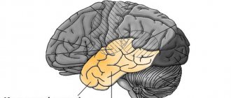
One third of the total mass of the human cerebral hemispheres is in the frontal lobes. If blood circulation is disrupted in this particular part of the brain, then, first of all, all cognitive (cognitive) processes suffer.
The main causes of impaired blood flow in the vessels of the frontal lobes of the brain are exacerbations of hypertension, atherosclerosis, some congenital pathological diseases of blood vessels, poor blood clotting, and a tendency to thrombus formation. All this leads to a stroke. Depending on its mechanism of action, a stroke can be ischemic or hemorrhagic. Further, in turn, the stroke leads to the development of frontal syndrome. It is important to note that stroke is not the only cause of this disease. However, symptoms may
Symptoms of frontal stroke
Frontal stroke most often manifests itself in the form of general cerebral symptoms:
- A person experiences acute pain in the front of the head, nausea and vomiting.
- Dizziness leads to loss of consciousness.
- A high body temperature may rise.
Specific symptoms of frontal stroke:
- Rudimentary reflexes appear: sucking, grasping, searching (in the case of extensive damage to the frontal lobes);
- A person loses the ability to control his own actions;
- The sense of self-awareness is lost;
- There are motor and speech disorders;
- The ability for abstract thinking and planning is lost;
- The functions of memory, attention and will of a person are impaired;
- The victim is unable to focus on anything, cannot solve complex problems, make logical connections, form concepts, etc.
Symptoms may also vary depending on the location of the damage. In case of a stroke on the left side of the frontal part of the brain, a person's verbal behavior is impaired. He is unable to quickly remember and name familiar objects, and cannot speak at a fast pace. If the damage occurs on the right side of the frontal part of the brain, then nonverbal fluency is impaired.
It is believed that lesions in the prefrontal region of the brain lead to disruption of a person’s executive functions. A person who has suffered a stroke of the frontal lobes may retain some motor functions, intelligence and perception, but at the same time the behavior and the very personality of the victim are distorted. Often such consequences are unnoticeable while the patient is still being treated in a medical facility. But over time, these deviations will become more and more apparent. The doctor’s task is to collect a complete history of the onset and course of the disease in order to subsequently correctly prescribe the necessary therapy.
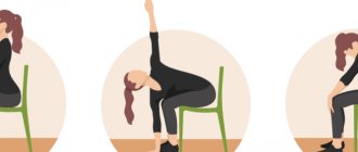
Frontal syndrome (Frontal lobe syndrome)
A mild degree of damage is manifested in a decrease in the patient’s interests, his inattention, and inactivity. Isolated frontal syndrome is not accompanied by paresis or sensory disorders. Habitual simple actions are completely preserved; difficulties arise when it is necessary to perform a complex multicomponent action or a given sequence of movements. Purposeful activity is interrupted by side impulsive actions. For example, seeing a bell button, the patient unconsciously presses it, implementing a habitual movement under the influence of a momentary impulse. Similarly, when cooking soup, the patient can put any inedible object that comes to hand into the pan.
Characteristic is “getting stuck” (perseveration) on performing a certain action: repeating a question, reading the same phrase, repeatedly squeezing a given hand, etc. The most demonstrative perseverations are when trying to draw a series of geometric figures according to a model. The first 2-3 figures can be displayed correctly, then the last figure is repeated. With more severe violations, an attempt to draw a circle leads to repeated repetition of the action with the loss of the ability to stop it independently.
With significant pathological processes, frontal syndrome occurs with astasia - a violation of the ability to maintain a certain body position (standing, sitting), abasia - the inability to walk. At the same time, in the prone position, movements are preserved in full. Disinhibition of oral automatisms is often observed, leading to the appearance of constant smacking and stretching of the lips with a tube. A grasping reflex is noted: the patient squeezes an object placed in his palm into a fist.
Severe apathetic-abulic syndrome is accompanied by a profound breakdown of voluntary motor activity. Patients are unable to initiate action, for example, when thirsty, they cannot make a request or take a nearby glass of water. Active speech is sharply reduced, answers to questions are monosyllabic, echolalia (repetition of the interlocutor’s phrases) is typical. A distinctive feature is the impossibility of both starting and stopping movement. Patients do not take the object offered to them; when placing it in their hand, they squeeze it tonically or repeatedly, unable to stop the action they have started. The automatic repetition of a motor act determines the tendency of patients to constantly fiddle with the edge of the bed, scratch the wall next to the bed, and fidget with their fingers.
Mental disinhibition syndrome is characterized by increased agitation, excessive speech production, and motor activity. Actions are aimed primarily at satisfying biological needs; there are no moral and ethical restrictions. Patients are euphoric, constantly making jokes, making puns, and fooling around. Behavior is often devoid of common sense and can be antisocial and aggressive. There is no criticism of one’s own condition.
Consequences of frontal stroke
The consequences of a frontal stroke can be expressed in the form of frontal syndrome. There are two types of this disease: abulic and disinhibited.
The abulic type leads to the loss of a person’s ability to think creatively, initiative, and curiosity. Often there is a disturbance in the emotional background, manifested in the form of apathy and indifference.
The disinhibited type of frontal syndrome is directly opposite to the abulic type - impulsive behavior occurs, a person loses common sense in their actions, and is unable to foresee the consequences of their actions. Memory and thinking are completely preserved. Thus, a person is able to characterize his actions in possible situations, but in practice he will act completely inadequately and unpredictably.
Clinical Brain Institute Rating: 4/5 — 10 votes
Share article on social networks
Symptoms of focal lesions of the cerebral hemispheres. Symptoms of a frontal lobe tumor
Similar symptoms can occur with damage of another etiology: vascular pathology, intoxication, so-called. endogenous psychoses, brain injuries, neuroinfections, etc.
The anterior parts of the frontal lobes, which are about 2 billion neurons, belong to the practically “silent” areas of the brain (in neurological terms). Presumably, it is the poles of the frontal lobes of the brain that perform or, on the contrary, are unable to perform the most complex functions when damaged, such as the integration of mental processes into a single psychological structure, self-awareness, development and functioning of personal structures, its higher emotions, aesthetic, intellectual and moral qualities, reflection and self-knowledge, goal setting and planning, forecasting and creativity, the formation of algorithms for intellectual activity, etc. In the literature there are descriptions of clinical cases when the so-called. “silent” tumors are manifested by schizophrenia-like symptoms (with disturbances in thinking in the form of its “slipping”, lack of focus, passing responses and fragmentation), loss of internal mental unity, i.e. discordance or schizis (Greek discordare - not to correspond; schizo - splitting ), development of disorders of self-perception, in particular depersonalization, etc.
With frontobasal tumors, disorders such as moria (euphoria, disinhibition, foolishness; Greek moria - stupidity), as well as pseudoparalytic syndrome (mania with absurd delusions of grandeur or depression with fantastic nihilistic delusions) have been described. Tumors of the convexital surface of the frontal lobes often manifest as apathy, aspontaneity (apathetic-abulic syndrome), lethargy, and stuporous states.
Convexital tumors in the premotor zone manifest themselves as adversive (Latin adversio - turning) and other epileptic seizures.
Tumors in Broca's speech area (posterior parts of the inferior frontal gyrus of the dominant hemisphere) cause motor aphasia (articulation disorders, agrammatism, telegraphic speech - phrases of 2-3 words, perseveration - stuck speech reaction, paraphasia - substitution of phonemes, syllables, words).
With tumors in the area of the anterior central gyrus, central paresis and paralysis occur on the other side of the body, and if both gyri are affected, paraparesis and paraparalysis occur (Greek paralyo - relax; paresis - weakening). Let us recall that central paralysis (paresis), which also occurs when the pyramidal tracts are damaged, is characterized by: 1) a reflex increase in muscle tone of the plastic type; 2) increased tendon and periosteal reflexes and, as a consequence, the appearance of clonus; 3) pathological and protective reflexes; 4) pathological synkinesis; 5) absence of muscle degeneration.
Irritation of the neurons of this gyrus is manifested by clonic convulsions, observed, in particular, with Kozhevnikov epilepsy, Jackson epilepsy (“motor” type of the disease), seizures in the form of nodding and pecking. Tonic convulsions may also occur, as, for example, with infantile spasms (infantile convulsions of the “eastern greeting”).
Tumors of the posterior third of the middle frontal lobe of the dominant hemisphere lead to the development of agraphia (a - prefix of negation; Greek grapho - to write) - loss of the ability to write due to loss of memory for the image of graphic signs.
A tumor in the area of the posterior part of the superior frontal gyrus leads to the development of cortical gaze palsy - loss of the ability to conjugately turn the head and eyes in the direction opposite to the localization of the lesion.
When the tumor is localized in the inferior frontal gyrus of the dominant hemisphere, dynamic aphasia occurs - loss of speech initiative, the urge to speak, up to mutism (Latin mutus - mute).
When the opercular region (located under the anterior and posterior central gyri) is damaged, oral apraxia develops (Latin operculum - tire; orum - mouth; a - prefix of negation; Greek praxis - action) - loss of the ability to voluntarily perform complex facial acts, for example, stick out the tongue , whistle, pretend to kiss. Irritation of neurons in this area is manifested by opercular automatisms - involuntary movements of chewing, slurping, smacking, swallowing, licking, etc.; hiccups may occur.
Parakinesis (Greek para - deviation from something, near, nearby; kinesis - movement), or Jacobi's symptom, is manifested by complex automated gestures that outwardly resemble expedient actions. These are, for example, rubbing, stroking, patting, feeling, grabbing. The disorder occurs when neurons in the frontal lobe of the brain (presumably located near the lower part of the anterior frontal gyrus) are stimulated. In children, “frontal” epileptic seizures have been described, manifested by unusual postures and other expressive acts, usually accompanied by loss of consciousness.
If the frontal cortex is damaged, frontal ataxia can also occur - impairment of standing and walking to the degree of astasia and abasia (Greek a - prefix of negation; ataxia - disorder; stasis - standing; basis - step). When standing, the patient sways, he leans to the sides or backwards. When walking, he loses his balance, especially when turning, when starting or stopping a movement, and sometimes his body leans backward. The gait is made “fox-like” - the feet are placed on the same line. In severe cases, gait apraxia occurs—loss of walking skill. There are no symptoms of cerebellar damage (nystagmus, dysmetria, etc.).
With frontal tumors, ideational and ideomotor apraxia can also occur - loss of practical skills caused by an intellectual disorder. In the first case, the patient loses the ability to draw up a plan or program for upcoming spontaneous activity (automated skills are preserved). In the second case, the patient loses the ability to perform actions on instructions from the outside (for example, at the doctor’s request, clench a fist, light a match). The disorder occurs when the premotor cortex of the dominant hemisphere is damaged.
What is described here represents only a small part of the more or less studied disorders that occur when the frontal lobes are damaged and their connections with other brain structures. The same should be said about the symptoms of damage to other areas of the cerebral cortex.
Return to Contents
Programs:
Assessment of rehabilitation potential
Movement restoration
Rehabilitation after stroke
Restoration of cognitive functions
Early (resuscitation) rehabilitation
ADHD symptoms
Parents often get angry with their child, but this is strictly forbidden! Why? I will try to explain it to you from the point of view of neuroscience.
1. Children in this group have hypofunction of the frontal lobes of the brain (the frontal lobes are responsible for motivation, planning, goals, goal-directed behavior, and also have inhibitory functions).
2. It was found that in such children the ratio of central nervous system neurotransmitters (dopamine, serotonin, norepinephrine), as well as N-acetylaspartate, glutamate/glutamine, etc. is disturbed.
3. When conducting an EEG, indirect signs of disorders in ADHD are often recorded: more slow waves and fewer beta waves compared to the norm, the activation reaction is often not expressed.
That is, it is clear that the child is not to blame for this, he is endowed with such a brain and he has to live with such a controlling central nervous system, and the fact that we get irritated, shout, humiliate and punish him will not help, but will only aggravate the problem.
Hemianopsia - symptoms and treatment
Clinical manifestations will depend on which part of the chain is affected. This will manifest itself in pathological changes in visual fields. It is immediately necessary to clarify that the field of view is the space within which, with a fixed gaze, objects are visible simultaneously.
Every person has central and peripheral vision. The central part of the retina, the macula, is responsible for the central part, and the rest of the retina is responsible for peripheral vision. Peripheral vision is very important. Thanks to him, a person orients himself in space. If peripheral vision is lost, even with preserved central (primary) acuity, it is very difficult for a person to navigate in space: he stumbles upon every object that does not fall into the point of fixation of the gaze. It is difficult for such people to work and move, which significantly reduces the quality of life [4].
The peripheral field of vision has its own boundaries; depending on the direction of view, it varies from 50° to 90°. Changes in visual fields play a key role in topical diagnosis of the level of damage and help to guess where the lesion is located. The lesion may affect different components of the visual analyzer.
Optic nerve damage
Complete damage to the optic nerve causes complete blindness of the eye (amaurosis). With partial damage, partial loss of function develops - subatrophy, which leads to changes in visual fields, the appearance of scotomas (blind sectors or islands in the visual field)
Optic chiasm lesion
The topographical features of the chiasm explain its vulnerability in various intracranial pathologies (pituitary tumors, dilatation of the third ventricle, increased intracranial pressure, sclerosis of the cerebral part of the internal carotid artery, etc.). Total damage to the chiasm causes complete bilateral blindness.
"Like a horse in blinkers"
More often (77% of cases), the chiasm is partially damaged [12]. As a rule, this occurs when a large tumor of the pituitary gland, growing upward, damages the diaphragm of the sella turcica and compresses the fibers in the middle section of the decussation, that is, it affects the crossing nerve fibers from the inner nasal halves of the retinas of both eyes. In this case, the external (temporal) visual fields will fall out and so-called temporal , or bitemporal, hemianopsia will occur. This type refers to heteronymous hemianopsia, which means loss of different halves of vision in both eyes. According to other sources, with pituitary adenoma, also with its upward growth, the development of bitemporal hemianopia is observed in 50-52% of cases [11]. Patients in this case feel like a “horse in blinders” because they are deprived of peripheral vision [2].
"The nose is missing"
If only the outer parts of the chiasm are damaged (for example, with aneurysms of the carotid arteries), the temporal halves of the retinas of both eyes will be dark. There will also be a different name, but already binasal hemianopsia with loss of internal visual fields. Many patients in this case cannot see their nose and describe the appearance of a black spot in front of the nose.
Damage to the cortical visual analyzer
Much more common are homonymous hemianopsias, which appear with damage to the optic tracts, thalamus, internal capsule in its posterior section and the occipital lobe [2][3].
“I see in halves”
Starting from the optic tract, the conduction and perception of irritation occurs in the same way: in the right paths of the optic tract - from the right halves of the retinas of both eyes, in the left - from the left. With disturbances in this zone, homonymous hemianopsia of the opposite visual fields occurs. For example, a lesion on the left causes right-sided hemianopsia of the same name, etc.
When both cuneus are damaged, lower hemianopsia occurs, while the upper one develops when the lingual gyrus (gyrus linguales) is damaged. However, they are rare.
If the cortical projection visual area or the visual pathways leading to it are not completely damaged, partial hemianopsia may appear. Thus, if the left cuneus is damaged, only the upper left quadrants of the retina will be “blind”, and, accordingly, only the lower right quadrants will fall out of the visual field.
Damage to the cerebral cortex
If the pathological process affects the calcarine groove, the patient experiences visual hallucinations in the form of simple photopsies (moving shiny dots, lines, figures) in opposite fields of vision. When the process is localized in the area of the superior-lateral surface, visual agnosia occurs—the inability to recognize objects [5].
b) Syndrome of disturbance of spatial synthesis
Also known as “ TPO syndrome ” is a syndrome of damage to the tertiary temporo-parieto-occipital cortex, which provides simultaneous (simultaneous) analysis and synthesis at a higher supramodal level (“quasi-spatial” according to Luria).
What is the manifestation of damage to the SRW zone?
Damage to the TPO zone manifests itself in disturbances in orientation in external space (especially on the right - left), defects in the spatial orientation of movements and visual spatial actions (constructive apraxia).
In visual-constructive activity, lateral differences are observed, which are easily detected in tests for drawing (or copying) various objects.
Significant differences occur when drawing (copying) real objects (house, table, person) and schematic images (cube or other geometric structures).
At the same time, it is important to evaluate not only the final result of performing a visual-constructive task, but also the dynamic characteristics of the execution process itself.
In the process of drawing (copying), patients with damage to the TPO zone of the right hemisphere of the brain perform a drawing, first depicting its individual parts, and only then bringing it to the whole.
With left-hemisphere lesions, visual-constructive activity unfolds in the opposite direction: from the whole to the details. At the same time, patients with damage to the right hemisphere tend to draw realistic parts of the picture (hair, a collar on a person, crossbars at a table, curtains, a porch at a house, etc.), while patients with left hemisphere tend to draw schematic images.
With right-hemisphere lesions, visual-constructive activity suffers more deeply, as evidenced by a violation of the integrity of the copied or independently depicted picture. Often parts are taken outside the contour and “attached” to it in random places. Structural errors such as lack of closure of the figure, violation of symmetry, proportions, and the relationship between the part and the whole are quite often observed. The presence of a sample not only does not help patients with damage to the right hemisphere (unlike those with left hemisphere), but often complicates and even disorganizes visual-constructive activity.
In addition to the listed symptoms, when the SRW zone is damaged, the following symptoms appear:
- agraphia,
- mirror copying,
- acalculia,
- finger agnosia,
- speech disorders (“semantic aphasia”, “amnestic aphasia”).
Violations of logical operations and other intellectual processes are noted. Patients are characterized by difficulties in operating with logical relationships, which require, for their understanding, the correlation of the elements included in them in some conditional, non-visual space (quasi-space).
The latter include specific grammatical constructions, the meaning of which is determined by the endings of words (father’s brother, brother’s father), ways of their arrangement (the dress touched the oar, the oar touched the dress), prepositions reflecting the turn of events in time (summer before spring, spring before summer), discrepancy between the actual course of events and the order of words in a sentence (I had breakfast after reading the newspaper), etc.
Intellectual disorders are manifested by disturbances in visual-figurative thought processes (such as mental manipulation of three-dimensional objects or tasks on “technical” thinking). Such patients cannot read a technical drawing or understand the structure of a technical mechanism.
The main manifestations also include disorders associated with operations with numbers (arithmetic problems).
Understanding of numbers is associated with a rigid spatial grid of placing the digits of units, tens, hundreds (104 and 1004; 17 and 71); operations with numbers (counting) are possible only if the scheme of the number and the “vector” of the operation performed are kept in memory (addition - subtraction; multiplication - division).
Solving arithmetic problems requires understanding the conditions containing logical comparative constructions (more - less by so many times, so many times, etc.).
All of these disorders are especially pronounced in left-sided lesions (in right-handed people).
With right-sided lesions in TPO syndrome there are no phenomena of semantic aphasia; Disturbances in counting and visual-figurative thinking become somewhat different.