Brain and spinal cord
Spinal cord
It is a nerve cord lying in the spinal canal formed by the vertebrae. Extends from the foramen magnum to the lumbar spine. At the top it passes into the medulla oblongata, at the bottom it ends with a conical point with a terminal filament.
The spinal cord is covered by several membranes: dura mater, arachnoid and pia mater. Between the arachnoid and soft membranes, cerebrospinal fluid circulates - cerebrospinal fluid, which surrounds the spinal cord and takes an active part in the metabolism of the spinal cord.
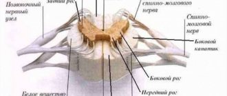
In cross section, the spinal cord (SC) resembles a butterfly. In the center is the gray matter, consisting of the cell bodies of neurons. At the periphery there is white matter, which is formed by the processes of neurons.
In the gray matter of the SC, there are two anterior projections (anterior horns), two lateral ones (lateral horns) and two posterior ones (posterior horns). In the next article we will study reflex arcs, so this knowledge will be very useful to us. The horns of the gray matter contain neurons that are part of the reflex arcs.
Numerous nerve fibers approach the posterior horns of the spinal cord, which unite to form bundles - the dorsal roots. Numerous nerve fibers emerge from the anterior horns of the spinal cord and form the anterior roots.
The white matter consists of numerous nerve fibers, bundles of which form cords. The spinal cord pathways are divided into ascending - from receptors to the brain, and descending - from the brain to effector organs. 31 pairs of spinal nerves arise from the spinal cord.
The spinal cord has two important functions:
- Reflex
- Conductor
Due to the bodies of neurons, which are located in the gray matter of the spinal cord and are part of the reflex arcs that provide reflexes.
Due to the presence of white matter in the spinal cord, which includes numerous nerve fibers that form bundles and cords around the gray matter.
Brain and its parts
We move on to the study of the human brain, the complex main organ of the central nervous system, located in a reliable bone container - the skull. The average brain weight ranges from 1300 to 1500 grams.
Let me note that the weight of the brain has nothing to do with intellectual abilities: for example, Albert Einstein’s brain weighed 1230 grams - less than that of the average person. Intelligence is rather determined by the complexity and ramification of the neural networks of the brain, but not by mass.
The human brain is divided into five sections: medulla oblongata, posterior (pons and cerebellum), middle, intermediate and terminal. The most ancient sections - the medulla oblongata, posterior and middle - form the brain stem, which resembles the structure of the spinal cord. Sometimes the intermediate section is also referred to as the brainstem. 12 pairs of cranial nerves arise from the brain stem.
The telencephalon differs from the structure of the brain stem; it is a huge accumulation (about 16 billion) of neurons that form the cerebral cortex (CCH). Neurons are arranged in several layers, their processes form thousands of synapses with other neurons and their processes. The centers of higher nervous activity - memory, thinking, speech - are located in the KBP.
We begin a fascinating journey through the parts of the brain. It is fundamentally important for you to separate and remember the functions of the various departments; for this, be sure to use your imagination!)
- Medulla
- Hindbrain (pons and cerebellum)
- Midbrain
- Diencephalon
- Finite brain
The most ancient part of the brain. Remember that it regulates vital functions: the cardiovascular system, respiratory and digestive processes. The centers of protective reflexes - vomiting, sneezing, coughing - are concentrated here.
The pons Varoliev performs a conductive function: all descending and ascending nerve pathways pass through the bridge. It also controls the work of the facial and chewing muscles of the face and the lacrimal gland.
The cerebellum has its own hemispheres connected to each other. The cerebellar cortex is formed by gray matter, the subcortical nuclei are surrounded by white matter.
The cerebellum takes part in the coordination of voluntary movements, helps maintain body position in space, regulates tone and balance. Thanks to the cerebellum, our movements are clear and smooth.
In the midbrain there are the superior (anterior) and inferior (posterior) tubercles of the quadrigeminal. The upper tubercles of the quadrigeminal are responsible for the visual orientation reflex, and the lower ones are responsible for the auditory orientation reflex.
What is the visual orientation reflex expressed in? Imagine walking into a dark room. In her corner the screen shines comfortably, the website (of course) Studarium is visible =) And then the visual orientation reflex begins: You move your eyes, turn your head in the direction of the source of intellectual light. At the same time, do not forget to regulate the size of the pupil and the accommodation of the eyes - all this is a visual orientation reflex.
The auditory orientation reflex is also necessary for us. It’s good if, while reading the textbook now, you are in silence. Suddenly your phone starts ringing: you immediately stop reading and head towards the source of the sound - the phone. Thanks to this orienting reflex, we can determine the location of the sound source relative to us (left, right, behind, in front).
The midbrain also performs a conductor function and is involved in the regulation of muscle tone and body posture.
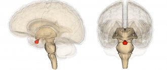
Let me remind you that the hypothalamus we studied, the associated pituitary gland, pineal gland and thalamus belong to the diencephalon. You know that the hypothalamus controls the pituitary gland, the conductor of the endocrine glands, therefore the functions of the hypothalamus are: regulation of the metabolism of proteins, fats and carbohydrates, as well as water-salt metabolism.
In addition, the hypothalamus controls the sympathetic and parasympathetic systems, regulates body temperature, and is responsible for sleep-wake cycles. The hypothalamus contains the centers of hunger and satiety.
Consists of subcortical structures and cerebral cortex (CBC). The surface of the KBP reaches an average of 1.5-1.7 m2. Such a large area is due to the fact that the CBP forms convolutions - elevations of the brain matter, and grooves - depressions between the convolutions.
Cerebral cortex
The cortex has several layers of cells, between which numerous branched connections are formed. Despite the fact that the cortex functions as a single mechanism, its different parts analyze information from different peripheral receptors, which I.P. Pavlov called the cortical ends of analyzers.
The cortical representation of the visual analyzer is located in the occipital lobe of the CBP; it is in connection with this that, when falling on the back of the head, a person sees “sparks from the eyes” when the neurons of this lobe are excited mechanically, as a result of the impact.
The cortical representation of the auditory analyzer is located in the temporal lobe of the cerebral cortex.
Remember that the cortical representation of the motor analyzer - the motor zone - is located in the anterior central (precentral) gyrus, and the representation of the skin analyzer - the sensory zone - is in the posterior central (postcentral) gyrus.
Think about it! When making any voluntary (conscious) movement, a nerve impulse arises precisely in the neurons of the precentral gyrus, from where it begins its long journey through the brain stem, spinal cord and finally reaches the effector organ.
Impulses from skin receptors reach the neurons of the postcentral gyrus - the sensory department, thanks to which we receive information from them and are aware of our own sensations.
The number of neurons in these convolutions allocated for different organs is not the same. Thus, the projection area of the fingers of the hand takes up a lot of space, making fine movements of the fingers possible. The projection area of the torso muscles is much smaller than the area of the fingers, since the movements of the torso are more uniform and less complex.
The areas of the brain that we studied, in which the transformation and analysis of incoming information occurs, are called associative zones of the CBP. These zones connect different parts of the CBP, coordinate its work, and play a crucial role in the formation of conditioned reflexes.
Our conscious activity lies within the framework of the cerebral cortex: any conscious movement, any sensation (temperature, pain, tactile) - everything has representations in the CBP. The cortex is the basis for communication with the external environment and adaptation to it. QBP also lies at the foundation of the thinking process. In general, you understand how highly you should value it and how well you should know this topic 
You've probably heard that the right and left hemispheres are functionally different. The left hemisphere contains the mechanisms of abstract thinking (language abilities, analytical thinking, logic), and the right hemisphere contains the mechanisms of concrete figurative thinking (imagination, parallel processing of information). With injuries or damage to the left hemisphere, speech may be impaired.
Diseases
Depending on the level of damage to the spinal cord during trauma, the picture of neurological disorders manifests itself differently. The higher the level of damage, the more nerve pathways are “cut off” from the brain. So, for example, with a lumbar injury, arm movements are preserved, but with a cervical injury, arm movements are impossible.
Sometimes after a stroke (bleeding in brain tissue) or injury, paralysis (complete lack of movement) develops on one side of the body. Knowing the anatomy, you can conclude: if movements are lost in the right arm and leg, then the stroke occurred on the left.
Why does this pattern exist? The fact is that the nerve fibers running from the precentral gyrus to the working organs - the muscles - form the so-called physiological cross at the border of the medulla oblongata and the spinal cord. That is, to put it simply: part of the nerves that came from the left hemisphere pass to the right side and vice versa - nerves from the right hemisphere pass to the left side.
© Bellevich Yuri Sergeevich 2018-2021
This article was written by Yuri Sergeevich Bellevich and is his intellectual property. Copying, distribution (including by copying to other sites and resources on the Internet) or any other use of information and objects without the prior consent of the copyright holder is punishable by law. To obtain article materials and permission to use them, please contact Yuri Bellevich
.
What does the human brain look like?
Brain anatomy is a relatively young science, as it was banned for a long time due to laws prohibiting the dissection and examination of human organs and the head.
The study of the topographic anatomy of the brain in the head area is necessary for accurate diagnosis and successful treatment of various topographic anatomical disorders, for example: skull injuries, vascular and oncological diseases. To imagine what a human GM looks like, you first need to study their appearance.
In appearance, the GM is a yellowish gelatinous mass enclosed in a protective shell, like all organs of the human body, they consist of 80% water.
The large hemispheres occupy almost the volume of this organ. They are covered with gray matter or cortex - the highest organ of human neuropsychic activity, and inside - with white matter, consisting of processes of nerve endings. The surface of the hemispheres has a complex pattern, due to convolutions going in different directions and ridges between them. Based on these convolutions, it is customary to divide them into several sections. It is known that each of the parts performs specific tasks.
In order to understand what a person's brain looks like, it is not enough to examine its appearance. There are several study methods that help to study the brain from the inside in section.
- Sagittal section. It is a longitudinal incision that passes through the center of a person’s head and divides it into 2 parts. It is the most informative research method; it is used to diagnose various diseases of this organ.
- The frontal section of the brain looks like a cross section of the large lobes and allows you to see the fornix, hippocampus and corpus callosum, as well as the hypothalamus and thalamus, which control the vital functions of the body.
- Horizontal section. Allows you to examine the structure of this organ in the horizontal plane.
The anatomy of the brain, as well as the anatomy of the human head and neck, is a rather difficult subject to study for a number of reasons, including the fact that their description requires studying a large amount of material and having good clinical training.
BRAIN RESEARCH
Brain research is difficult for two main reasons. Firstly, direct access to the brain, which is well protected by the skull, is not possible. Secondly, brain neurons do not regenerate, so any intervention can lead to irreversible damage.
Despite these difficulties, research on the brain and some forms of its treatment (primarily neurosurgery) have been known since ancient times. Archaeological finds show that already in ancient times man performed craniotomy to gain access to the brain. Particularly intensive brain research was carried out during periods of war, when a variety of traumatic brain injuries could be observed.
Brain damage as a result of a wound at the front or an injury received in peacetime is a kind of analogue of an experiment in which certain areas of the brain are destroyed. Since this is the only possible form of “experiment” on the human brain, experiments on laboratory animals became another important method of research. By observing the behavioral or physiological consequences of damage to a particular brain structure, one can judge its function.
The electrical activity of the brain in experimental animals is recorded using electrodes placed on the surface of the head or brain or inserted into the brain substance. In this way, it is possible to determine the activity of small groups of neurons or individual neurons, as well as to detect changes in ion flows across the membrane. Using a stereotactic device, which allows you to insert an electrode into a certain point of the brain, its inaccessible deep parts are examined.
Another approach is to remove small sections of living brain tissue, then maintain it in the form of a slice placed in a nutrient medium, or the cells are isolated and studied in cell cultures. In the first case, it is possible to study the interaction of neurons, in the second - the vital activity of individual cells.
When studying the electrical activity of individual neurons or their groups in different areas of the brain, the initial activity is usually recorded first, then the effect of a particular influence on cell function is determined. Another method uses an electrical impulse through an implanted electrode to artificially activate nearby neurons. This way you can study the effect of certain areas of the brain on other areas of the brain. This method of electrical stimulation has proven useful in the study of brainstem activating systems passing through the midbrain; it is also used when trying to understand how learning and memory processes occur at the synaptic level.
Already a hundred years ago it became clear that the functions of the left and right hemispheres are different. The French surgeon P. Broca, observing patients with cerebrovascular accident (stroke), discovered that only patients with damage to the left hemisphere suffered from speech disorders. Subsequently, studies of hemispheric specialization were continued using other methods, such as EEG recording and evoked potentials.
In recent years, sophisticated technologies have been used to obtain images (visualization) of the brain. Thus, computed tomography (CT) has revolutionized clinical neurology, making it possible to obtain intravital detailed (layer-by-layer) images of brain structures. Another imaging technique, positron emission tomography (PET), provides a picture of the metabolic activity of the brain. In this case, a person is injected with a short-lived radioisotope, which accumulates in various parts of the brain, and the more, the higher their metabolic activity. Using PET, it was also shown that speech functions in the majority of those examined were associated with the left hemisphere. Because the brain operates using a huge number of parallel structures, PET provides information about brain function that cannot be obtained using single electrodes.
As a rule, brain studies are carried out using a complex of methods. For example, the American neurobiologist R. Sperry and his colleagues, as a therapeutic procedure, performed transection of the corpus callosum (a bundle of axons connecting both hemispheres) in some patients with epilepsy. Subsequently, the specialization of the hemispheres was studied in these split-brain patients. It was found that the dominant (usually left) hemisphere is primarily responsible for speech and other logical and analytical functions, while the non-dominant hemisphere analyzes the spatiotemporal parameters of the external environment. So, it is activated when we listen to music. The mosaic pattern of brain activity suggests that numerous specialized areas exist within the cortex and subcortical structures; the simultaneous activity of these areas supports the concept of the brain as a parallel processing computing device.
With the advent of new research methods, ideas about brain function are likely to change. The use of devices that make it possible to obtain a “map” of the metabolic activity of various parts of the brain, as well as the use of molecular genetic approaches should deepen our knowledge of the processes occurring in the brain. See also NEUROPSYCHOLOGY.
What, according to scientists, constantly arises in the human brain
Experts who study the psychological capabilities of the brain believe that the performance of cognitive and mental functions occurs as a result of biochemical currents, however, this theory is currently being questioned because this organ is a biological object and the principle of mechanical action does not allow us to fully understand its nature.
The brain is a kind of steering wheel of the whole organism, performing a huge number of tasks every day.
The anatomical and physiological features of the structure of the brain have been the subject of study for many decades. It is known that this organ occupies a special place in the structure of the human central nervous system (CNS), and its characteristics are different for each person, so it is impossible to find 2 people who think absolutely alike.
Cortical areas
On the surface of the hemispheres there are permanent grooves dividing them into 5 lobes. The frontal lobe (lobus frontalis) lies in front of the central sulcus (sulcus centralis). The occipital lobe extends from the central to the parieto-occipital sulcus (sulcus parietooccipitalis).
Frontal lobe regions
The main motor area is located in front of the central sulcus, where there are pyramidal cells, the axons of which form the pyramidal (cortical) path. These paths provide precise and comfortable movements of the body, especially the forearms, fingers, and facial muscles.
Premotor cortex. This area is located in front of the primary motor area and controls more complex free activity movements that depend on sensory feedback - grasping objects, moving over obstacles.
Broca's speech center is located in the lower part, usually the left or dominant hemisphere. Broca's center in the left hemisphere (if it is dominant) controls speech, in the right hemisphere it supports the emotional coloring of the spoken word; this area is also involved in short-term memory for words and speech. Broca's center is associated with the preferential use of one hand for work - left or right.
The visual area is the motor part that controls the required rapid eye movements when viewing a moving target.
The olfactory region is located at the base of the frontal lobes and is responsible for the perception of smell. The olfactory cortex connects to the olfactory areas in the lower centers of the limbic system.
The prefrontal cortex is a large area of the frontal lobe responsible for cognitive functions: thinking, perception, conscious storage of information, abstract thinking, self-awareness, self-control, perseverance.
Areas of the parietal lobe
The sensitive area of the cortex is located just behind the central sulcus. Responsible for the perception of general bodily sensations - the perception of skin (touch, warmth, cold, pain), taste. This center is capable of localizing spatial perception.
Somatosensitive area - located behind the sensitive one. Participates in recognizing objects based on their shape based on previous experience.
Occipital lobe regions
The main visual area is located at the end of the occipital lobe. It receives visual information from the retina and processes information from both eyes together. Here the orientation of objects is perceived.
Associative visual area - located in front of the main one, cooperates with it to determine the color, shape, and movement of objects. It also communicates with other parts of the brain through the anterior and posterior pathways. The anterior tract runs along the lower edge of the hemispheres and is involved in word recognition during reading and face recognition. The posterior pathway passes into the parietal lobe and is involved in spatial connections between objects.
Temporal lobe areas
The hearing zone and vestibular region are located in the temporal lobe. There is a distinction between the main and associative areas. The main one perceives volume, pitch, rhythm. Associative – based on memorizing sounds and music.
Speech area
The speech area is a broad area associated with speech. The left hemisphere is dominant (in right-handed people). To date, 5 areas have been identified:
- Broca's area (speech formation);
- Wernicke's area (speech understanding);
- lateral prefrontal cortex in front of and below Broca's area (speech analysis);
- area of the temporal lobe (coordination of auditory and visual aspects of speech);
- internal share – articulation, recognition of rhythm, voiced word.
The right hemisphere is not involved in the speech process in right-handed people, but works on the interpretation of words and their emotional connotation.
How does the human brain work?
Scientists all over the world study the brain, its structure and the functions it performs. Over the past few years, many important discoveries have been made, however, this part of the body remains incompletely studied. This phenomenon is explained by the difficulty of studying the structure and functions of the brain separately from the skull.
In turn, the structure of the brain structures determines the functions that its departments perform.
It is known that this organ consists of nerve cells (neurons) connected to each other by bundles of filamentous processes, but how their interaction occurs simultaneously as a single system is still unclear.
A diagram of the structure of the brain, based on the study of a sagittal section of the skull, will help to study the sections and membranes. In this figure you can see the cortex, the medial surface of the cerebral hemispheres, the structure of the trunk, cerebellum and corpus callosum, which consists of the splenium, trunk, genu and beak.
The brain is reliably protected externally by the bones of the skull, and internally by 3 meninges: the hard arachnoid and the soft. Each of them has its own device and performs specific tasks.
- The deep soft membrane covers both the spinal cord and the brain, while it extends into all the cracks and grooves of the cerebral hemispheres, and in its thickness there are blood vessels that feed this organ.
- The arachnoid membrane is separated from the first by a subarachnoid space filled with cerebrospinal fluid (cerebrospinal fluid), which also contains blood vessels. This shell consists of connective tissue, from which thread-like branching processes (cords) extend; they are woven into the soft shell and their number increases with age, thereby strengthening the connection. Between them. Villous outgrowths of the arachnoid membrane protrude into the lumen of the sinuses of the dura mater.
- The hard shell, or pachymeninx, consists of connective tissue and has 2 surfaces: the upper one, saturated with blood vessels, and the inner one, which is smooth and shiny. This side of the pachymeninx is adjacent to the medulla, and the outer side is adjacent to the cranium. Between the dura mater and the arachnoid membrane there is a narrow space filled with a small amount of liquid.
About 20% of the total blood volume circulates in the brain of a healthy person, which enters through the posterior cerebral arteries.
The brain can be visually divided into 3 main parts: 2 cerebral hemispheres, brainstem and cerebellum.
Gray matter forms the cortex and covers the surface of the cerebral hemispheres, and a small amount of it in the form of nuclei is located in the medulla oblongata.
In all parts of the brain there are ventricles, in the cavities of which the cerebrospinal fluid that is formed in them moves. In this case, fluid from the 4th ventricle enters the subarachnoid space and washes it.
Brain development begins while the fetus is in utero, and it is finally formed by the age of 25.
How does the human brain work?
The work of the human brain does not stop even in sleep; it is known that people in a coma also have some parts functioning, as evidenced by their stories.
The main work of this organ is carried out with the help of the cerebral hemispheres, each of which is responsible for a specific ability. It has been noted that the hemispheres are unequal in size and function - the right side is responsible for visualization and creative thinking, usually larger than the left side, responsible for logic and technical thinking.
It is known that men have a larger brain mass than women, but this feature does not affect mental abilities. For example, Einstein's figure was below average, but his parietal area, which is responsible for cognition and the creation of images, was large, which allowed the scientist to develop the theory of relativity.
Some people are endowed with super abilities, this is also the merit of this organ. These features manifest themselves in high speed of writing or reading, photographic memory and other anomalies.
One way or another, the activity of this organ is of great importance in the conscious control of the human body, and the presence of the cortex distinguishes humans from other mammals.