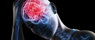
Mechanical damage to the human skull along with its contents, which is manifested by established neurological symptoms, is called traumatic brain injury. Timely first aid for traumatic brain injuries helps save the life and many functions of the victim’s body, so it is extremely important to know all the basics of behavior in such a situation.
Main causes and types of injuries in the skull area
The pathologies under study can arise as a result of many factors, for example, during road traffic accidents, human falls from a height, at work and at home, during active sports.
Content:
- Main causes and types of injuries in the skull area
- Clinical course of the pathology
- First aid
The main types of traumatic brain injuries include closed, open and penetrating injuries to the head. The closed type of such injuries include those in which the aponeurosis remains without damage, but there are injuries or bruises to the head in the soft tissue section. Open craniocerebral injuries are characterized by damage to the aponeurosis and integrity of the skin on the head. Experts consider penetrating injuries to be those that result in disruption of the integrity of the dura mater of the brain.
Combined trauma - symptoms and treatment
A distinctive feature of the treatment of combined trauma is the awareness of its multicomponent nature and severity. It requires the intervention of doctors of narrow specialties, the vigilance of the responsible surgeon and his readiness to expand and supplement the diagnosis at any time.[16]
The intervention of doctors is simultaneous, with priority in compensating for violations of vital functions and life-threatening conditions. This is a collective and well-coordinated work, which is focused on the department for anti-shock measures. Such a department differs from the structures of a medical institution in its expanded capabilities of equipment, movement, convenient layout of the diagnostic unit, focus on the mass of admissions and adaptation to the needs of intensive care.[10]
Characteristic is the simultaneous (simultaneous) work of surgical teams with one patient to carry out emergency operations in different anatomical areas.[17]
The stages of treatment, compliance with the volume of assistance and its timeliness are very important. The organization of medical care for combined trauma is guided by the canons of disaster medicine with the principles of evacuation of victims.
There is a concept called “damage control” that all emergency physicians are familiar with. According to this approach, the victim receives only the minimum amount of medical care that is necessary for his condition.[17] The idea is to provide types of assistance in stages, if necessary - delayed, due to the impossibility of solving all the patient’s problems at once.
Treatment begins with the relief of the most threatening disorders.[20]
Traumatic shock and failure of body systems during combined trauma in themselves pose a great risk to the patient’s life. Each additional manipulation, operation and medical intervention greatly increases the load on the body and aggravates the shock.
Despite the therapeutic effect, any medical intervention also brings stress to the body, including when anesthesia or rough influences are necessary. The benefits of an intervention must outweigh the possible harm of the intervention itself. In order not to aggravate the condition of an unstable patient, only those simple and reliable manipulations are performed that will save his life. Everything else that needs to be done to radically solve the problem is put off “for later.” You can continue when the shock is compensated and the patient is able to tolerate further treatment.[10]
To an outsider, it may seem that a more logical treatment tactic is to “get the job done,” that is, to try to completely heal the traumatic injury. However, this is only true for isolated trauma. In the case of a combined injury, such a race for the ideal result is a gamble that can take the patient’s life.
Algorithms are used according to which, first of all, the pathological processes that cause shock are eliminated: pain, bleeding, impaired ventilation of the lungs. All necessary operations are performed in a minimal volume and using simple technology. In the future, with a good prognosis and stabilization of the condition, doctors postpone operations that become more complicated in terms of volume and nature.[6]
Clinical course of the pathology
More often than others, with traumatic brain injuries, a concussion occurs without macrostructural lesions. Brain damage at the cellular level is reversible and treatable. The main symptoms of a concussion are loss of consciousness for a short time, nausea and vomiting, dizziness, headaches, double vision, sweating, memory loss. From a neurological point of view, there is a violation of symmetry in tendon reflexes, small-scale nystagmus, which under normal conditions disappears a week after the injury. If the victim undergoes a computer or magnetic resonance imaging scan, they will not reveal any pathologies.
With traumatic brain injuries, brain contusion can occur. This pathology leads to hemorrhages and destruction (gross macrostructural disorders) in the brain, accompanied by subarachnoid hemorrhage. Often a brain contusion is accompanied by a fracture of the skull bones. All this together can lead to swelling of the brain.
Manifestations of brain contusion by severity:
- Mild degree - the patient’s loss of consciousness can last up to 20 minutes, after which dizziness, vomiting and nausea, headaches, anterograde or retrograde amnesia occur. Vital functions remain unchanged; cardiovascular changes in the form of hypertension or bradycardia may occur. Neurological symptoms include pyramidal insufficiency, clonic nystagmus, and slight anisocoria.
- In the middle stage, the patient’s unconscious state lasts for several hours, after which severe memory loss occurs, repeated vomiting occurs, and mental health is impaired. Vital functions are impaired, persistent bradycardia, tachypnea with preservation of airway patency, hypertension. From a neurological point of view, nystagmus, imbalance in tendon reflexes and muscle tone, and various meningeal signs are observed. Focal manifestations in the form of ocular and pupillary disorders, changes in speech, and paresis of the limbs are also obvious.
- In severe cases, the patient falls into a coma, which, if not fatal, can last several weeks. Severe violations of vital functions pose a threat to life. The movements of the eyeballs are floating, the rhythm and frequency of breathing are disturbed, bilateral miosis or mydriasis, and convulsions occur. With such traumatic brain injuries, there are fractures of the skull bones in combination with massive subarachnoid hemorrhage.
Compression of the brain substance may occur due to intracranial hematomas that form above or below the soft membrane.
Clinically, compression can manifest itself in the same way as a bruise, but it has a very dangerous consequence. After the victim returns to consciousness, he usually feels better, but this does not last long, until the intracranial hematoma grows in size and begins to compress the brain. After this, the person falls into a coma again, and the prognosis for this state is rarely satisfactory. The functioning of the respiratory and vascular centers is disrupted.
According to the nature and severity of damage to the brain substances, they are distinguished:
Brain concussion
Develops more often with closed craniocerebral injury. It is characterized by functional disorders without morphological changes: loss of consciousness, nausea or vomiting, headache, retrograde amnesia. These disorders are temporary and reversible.
Brain contusion
This type of injury is classified as severe injury. It is characterized by focal disorders associated with damage to brain tissue and can occur not only at the site of application of the traumatic force, but also on the opposite side. As a result, severe circulatory disorders are possible, accompanied by cerebral edema and increased spinal pressure.
Brain compression
It is a severe complication of skull damage. Occurs with intracranial hematoma (bleeding from intracranial vessels), cerebral edema, traumatization by bone fragments of the cranial vault and foreign body, pneumocephalus (accumulation of air in the cranial cavity).
Diffuse axonal brain injury
It is characterized by a long-term (up to 2-3 weeks) comatose state, pronounced brainstem symptoms. There are disturbances in the frequency and rhythm of breathing. Autonomic disorders are pronounced.
Clinical examination of the patient
Recommendations for assessing response in neurocognitive disorders
- Adequate stimulation is carried out only when it is certain that the patient's level of wakefulness is maximum.
- It is necessary to eliminate the causes that cause quantitative disorders of consciousness (taking sedatives, epileptic seizures).
- Attempts to elicit a response to verbal stimulation should not be aimed at eliciting movements, which are often performed reflexively.
- Research into the ability to carry out commands should include the types of motor activity available to the patient.
- Many different behavioral responses must be examined using a diverse range of stimuli.
- The study should be carried out in a calm environment.
- Initial estimates should be verified repeatedly using multiple re-assessments and acceptable response measurement approaches.
- Specific quantification techniques and tools may be useful.
A systematic approach to assessing patients with impaired consciousness includes the following steps:
Assessment of the functioning of the brain stem and other subcortical formations
- Pupillary reactions, blink reflex to visual stimuli.
- Eye movements, gaze deviations.
- Oculovestibular reflexes (oculocephalic reflex, calorimetric test).
- Corneal reflexes.
- Vomiting reflex.
- Breathing pattern.
- Decerebrate postures.
- Other postural reflexes, assessment of muscle tone.
Assessment of cortical functions
- Evaluation of voluntary actions
- Purposeful complex movements (including isolated motor control involving the cortex) compared with postural (decortication, decerebrate) or reflexive or stereotyped, monotonous (subcortical structures mediated) movements. Note: Pilon & Sullivan note different profiles of postural tone relationships for VS and SMS. Patients with VS exhibit classic decerebrate, decortication, and hypotonic postural features, whereas patients with SMS exhibit symmetrical or asymmetrical global flexor postures. However, there is no characteristic reflex posture with absolute diagnostic significance for separating these two groups.
- Involuntary vocal and verbal activity.
- Eye movements (signs of fixation of gaze or tracking of an object, differentiated from involuntary eye movements not related to the stimulus, or fixed gaze); responses to artificial or natural external stimuli.
- Tracking or fixating the gaze on various stimuli (use objects that do not make sounds: family photos, pictures of faces, money, mirror)
- Verbal stimulation (patient's name, commands, greeting): Begin with simple commands that encourage actions within the patient's ability.
- Eye commands: “look here”, “blink twice”.
- Commands for limbs: “show thumb”, “show 2 fingers”, “raise your hand”.
- Commands for the oral muscles: “open your mouth”, “show your tongue”
- Commands for the whole body or axial muscles: “turn your head”, “lean forward”.
- Ask the patient to stop the movement or hold the limb to highlight spontaneous repetitive movements.
- Painful stimulation.
- Assess the localization and purposefulness of defensive reactions, compare them with reflexive and generalized, stereotypical movements, evaluate facial expression
- Responses to random external stimuli.
- Assess attempts to grasp or perform actions with objects surrounding the patient (clothing, objects at hand).
- Observe changes in facial expression as you respond to stimuli such as a familiar voice, dialogue, pictures, music, etc.
- Notice attempts at purposeful movement in bed, in a chair, and while walking.
- Note gestures when communicating emotionally (yes/no signals).