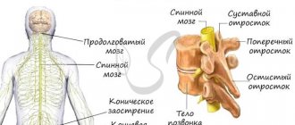
General information
Formed from the anterior end of the primary neural tube.
In embryogenesis, it is divided into 2 parts, one of which gives rise to the telencephalon, the second - the intermediate brain. According to Alexander Luria’s model, it consists of 3 blocks:
- Block regulating brain activity levels. Ensures the implementation of certain types of activities. Responsible for emotional reinforcement of activity based on predicting its results (success - failure).
- Block for receiving, processing and storing incoming information. Participates in the formation of ideas about ways to implement activities.
- Block of programming, regulation and control over the organization of mental activity. Compares the resulting result with the original intent.
The forebrain takes part in the work of all blocks. Based on information processing, it controls behavior. Administrator of higher psychological functions: perception, memory, imagination, thinking, speech.
Anatomy
The structure of a living individual is not easy to describe. Especially such a component as the brain. This universe that exists in everyone continues to hide its secrets. But this does not mean that they are not worth understanding.
Development
The forebrain is formed at 3-4 weeks of prenatal development. By the end of the 4th week of embryogenesis, the telencephalon, diencephalon, and the cavity of the third ventricle are formed from the forebrain.
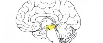
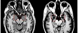
Diencephalon
It consists of the thalamic and hypothalamic regions, which are located on the sides of the third ventricle between the hemispheres and the midbrain.
The thalamic region unites:
- The thalamus is an ovoid formation located deep under the cerebral cortex. The oldest, largest (3-4 cm) formation of the diencephalon;
- The epithalamus is located above the thalamus. It is famous for the fact that it contains the pineal gland. Previously, it was believed that the soul lived here. Yogis associate the pineal gland with the seventh chakra. By awakening the organ, you can open the “third eye”, becoming clairvoyant. The gland is tiny, only 0.2 g. But the benefits for the body are enormous, although previously it was considered a rudiment;
- subthalamus - a formation located below the thalamus;
- metathalamus - bodies located in the posterior part of the thalamus (previously considered a separate structure). Together with the midbrain, they determine the work of the visual and auditory analyzers;
The hypothalamic region includes:
- hypothalamus. Located under the thalamus. Weighs 3-5 g. Consists of specialized groups of neurons. Connected with all departments. Controls the pituitary gland;
- the posterior lobe of the pituitary gland is the central organ of the endocrine system, weighing 0.5 g. Located at the base of the skull. The posterior lobe, together with the hypothalamus, forms the hypothalamic-pituitary complex, which controls the activity of the endocrine glands.
Finite brain
Unites:
- cortical hemispheres. The bark appeared late in the development of the animal world. Occupies half the volume of the hemispheres. Its surface can exceed 2000 cm2;
- corpus callosum - a nerve tract connecting the hemispheres;
- striped body. Located on the side of the thalamus. On a section it looks like repeating stripes of white and gray matter. Promotes regulation of movements, motivation of behavior;
- olfactory brain. Unites structures that differ in purpose and origin. Among them is the central section of the olfactory analyzer;
Evolutionary development
The modern school biology course covers topics from simple to complex. First we talk about cells, protozoa, bacteria, plants, fungi. Later there is a transition to animals and humans. To some extent, this reflects the hypothetical course of evolution. Looking at the structure of, for example, worms, it is easy to notice that it is much simpler than that of humans or higher animals. But these organisms have something important - a nerve ganglion that performs the functions of the brain.
The nervous system is generally extremely complex. It includes not only the brain and spinal cord, but also numerous processes consisting of special cells, as well as all the sensory organs. Thanks to this system, human life is possible in the form it exists. And, of course, the main organ in it is still the brain, which even in itself has a rather complex structure.
Anatomical features
Intermediate
The thalamus is egg-shaped and gray-brown in color. Structural unit - nuclei, which are classified according to functional and compositional characteristics.
The epithalamus consists of several units, the most famous of which is the grayish-reddish pineal gland.
The subthalamus is a small region of gray matter nuclei connected to white matter.
The hypothalamus consists of nuclei. There are about 30 of them. Most are paired. Classified by location.
Posterior lobe of the pituitary gland. The pituitary gland is a round-shaped formation, the location is the pituitary fossa of the sella turcica.
Finite
Unites the hemispheres, corpus callosum and striatum. The largest department by volume.
The hemispheres are covered with gray matter 1-5 mm thick. The mass of the hemispheres is about 4/5 of the mass of the brain. Convolutions and grooves significantly increase the area of the cortex, containing billions of neurons and nerve fibers arranged in a certain order. Underneath the gray matter lies white matter—the processes of nerve cells. About 90% of the cortex has a typical six-layer structure, where neurons are connected through synapses with each other.
From the point of view of phylogenesis, the cerebral cortex is divided into 4 types: ancient, old, intermediate, new. The main part of the human cortex is the neocortex.
The corpus callosum is shaped like a wide strip. Consists of 200-250 million nerve fibers. The largest structure connecting the hemispheres.
PsyAndNeuro.ru
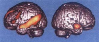
Based on the results of a study of more than 2000 images of the brain, data were obtained indicating the presence of highly reproducible sex differences in the volume of some of its parts. It turned out that the pattern of sex differences in brain volume is consistent with the patterns of gene expression on the sex chromosomes, which were obtained from the results of post-mortem studies of the cerebral cortex. Thus, it can be assumed that sex chromosomes play a role in the formation of sex differences in brain anatomy or in the maintenance of these differences. The results of the study, conducted at the National Institute of Mental Health (NIMH), were published in the Proceedings of the National Academy of Sciences.
The study authors believe that increasing knowledge about sex differences in brain structure is important for understanding the significance of already established sex differences in cognition, behavior and risk of mental illness. “We were inspired by new animal data on sex differences and wanted to try to bridge the existing gap between animal data and our models of sex differences in the human brain,” says co-author Armin Raznahan, M.D., Ph.D. .
It has long been known that there are stable sex differences in subcortical brain structures in mice. Some studies have attributed these anatomical differences primarily to the influence of sex hormones and have emphasized a gonadocentric explanation for sex differences arising during brain development. However, recent studies in mice have found consistent sex differences in cortical structures, and gene expression data suggest that sex chromosomes play a role in the formation of these anatomical sex differences. But while the mouse brain is similar to the human brain in many ways, it was unclear how the findings would apply to humans.
To explore the neurobiological basis of sex differences in the human brain, Raznahan, Siyuan Liu, Ph.D. et al first analyzed neuroimaging data collected by the Human Connectome Project (HCP). After processing data from 976 healthy adults aged 22 to 35 years, consistent sex differences were identified in the volume of certain cortical structures. On average, women had relatively larger cortical volumes in the medial and lateral prefrontal areas, orbitofrontal areas, superior temporal areas, and lateral parietal areas. In men, on average, relatively greater cortical volume was found in ventral temporal regions and occipital regions, including the pole temporal lobe, fusiform gyrus, and primary visual cortex.
Then Liu et al. used two complementary approaches to test the reproducibility of the results. The researchers first conducted 1,000 comparisons, randomly splitting the HCP database into two halves and comparing results in both halves. The results of these comparisons confirmed that the pattern of sex differences in cortical volume is stable. In a second step, the researchers quantified the reproducibility of the HCP data using an independent neuroimaging database from the UK Biobank. Although the two databases differed significantly in both demographic characteristics and methodological approaches, the researchers found that the overall pattern of sex differences in cortical volume in the two databases was largely consistent.
Then Liu et al. compared the anatomical data obtained with publicly available maps of gene expression in the brain, which were compiled based on 1317 postmortem tissue samples from 6 people. It turned out that the spatial distribution of sex differences in cortical volume largely coincides with the spatial distribution of sex chromosome gene expression in the cortex. In particular, cortical areas with relatively high expression of sex chromosome genes are generally thicker in men than in women.
This correspondence with cortical sex chromosome gene expression is also consistent with previous findings from mouse studies and suggests that sex differences in brain anatomy may be at least in part due to genetic mechanisms conserved throughout mammalian evolution. It can also be assumed that sex differences in cortical thickness may be caused by genes located on the X and Y sex chromosomes.
“Men and women differ in many genetic and environmental factors that can potentially influence brain development. Because human experiments are challenging, we often rely on observational data to identify possible genetic or environmental factors influencing sex differences in the brain, says Raznahan. “The fact that we obtained a high level of reproducibility of anatomical sex differences across different groups of men and women, and saw an association between these differences and sex chromosome gene expression, suggests that these differences are likely not primarily due to the environment.”
The researchers also compared the anatomical results with data obtained from more than 11,000 functional neuroimaging studies. It turned out that the areas of the brain where sex differences are observed according to the HCP database, and the areas of the brain responsible for processing faces according to functional neuroimaging studies, largely overlap spatially.
Taken together, these results shed light on mechanisms that may influence sex differences in brain anatomy and point to genetic factors that may contribute to sex differences in behavior and the prevalence of brain disease. Using these correlation data as a roadmap, future studies will be able to more effectively explore the causes and consequences of sex differences in the human brain.
Author of the translation: Lafi N.M.
Source: Integrative structural, functional and transcriptomic analyzes of sex-biased brain organization in humans. Proceedings of the National Academy of Sciences
(2020). DOI: 10.1073/pnase.1919091117
Functions
Mission – organization of mental activity.
Intermediate
Participates in coordinating the work of organs, regulating body movement, maintaining temperature, metabolism, and emotional background.
Thalamus . The main task is to sort information. It works like a relay - it processes and sends data coming from receptors and pathways to the brain. The thalamus affects the level of consciousness, attention, sleep, wakefulness. Supports speech functioning.
Epithalamus . Interaction with other structures occurs through melatonin, a hormone produced by the pineal gland in the dark (therefore, it is not recommended to sleep in the light). A derivative of serotonin - the “happiness hormone”. Melatonin is a participant in the regulation of circadian rhythms, being a natural sleep aid, it affects memory and cognitive processes. It affects the localization of skin pigments (not to be confused with melanin), puberty, and suppresses the growth of a number of cells, including cancer cells. Through connections with the basal ganglia, the epithalamus participates in the optimization of motor activity, and through connections with the limbic system, in the regulation of emotions.
Subthalamus . Controls the body's muscle responses.
Hypothalamus . Forms a functional complex with the pituitary gland and directs its work. The complex controls the endocrine system. The hormones it produces help cope with distress and maintain homeostasis.
Thirst and hunger centers are located in the hypothalamus. The department coordinates emotions, human behavior, sleep, wakefulness, and thermoregulation. Endorphins similar in action to opiates are found here, which help to endure pain.
Finite brain
Hemispheres
They act together with subcortical structures and the brain stem. Main destination:
- Organization of interaction of an organism with the environment through its behavior.
- Consolidation of the body.
Corpus callosum
The corpus callosum received attention after operations to dissect it in the treatment of epilepsy. The operations relieved seizures while changing a person’s personality. It was found that the hemispheres are adapted to work independently. However, to coordinate activities, information exchange between them is necessary. The corpus callosum is the main transmitter of information.
Striatum
- Reduces muscle tone.
- Contributes to the coordination of internal organ function and behavior.
- Participates in the formation of conditioned reflexes.
The olfactory brain contains centers that control the sense of smell.
Cerebral cortex
Head of mental processes. Controls sensory and motor functions. Consists of 4 layers.
The ancient layer is responsible for elementary responses (for example, aggression) characteristic of humans and animals.
The old layer is involved in the formation of attachment and laying the foundations of altruism. Thanks to the layer we are happy or angry.
The intermediate layer is a formation of a transitional type, since the modification of old formations into new ones is carried out gradually. Ensures the activity of the new and old cortex.
The neocortex concentrates information from subcortical structures and the brainstem. Thanks to it, living beings think, talk, remember, and create.
5 cerebral lobes
The occipital lobe is the central section of the visual analyzer. Provides visual pattern recognition.
Parietal lobe:
- controls movements;
- orients in time and space;
- provides perception of information from skin receptors.
Thanks to the temporal lobe, living things perceive a variety of sounds.
The frontal lobe regulates voluntary processes, movements, motor speech, abstract thinking, writing, self-criticism, and coordinates the work of other areas of the cortex.
The insula is responsible for the formation of consciousness, the formation of an emotional response and the maintenance of homeostasis.
Functions and structure of the diencephalon
Even knowing which parts of the brain are responsible for what, it is impossible to understand the work of the body without determining the function of the diencephalon. This part of the brain includes:
- thalamus;
- hypothalamus;
- pituitary;
- epithalamus.
The diencephalon is responsible for regulating metabolism and maintaining normal conditions for the functioning of the body.
The thalamus processes tactile and visual sensations. Detects vibration and responds to sound. Responsible for the change between sleep and wakefulness.
The hypothalamus controls heart rate, body thermoregulation, blood pressure, the endocrine system and emotional mood, produces hormones that help the body in stressful situations, and is responsible for feelings of hunger, thirst and sexual satisfaction.
The pituitary gland is responsible for sex hormones, maturation and development.
The epithalamus controls biological rhythms, secretes hormones for sleep and wakefulness, reacts to light when eyes are closed and secretes hormones for awakening, and is responsible for metabolism.