Structure of the spinal cord
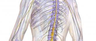
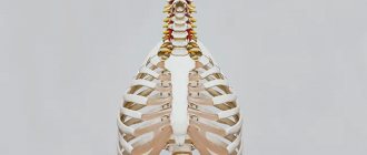
Before talking about the components, you should understand the system itself. The spinal cord is located in the spinal canal, and is surrounded by three boundaries in the form of membranes:
- Soft
- Cobweb
- Solid
It is in constant interaction with internal organs and the brain. Its length is approximately 40 centimeters for women and 45 for men, weighing about 30 grams. It’s not for nothing that there is an expression: the spool is small, but expensive. The psychological state, the functioning of internal organs, reflexes and motor activity of a person depend on this small link.
The components of the spinal cord are gray and white matter. The location of the first of them is the central departments, the second is the periphery. In the embryo, the spinal cord occupies almost the entire spinal canal, and with subsequent growth it moves upward.
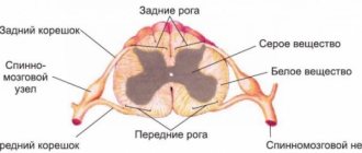
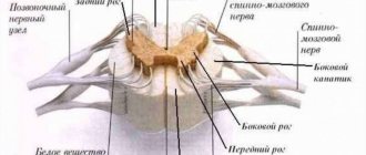
Looking at a cross-section of the human spinal cord (SC), you can see a central canal filled with brain fluid, gray matter in the shape of a butterfly (whose wings resemble horns) and white matter formed from paired cords - posterior, anterior and lateral.
Each of these sections performs a specific function that plays a vital role for a person. They ensure the passage and delivery of nerve impulses to various parts of the human body.
Symptoms
Symptoms of pathological processes develop over several years and can manifest themselves in two ways:
- Continuously evolving, with constant complication.
- During the period of progression of pathological changes, long periods of stabilization are observed without deterioration in the patient's health.
The clinical picture of the disease directly depends on the location of the pathological process in the brain and its type
Typically, symptoms worsen when your blood pressure is high for a long time.
In the initial stages, the clinical picture of leukoencephalopathy is characterized by:
- decreased mental performance;
- weakness in the limbs;
- absent-mindedness;
- slower reaction;
- apathy;
- tearfulness;
- clumsiness.
Further, sleep is disturbed, muscle tone increases, the person becomes irritable, susceptible to depression, feelings of fear, and phobias.
As a result, the patient becomes completely helpless, susceptible to epileptic seizures, unable to care for himself or control bowel and bladder emptying.
The disease develops quite quickly in most cases
First, attention is drawn to absent-mindedness, emotional lability, indifference, a tendency to depression, and the appearance of phobias. The patient loses the ability to pronounce words, concentrate and switch attention, quickly gets tired, cannot even analyze ordinary events of the day, and forgets the names of relatives. A progressive disease leads to sleep disturbances, irritability, increased muscle tone, and involuntary movements of the head and eyes
The patient's gait is disturbed
A progressive disease leads to sleep disturbances, irritability, increased muscle tone, and involuntary movements of the head and eyes. The patient's gait is disturbed.
It has been proven that leukoencephalopathy is caused by the presence of human polyomavirus. According to some data, about 80% of the world's population are carriers of the infection. But, despite this fact, cases of polyomavirus activation do not occur so often. Typically, the activation mechanism of the virus occurs under certain conditions, one and the main of which is a decline in the natural protective functions of the body, that is, immunity.
Important Registration of a disability group after a stroke
In addition to HIV infection and AIDS, predisposing causes for the development of this disease also include:
- leukemia and other blood pathologies;
- hypertension;
- tuberculosis;
- oncology;
- sarcoidosis;
- systemic lupus;
- rheumatoid arthritis;
- taking immunosuppressive drugs prescribed after organ transplantation;
- use of monoclonal antibodies;
- lymphogranulomatosis.
The disease has several forms:
- 1. Small focal leukoencephalopathy of vascular origin. It is a disease in which there is a chronic pathological condition of the blood vessels of the brain, which subsequently leads to damage to the cells of the white matter of the cerebral hemispheres. Predisposing factors to the development of this form of pathology is hypertension. Most often, this form is diagnosed in men whose age exceeds 55-60 years. Also at risk are people who have a genetic hereditary predisposition to the pathology. A complication of small focal leukoencephalopathy of vascular origin is the development of senile dementia.
- 2. Progressive multifocal encephalopathy. In this form of the disease, there is damage to the central nervous system by the virus, which, in turn, results in the destruction of the white matter of the brain. In most cases, progressive multifocal encephalopathy occurs against the background of human immunodeficiency. This pathology is one of the most dangerous, as its consequence can be sudden death.
- 3. Periventricular form. Develops against a background of chronic lack of oxygen and ischemia. The result of such circumstances is damage to the subcortical structures of the brain, mainly its trunk, the cerebellum and those parts that are responsible for motor activity. The periventricular form is characteristic of newborn babies and can lead to the development of cerebral palsy.
What is gray matter?
Because the gray matter surrounds the central canal, it is called the intermediate canal. It is presented in the form of two vertical strands of irregular geometric shape, located on both sides of the central channel, called the left and right gray pillars. These elements are connected to each other by a thin plate (adhesion), surrounding and protecting the canal on all sides, adjacent from above to the fourth ventricle of the brain, ending below with the terminal ventricle called Krause.
The basic structure of the gray matter of the SC is clearly represented in the form of horns:
- The anterior ones are the location of the largest neurons, forming five main clusters or nuclei. These clusters provide motor activity of the human body, abdominal and thoracic vibrations during breathing.
- The structure of the posterior pillars (horns) is heterogeneous. It consists of axons and neurons, which are multi-polar cells. Each of them occupies a specific place on the territory of the CM. The processes of these neurons extend to the corresponding adjacent segments of the anterior layer.
- The lateral horns are responsible for the functionality of the autonomic nervous system (ANS) and have protrusions in the areas of the eighth cervical and second lumbar vertebrae. The upper (cervicothoracic) region regulates the functioning of the heart, digestive organs, sweat glands and blood vessels. Lower (lumbosacral) – defecation, urination, erection and ejaculation.
The nerve centers of the SC are segments with neurons located in them that affect all functioning receptors and internal organs. It is quite clear how important they are for the full life of the body.
What is the composition of gray matter?
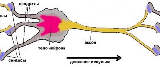
Gray matter is formed from nerve cells, of which there are more than thirteen million (including glia and processes). All neurons grouped into nuclei or plexuses that make up the gray matter of the SC are divided into three main groups:
Internal cells with numerous processes that do not extend beyond the boundaries of the substance itself and provide interaction (forming synapses) with other neurons of the SC.
Root cells (the largest motto- and multi-neurons of the ANS), the neurons of which protrude beyond the boundaries of the gray matter and innervate skeletal muscles.
Tufted neurons forming many pathways leading to the brain. These cells got their name due to their ability to form bundles, which are a kind of switches connecting SC segments.
So, the spinal cord includes neuroglia, ganglion nerve cells, pulpal and non-pulpate nerve fibers. These cells are grouped into nuclei and are located in segments along the entire length of the spinal cord and its primary three-membered reflex arc.
Each segment of gray matter is separated by barriers in the form of Rexed plates that perform specific functions:
- Reception of impulses from the dorsal roots and their transmission to the spinothalamic tract.
- Accompaniment of impulses (afferents) of temperature and pain sensitivity.
- Processing information from the muscular system.
- Receiving and transmitting brain impulses.
- Innervation of the muscles of the limbs, body and internal organs.
Disturbances in the functioning of any of the links in this system lead to serious consequences, including paralysis, partial or complete.
Characteristics of the gray matter of the spinal cord
, the gray matter of the spinal cord is represented by an accumulation of bodies and dendrites of nerve cells, which in functional terms can be intercalary or efferent neurons. Gray matter occupies a central position
and has the appearance of
a butterfly with spread wings
(or the letter H).
Clusters of nerve cell bodies in certain areas of the gray matter form its nuclei , which are classified according to functional principles
into the following groups:
Ø sensitive , predominantly formed by secondary afferent neurons
(a type of interneuron) that receive sensitive information from the primary afferent neurons of the spinal ganglia and conduct it further towards
the brain (i.e. the axons of the neurons of these nuclei form ascending pathways to the brain ). Due to the fact that the axons of sensory neurons of the spinal ganglia enter the spinal cord as part of the dorsal roots of the spinal nerves, secondary afferent neurons forming the sensory nuclei are usually located in the dorsal horns and the intermediate zone
of the gray matter of the spinal cord
Ø motor , formed mainly by a cluster of motor efferent neurons (motoneurons)
gray matter of the spinal cord, are located
in the anterior horns of the gray matter
, and their
axons leave the spinal cord as part of the anterior roots of the spinal nerves and are directed to certain skeletal muscles
Ø vegetative , formed by a cluster of vegetative efferent neurons
, are represented by two varieties:
sympathetic nuclei (lateral intermediate nucleus, located in the lateral horns of the gray matter of the spinal cord) and parasympathetic nuclei (lateral intermediate nucleus, located in the intermediate zone of gray matter of the II-IV sacral segments of the spinal cord), the axons of the neurons of these nuclei emerge from spinal cord
as part of the anterior roots and
are directed to the vegetative nodes
Ø intercalary intraspinal nuclei , formed by a cluster of intercalary intraspinal neurons
,
transmitting information within the spinal cord itself ; laid, as a rule, at the base of the posterior horns of the spinal cord
.
According to a newer classification of the gray matter of the spinal cord, proposed by Rexed, there are 10 sequentially located plates in it, and the numbering begins from the apex of the dorsal horns. The plates of gray matter of the spinal cord include predominantly some functionally specific neurons, and therefore specialize in performing certain functions. For example, the first plate of Rexed corresponds to the substantia gelatinous substance ( gelatinous substance of Rolland ), which contains both intercalary intraspinal and predominantly secondary afferent neurons, the axons of which form the ventral spinothalamic tract of the opposite side (path of tactile sensitivity).
Brief characteristics of the nuclei and plates of gray matter spinal cord is shown in Figure 5.
Fig.5. Topography of nuclei and plates of gray matter of the spinal cord
1 – border (terminal) Lissauer zone, neurons of this zone receive afferent information from primary afferents (axons of pseudounipolar neurons of the spinal ganglia) and transmit it to other interspinal or motor neurons of the spinal cord. Some neurons of the border zone take part in the formation of the ventral spinothalamic tract of the opposite side (pathway of tactile sensitivity)
Spongy zone
plate I corresponds to the substantia ( substantia gelatinosa of Rolland) ( 3 ), the latter is represented by both intercalated intraspinal and predominantly secondary afferent neurons, the axons of which form the ventral spinothalamic tract of the opposite side (pathway of tactile sensitivity)
II, III, IV plates correspond to the apex and central part of the posterior horns; both intercalary intraspinal and secondary afferent neurons lie here. The bodies of these neurons, grouping, form two nuclei: the intrinsic nucleus of the dorsal horns (5) and the reticular nucleus (4). The first is related to the formation of the lateral spinothalamic tract of the opposite side (the path of pain and temperature sensitivity). The axons of the neurons of the retinal nucleus are directed to the reticular formation of the spinal cord.
V, VI plates correspond to the base of the posterior horns. The neurons of these plates are related to the processing of proprioceptive information coming both directly from the primary afferents and from the intercalated intraspinal neurons of the I-IV plates and the further transmission of information to the neurons of the VII-IX plates. The bodies of the secondary afferent neurons of this plate form two nuclei: the thoracic nucleus ( Clark's nucleus (6), located at the level of all thoracic and upper lumbar segments) and the commissural nucleus (7), separating the thoracic nucleus from the posterior cords of the white matter of the spinal cord. The neurons of Clark's nucleus are related to the formation of the dorsal spinocerebellar tract on their side , and some of the axons of the commissural nucleus are sent to the dorsal cord .
Plate VII corresponds to the intermediate zone and the base of the posterior horns. This area mainly contains intercalary proprioceptive neurons , which transmit afferent information from other intercalary or directly primary afferent neurons to motor neurons of plates VIII-IX. Nerve fibers of the vestibulo- and reticulospinal tracts form synapses on the bodies and dendrites of the intercalary proprioceptive neurons of this plate.
Throughout the entire length of the spinal cord, plate VII contains the medial intermediate nucleus (8), represented by secondary afferent neurons, the axons of which take part in the formation of both the ventral spinocerebellar tract and the visceroceptive sensitivity pathway of the opposite side .
In plate VII in the lateral horns of the last cervical, all thoracic and upper two lumbar segments
lies
the lateral intermediate nucleus (9), formed by the bodies of the first efferent sympathetic neurons and representing the sympathetic autonomic center for the regulation of physiological functions in the body.
At the level of the II-IV sacral segments in the lateral region of the VII plate lies the Onufrovich nucleus , formed by a cluster of the first efferent parasympathetic neurons . Thus, the lateral intermediate nuclei, located at the level of the last cervical, all thoracic, upper two lumbar and II-IV sacral segments, represent the centers of the autonomic nervous system.
VIII, IX plates correspond to the central part and apex of the anterior horns of the gray matter of the spinal cord. Mostly motor neurons (although intercalary proprioceptive neurons are also found). Motor neurons VIII and IX plates, grouping, form 5 nuclei: dorso- (10) and ventro- (11) lateral , dorso- (12) and ventro- (13) medial and one central (14). On the bodies and dendrites of these neurons, both the primary afferent and intercalated proprioceptive neurons of laminae I-VII and the nerve fibers of the rubro- and corticospinal tracts form synapses. The axons of the neurons of these nuclei (motor nerve fibers) leave the spinal cord as part of the anterior roots and are directed to the skeletal muscles they innervate. It is believed that the neurons of the medial nuclei of the anterior horns are related to the innervation of the muscles of the trunk, and the lateral nuclei - the muscles of the limbs.
The X plate is a layer of gray matter surrounding the spinal canal, represented mainly by neuroglial cells.
Secrets of his education
The gray matter of the SC is formed from many neurons with processes, united into nuclei and without a myelin sheath. To be more precise, it contains:
- Long neural processes
- Short neural processes
- Neuron cell bodies
- Connective and accompanying tissue
- Blood vessels
Each group of neurons is responsible for specific functions:
Sensitive ones provide the perception of impulses coming from tissues and organs. Their receptors are located throughout the human body and are concentrated on the skin and internal organs. Only their processes penetrate into the spinal cord, and the neurons themselves are located outside it.
Motor muscles are responsible for conducting impulses to the muscles from the centers of the spinal cord or brain.
Intercalary neurons have a complex structure and are located between the processes of sensory and motor neurons. Their main task is to receive and process all incoming information.
The anterior horn of the gray matter of the spinal cord is the largest and widest and houses motor neurons. The location of the sensitive nerve cells is the dorsal horn, which ensures the functioning of the internal glands and organs. The smallest and weakest middle horn is inhabited by intercalary neurons; it is here that the initial processing of all signals coming from various zones of the body takes place.
Most cells in the spinal cord are designed to control, analyze, receive and transmit pain impulses. They are multipolar, which distinguishes them from white matter neurons of the SC.
In a few words, gray matter can be characterized as a collection of nerve fibers and neurons with processes up to 0.1 millimeters in diameter and up to one and a half meters long. Each of these elements has its own purpose and different functionality.
Functions and Features
Gray matter is located in the center of the spinal cord along almost the entire spinal column. At the levels of the lumbar and cervical regions it has a pronounced thickening, the presence of gray brain tissue is somewhat increased here. Thanks to this structure, human mobility and the ability to perform certain manipulations are ensured.
In addition, the SM performs a number of other important functions:
- Protective. The gray matter, like a membrane, is located around the central canal, protecting and circulating the cerebrospinal fluid (CSF), which delivers nutrients to tissues and nerve fibers.
- Regulatory. Most of the neurons and cells of the gray matter of the SC are capable of controlling many vital processes. They regulate the proper functioning of certain organs: kidneys, intestines, bladder, secretory glands and reproductive system.
- Conductor, which consists in ensuring information exchange between the brain and all systems, muscles, blood vessels and internal organs.
- Reflex. Many impulses received by the spinal cord are processed independently by it. At the same time, the load on the brain is reduced, responses occur without its participation and are called unconditioned reflexes. They play an important role, as they are sometimes created in an instant reaction (for example, to impulses of a warning and painful nature).
It turns out that the set of responsibilities assigned to the spinal cord is very large. This explains its complex structure and intricate structure.
Treatment and diagnosis
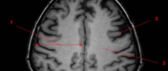
The main way to find multiple lesions is to visualize the medulla on magnetic resonance imaging. On layer by layer
Spots and pinpoint changes in tissue are observed in the images. MRI shows not only lesions. This method also reveals the cause of the lesion:
- A single lesion in the right frontal lobe. The change indicates chronic hypertension or a previous hypertensive crisis.
- Diffuse foci throughout the cortex appear when the blood supply is disrupted due to atherosclerosis of the cerebral vessels or.
- Foci of demyelination of the parietal lobes. It speaks of a disruption in the flow of blood through the vertebral arteries.
- Massive focal changes in the white matter of the cerebral hemispheres. This picture appears due to atrophy of the cortex, which forms in old age, from Alzheimer's disease or Pick's disease.
- Hyperintense lesions in the white matter of the brain appear due to acute disruption of the blood supply.
- Small foci of gliosis are observed in epilepsy.
- In the white matter of the frontal lobes, single subcortical foci are predominantly formed after a heart attack and softening of the brain tissue.
- A single focus of gliosis in the right frontal lobe most often appears as a sign of brain aging in older people.
Magnetic resonance imaging is also performed on the spinal cord, in particular on the cervical and thoracic regions.
Related research methods:
Visual and auditory evoked potentials. The ability of the occipital and temporal regions to generate electrical signals is tested.
Lumbar puncture. Changes in the cerebrospinal fluid are examined. Deviation from the norm indicates organic changes or inflammatory processes in the cerebrospinal fluid ducts.
A consultation with a neurologist and psychiatrist is indicated. The first studies the functioning of tendon reflexes, coordination, eye movements, muscle strength and synchrony of extensor and flexor muscles. The psychiatrist examines the patient’s mental sphere: perception, cognitive abilities.
Foci in the white matter are treated with several branches: etiotropic, pathogenetic and symptomatic therapy.
Etiotropic therapy is aimed at eliminating the cause of the disease. For example, if vasogenic lesions of the white matter of the brain are caused by arterial hypertension, the patient is prescribed antihypertensive therapy: a set of drugs aimed at lowering blood pressure. For example, diuretics, calcium channel blockers, beta blockers.
Pathogenetic therapy is aimed at restoring normal processes in the brain and eliminating pathological phenomena. Drugs are prescribed that improve blood supply to the brain, improve the rheological properties of blood, and reduce the need for oxygen in brain tissue. Vitamins are used. To restore the functioning of the nervous system, it is necessary to take B vitamins.
Important Abstract: blood-brain barrier
Symptomatic treatment eliminates symptoms. For example, for seizures, antiepileptic drugs are prescribed to eliminate foci of excitation. In case of low mood and lack of motivation, the patient is given antidepressants. If lesions in the white matter are accompanied by an anxiety disorder, the patient is prescribed anxiolytics and sedatives. If cognitive abilities deteriorate, a course of nootropic drugs is indicated - substances that improve the metabolism of neurons.
Didn't find a suitable answer? Find a doctor and ask him a question!