Amygdala
Admin
09.01.2019
Uncategorized
Amygdala
(amygdala, amygdala complex, (Latin) corpus amygdaloideum, from Greek αμυγδαλή, amygdalē - “almond”) - a paired almond-shaped group of nuclei located deep in each medial temporal lobe (MT) of the brain of complex vertebrates, including humans [1] . As numerous studies show, the amygdala plays a key role in processing and remembering emotional reactions. It is considered an integral part of the limbic system[2]. Moreover, anatomically, the amygdala[3], especially its central and medial nuclei,[4] are often considered part of the basal ganglia.
Internal structure of the amygdala
The amygdolar complex includes several different substructures that have different functional purposes. There are several options for their classification, with varying degrees of detail. Among the substructures of the amygdala, at least the basolateral complex of nuclei (or simply basolateral nuclei), the central, medial and cortical nuclei (or preamygdala cortex) are usually distinguished. The basolateral complex of the amygdala, in turn, is divided into the basolateral (lateral), basomedial (middle, or additional) and basoventral (lower) nuclei (see figure) [2][5][6].
The location and main nuclei of the human amygdolar complex (amygdala) using the example of a coronal section approximately through its middle. Designations of the amygdala nuclei in the figure: La
- Lateral nucleus,
BL
- Basolateral nucleus,
BM
- Basomedial nucleus,
BV
- Basoventral nucleus,
Ce
- Central nucleus,
Me
- Medial nucleus,
VCo
- ventral cortical nucleus (or preamygdala cortex).
In addition, around the amygdala are marked: Cl
- septum of the brain,
Hi
- Hippocampus
Ent
- Entorial cortex,
F
- endorchial cortical fold,
NbM
- basal ganglia of Meynert,
TrO
- optic tract,
V
- lateral cerebral ventricle.
Rat amygdala nuclei
We also present a diagram of the location of the nuclei of the (right) amygdala complex of the rat brain [7]. In this figure, all nuclei are divided into three groups: the central and medial nuclei are colored orange, the basolateral nuclei are colored green, and the cortical nuclei are colored brown. In addition, the nuclei called intercalary masses ( In
).
Designations in the figure: CEl
, central nucleus, lateral section;
CEm
, central nucleus, medial division;
CEc
, central nucleus, capsular compartment;
COa
, cortical nucleus, anterior region;
Cop
, cortical nucleus, posterior division;
BOT
, subject to the core of the olafactory tract;
Bi
, basal nuclei, intermedial section;
Bpc
, basal nucleus, parvocellular division;
BMmc
, basomedial nucleus, magnocellular division;
BMpc
, basomedial nucleus, parvocellular compartment;
Ld
, lateral nucleus, dorsal division;
Lvm
, lateral nucleus, ventromedial division;
Lvl
, lateral nucleus, ventrolateral division;
Md
, medial nucleus, dorsal division;
Mv
, medial nucleus, ventral division;
In
, intercalary masses (core);
Pir
, pear-shaped (piriform) bark. To simplify this diagram, the magnocellular region of the nucleus basalis, which is located more rostrally (anteriorly), is not shown, as is the rostral region of the medial nucleus, as well as the amygdalohippocampal region, which is located more caudally (posteriorly).
Main external relations
The lateral amygdala nucleus, which then sends impulses to the rest of the basolateral complex and the centromedial nuclei, receives signals from multiple sensory systems, including the neocortex, and is considered the main (multisensory) “input to the amygdala” [8].
The cortical nucleus of the amygdala (or preamygdala cortex) is involved in processing odors and responding to pheromones. Therefore, it receives signals from the Olafactor flask and Olafactor cortex.
Neuroanatomical structures associated with panic disorder, their connections and functions.
The Amygdala sends its impulses to the Hypothalamus to activate the sympathetic nervous system, to the reticular nucleus of the thalamus to accelerate reflexes, to the nuclei of the trigeminal nerve and facial nerve, as well as to the ventral tagmental area, locus coeruleus, and to the laterodorsal tagmental area to activate the flow of dopamine, norepinephrine and epinephrine.[5]
The diagram on the left shows the central role of the amygdala in panic personality disorder. [9]
Is it possible to remove the amygdala, and with it the fear?
If fear and anxiety ruin a person’s life, maybe you can simply remove the tonsil and live in peace, without worrying?
The amygdala's reputation in the media is clear: it is the locus of fear, anxiety and stress. In CT scanners, the amygdala area lights up like a light bulb in response to most tests in which people experience severe emotional discomfort. What happens if a person is left without an amygdala?
In medicine, in fact, there are cases of tonsil removal: the indication was either severe forms of epilepsy or uncontrolled aggression - this sometimes occurs in patients with mental disorders. The first operations were carried out back in the 1960s - oil was injected into the brain to destroy the tonsil. Since then, hundreds of patients around the world have undergone amygdalotomy (that is, removal of the amygdala). Although there are now many other techniques for treating mental disorders and abnormalities, amygdalotomy has not yet disappeared from practice (the same applies to witch hunts - it is still (!) practiced in some African and Asian countries - although it would seem) .
So, in 2021, in China, surgeons removed the tonsil from a girl with mental retardation, psychiatric symptoms and aggression - and even cheerfully offered to continue research on the prospects of such interventions in controlled studies with a large number of participants. However, these cases are difficult to correlate with normal people - very specific indications for such an operation.
Interestingly, there are also cases of “natural amygdalotomy”. Urbach-Wiethe disease is a very rare recessive genetic disease, with even fewer known cases than patients with surgically removed tonsils. In patients with Urbach-Wiethe disease, degradation of the amygdala occurs without any outside intervention. Patients usually experience dermatological and neurological symptoms: their skin and mucous membranes thicken, wounds heal poorly, and in another 50-75% of cases, the substance inside the temporal lobes becomes calcified - and this leads to damage in the tonsil and surrounding areas.
Patient S.M. is one of a string of famous neurological patients, and she suffers from Urbach-Wiethe disease. Many things that frighten people do not bother S.M. at all: she is indifferent to horror films, spiders, snakes and even death threats. CM. described to researchers a case when an unknown person put a knife to her throat in a dark park, threatening to “kill the bitch.” She calmly answered him something, and her composure, apparently, scared off the would-be killer: he let her go, and she calmly went about her business.
It turned out, however, that patients with a damaged amygdala are quite capable of experiencing fear, moreover, when inhaling air with a high content of carbon dioxide S.M. I experienced something similar to a panic attack. She grabbed the experimenter's hand, screamed and was extremely frightened: a wildly beating heart, wide pupils, goosebumps all over her body and tense muscles - clear signs of extreme fright were evident.
The way S.M. reacted far exceeds the reaction of ordinary people to inhaling CO₂; Moreover, after several repetitions, a person most often gets used to the effect of carbon dioxide and stops panicking, while S.M. the reaction to CO₂ remained extremely acute and inadequate to the threat. She later described the experience as the worst thing that had ever happened to her - the killer in the park was nothing compared to inhaling carbon dioxide. Two other patients with an absent amygdala also experienced panic when inhaling CO₂ and also described their sensations as completely new and completely terrible.
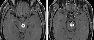
Non-invasive amygdatotomy on brain scans. Circled in red is the area of the amygdala, which atrophies in patients with Urbach-Wiethe disease. On the left is the norm, on the right is patient S.M.
It turns out that removing the amygdala does not eliminate fear - although at least it dulls the reaction to many “fearful” stimuli for a person, it does not protect against other “non-social” sources of fear. The researchers hypothesize that patients are protected from external sources of fear that people recognize based on what they hear and see. In the case of carbon dioxide, fear acts from the inside: dissolving in the blood, CO₂ directly affects the structures of the brain stem, and its action does not need to be interpreted by the amygdala. Researchers believe that the amygdala can normally regulate the strength of the threat response and suppress panic. This assumption is also supported by the fact that in one study, atrophy and weak activity in the amygdala were found in people with panic disorder.
It is possible that the amygdala is important not only so that we can learn to fear those things that are dangerous. It also helps us stop being afraid of things that no longer pose a threat. Perhaps it was thanks to this that Alex Honnold was able to train his amygdala the same way he trained his muscles - the main thing is not to panic or twitch, and then everything will most likely end with a happy ending.
Link.
Basic internal communications
Basic connections of the Amygdala nuclei. The power of the arrows corresponds to the power of the connections.
We will present the general scheme of internal connections of the amygdala complex of primates according to the work [8]. As already noted, the lateral nucleus (its dorsal part) receives many connections from the sensory cortex and sends its connections to almost all other nuclei of the amygdala, although the latter differ in their power. The strongest projections of the lateral nucleus go to all sections of the basal ganglia and preamygdaloid cortex. It is important to note that its projections into the central nucleus of the amygdala are relatively weak relative to, for example, connections with the basal ganglia.
Connections of the amygdala nuclei
Note again that, in general, the lateral nucleus is the main input to the amygdala. There, information is processed and transmitted basally - medially and further to the central nucleus, which can be called the main output instance of the amygdaloid complex. At the moment, intraamygdaloid connections have been studied in detail for rats, and there is also data for cats and primates.
Let us give as an example a diagram [7] of the main internal and internuclear connections for the amygdala of rats (see Fig., designations as before) obtained from anatomical studies. Most of these connections are glutamatergic. In [7], intranuclear connections are discussed in detail using the example of the lateral, basal and central nuclei of the amygdala. However, it is noted that the neurons of the basal and lateral nuclei have extensive dendritic trees located in neighboring sections of the same nuclei. The psychophysiological significance of such connections remains unclear.
Functions
The amygdala of the brain specializes in the perception of emotions and is activated when a person experiences strong emotional experiences. The main function is to prepare the body for a “fight or flight” response. The functions of the amygdala are distributed based on its structure. The amygdala structure consists of nuclei of different types, divided mainly taking into account functional differences:
- Basolateral. Associated with emotional manifestations. An input structure that receives diverse sensory-type information from the cortical regions and thalamus.
- Central. Consists of lateral and medial sections. The medial section projects incoming information to the brain stem and hypothalamus, which causes the occurrence of autonomic and motor reactions appropriate to the situation.
- Cortical (cortical). Reacts to taste sensations.
- Medial. Associated with the olfactory system.
Activation of the basolateral, medial and central nuclei gives an anxiogenic (state of horror, fear) effect. Inhibition of the activity of these structures is accompanied by an anxiolytic (suppression of anxiety) effect. Dysfunction of the tonsils underlies the development of anxiety-phobic disorders.
The coordinated interaction of the amygdala nuclei allows you to promptly recognize danger and respond adequately to it. For example, the unpleasant smell and taste of spoiled foods make a person experience a feeling of disgust and hostility, which protects against food poisoning.
The reaction to the unpleasant odor of waste products is the desire to withdraw and avoid contact, which prevents infection by pathogenic microorganisms. Close interaction with the hippocampus involves fixing in memory memories of experienced events that provoked a feeling of horror, strong fear, which subsequently affects the occurrence of an appropriate reaction to such stimuli.
If an individual repeatedly encounters similar situations, the corresponding images stored in long-term memory activate the amygdala complex. As a result of the perception of external stimuli that are associated with the experienced traumatic situation, the body mobilizes efforts to avoid troubles.
The amygdala complex is involved in the formation of the reaction of aggression, affective (sensual, emotional) perception, experience and manifestation of emotions. The tasks of the amygdala complex include emotional assessment of stimuli that are primarily processed in cortical structures. Stimuli containing an emotional component are better recorded in memory.