Motor speech area of the cerebral cortex, Broca's area
The motor speech area is an area of the cerebral cortex located in the posterior
section of the inferior frontal gyrus of the dominant hemisphere anterior to the cortical centers of movements of the lips, larynx, and tongue. When the motor speech area is damaged, motor alalia or motor aphasia occurs (motor alalia or motor aphasia syndrome).
Sensory speech area of the cerebral cortex, Wernicke's center
The sensory speech area of the cerebral cortex is an area of the cerebral cortex in the posterior part of the superior temporal gyrus of the dominant hemisphere. The term "sensory" comes from the Latin word "sensus", which means "feeling, perception, sensation". A synonym for the sensory speech area is Wernicke's center, Wernicke's area. When the sensory speech area is damaged, sensory alalia syndrome or sensory aphasia occurs.
When the sensory and motor areas of the cerebral cortex are damaged, sensorimotor alalia or sensorimotor aphasia occurs simultaneously.
What does FOXP2 gene say?
All FOX genes regulate normal embryonic development, and FOXP2 is no exception. The expression of this gene is increased in the precursor cells of brain neurons, and when FOXP2 is turned off, their occurrence is suppressed [3]. One of the ways that FOXP2 regulates cell maturation is through its control over the activity of the SRPX2 (sushi repeat-containing protein X-linked 2) gene, which encodes the structure of the peroxiredoxin protein. Through this gene, FOXP2 controls the formation of synapses (synaptogenesis), and reduced SRPX2 activity leads to impaired synaptogenesis and auditory communication in mice [4].
During the evolutionary process, DNA can change randomly, that is, mutations occur in the molecule. Substitutions in the nucleotide sequence, in which the structure of the protein does not change, are called synonymous. If a replacement in DNA leads to the appearance of a new amino acid in a protein, then such a replacement is considered nonsynonymous and, as a rule, leads to a change in the function of the protein. When studying the molecular evolution of FOXP2, interesting circumstances were revealed [5]. This gene is one of the most conserved in human DNA, and the greatest changes in FOXP2 within the primate group occurred after the divergence of the evolutionary lines of humans and chimpanzees, our closest relatives. Rhesus macaques, gorillas and chimpanzees had only synonymous DNA substitutions, and only orangutans had one non-synonymous substitution (Fig. 2). The highly conserved structure of the gene is associated with the many functions that it regulates and their importance for the developing organism. If the mutation of FOXP2 resulted in forms of the protein it encodes that did not fully perform the necessary functions, this led to improper development of the embryo and its death. Such mutations could not be passed on to the next generation. Two nonsynonymous substitutions that arose in humans in the FOXP2 gene apparently gave our ancestors a serious advantage and became established in the Homo sapiens genome.
Figure 2. Evolution of the FOXP2 gene. The numbers indicated through the line represent the number of substitutions (mutations) in the DNA sequence: the number of non-synonymous substitutions is given before the line, and synonymous ones after the line. In humans, for example, in comparison with chimpanzees, only two substitutions occurred, but both were nonsynonymous, that is, they led to a qualitative change in the gene. At the same time, 131 synonymous substitutions and only one nonsynonymous substitution occurred in mice.
website www.evolutionpages.com
Treatment of damage to speech zones in Saratov, how to improve the functioning of speech zones
Sarklinik provides comprehensive conservative treatment of the speech areas of the cerebral cortex in children (boys and girls), adolescents (boys and girls), adults (men and women) in Saratov, Russia. At the first appointment, the doctor will tell you what reflex massage of speech zones is, what new advanced methods exist for restoring speech disorders; how to treat pathology of the motor and sensory speech zones, how to restore and improve speech. What happens when a pulsed magnetic field is applied to the speech centers in the cerebral cortex in Russia, does this technique always help? What new methods exist for stimulating speech centers that give 95% results? How to really improve the work of the Wernicke center, how to activate the Wernicke area? What is Broca's center? How to increase a child’s vocabulary in Russia? How to improve the impressive (sensory) and expressive (motor) speech of infants, preschoolers, and schoolchildren? Where to treat mental retardation, and what to do if speech zones are disrupted, how to treat speech disorders in Saratov . On the sarclinics website you can ask the doctor a question online and read reviews about the treatment of children.
"Bio/mol/text"-2015
This work was published in the “Best Review Article” category of the “bio/mol/text” competition 2015.
The sponsor of the nomination “Best article on the mechanisms of aging and longevity” is the Science for Life Extension Foundation. The audience award was sponsored by Helicon.
Sponsors of the competition: laboratory of biotechnological research 3D Bioprinting Solutions and studio of scientific graphics, animation and modeling Visual Science.
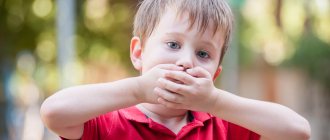
In the late 1980s, teachers at a school in west London noticed that seven children who had speech problems were growing up in the same family. This family (in the scientific literature it appears under the name “KE family”) was of Pakistani origin, and upon closer examination of its members, it turned out that in three generations of this family there were people with speech problems (Fig. 1). They had difficulty pronouncing words, and sometimes words were replaced by words that sounded similar. If they spoke Russian, then, for example, instead of the word “oven” they would pronounce “flow”. Mild, mild disorders and more severe forms of speech disorders that seriously impede communication were discovered in the family.
Figure 1. Family tree of the KE family. In three generations of the family, people were found to have speech problems of varying severity (black shaded figures). These were representatives of both sexes: men (squares) and women (circles).
[2]
Given that speech problems were passed down from generation to generation, doctors who studied the KE family suggested that some kind of genetic disorder underlies these disorders. Difficulties with speech arose in representatives of both sexes, which means that the “culprit” gene was not located on the sex chromosomes (X or Y), but on autosomes. As a result, a team of geneticists from Oxford was able to determine that the desired gene was located on chromosome 7 [1]. The KE family has also been studied by linguists, for example, Myrna Gopnik from Canada. They suggested that speech disorders in the family are caused by a mutation in the “grammatical gene,” which is responsible for syntactically and grammatically correct construction of phrases. It was later found that representatives of the studied family had problems not only with syntax and articulation, but also generally experienced difficulties in controlling the tongue and lips [2]. This disorder was later called verbal dyspraxia. The KE family's brains were unable to accurately control their lips and tongue, causing words to not be pronounced correctly (see sidebar).
Treatment of speechopathy in Saratov, how to treat speechopathy in Russia
Sarklinik provides speech therapy treatment in Saratov , treatment of speech pathologies, speech disorders in children, boys, girls, teenagers in Russia. Sarklinik knows how to treat logopathies, how to cure logopathies, how to get rid of logopathies.
Treatment of alalia, treatment of aphasia, treatment of stuttering, treatment of speech delay, treatment of speech development disorder.
Sign up for a consultation. There are contraindications. Specialist consultation is required.
Photo: (©) Bioraven | Dreamstime.com\Dreamstock.ru
Related posts:
Which clinic best treats nocturnal enuresis in children in Russia
Mental retardation in children: moderate, mild, severe, profound, mental retardation, dementia, treatment
Why is my child doing poorly at school? I am doing very poorly, what should I do?
Wet bed, bed, the child is sleeping soundly and the described bed
Apgar scale, assessment of the newborn on the Apgar scale, points
Comments ()
8.2. Disturbance of the speech sphere with damage to the left and right hemispheres of the brain and with damage to the midline structures
Below we will present the results of a neuropsychological examination of the speech and perceptual spheres of children conducted by E. G. Simernitskaya (1985). These data represent the result of one of the most complete and comprehensive neuropsychological examinations of children with organic brain lesions.
The data were obtained from an examination of 130 children with lesions of the left hemisphere, 90 people with lesions of the right hemisphere and 76 people with lesions of the diencephalic region aged 6 to 16 years.
A study by E. G. Simernitskaya showed that the deficiency of speech functions was most clearly manifested when using special neuropsychological tasks that assessed the state of auditory-verbal memory.
Analysis of auditory-verbal memory impairments in the examined children showed that with lesions of the left hemisphere in children, a central place in the structure of the observed disorders is occupied by a narrowing of the auditory-verbal memory volume.
With lesions of the right hemisphere, the symptom of impaired reproduction of a given order of elements comes first.
With lesions of the diencephalic region, the central place is occupied by the symptom of pathological inhibition of traces under conditions of interference.
8.2.1. Disturbance of the auditory-speech sphere with damage to the left hemisphere of the brain
Damage to the left hemisphere was characterized by a low frequency of aphasia (17% of children versus 67% of adults), although it is impossible to speak of complete preservation of speech function. Speech was completely preserved in children in 12% of cases, and in cases of damage to the temporal region of the left hemisphere - only in 7% of cases.
Speech disturbances were recorded especially often during epileptic seizures. This could be: loss of the ability to speak independently, impaired understanding of addressed speech, distortion of one’s own speech, which became incomprehensible to others, forgetting words and names of objects.
In a neuropsychological study outside the acute period, speech disorders were not detected in these children.
When studying auditory-verbal memory, the symptom of narrowing the volume of reproduction was observed in first place (in 86% of children); it was observed and manifested itself to a greater extent when memorizing words that were not related in meaning.
For example, when two groups of three words in each are presented for memorization, the reproduction looks like this (stimulus words - ball, floor, bread, leaf, knife, eye):
1 reproduction - ball, eye, nose;
2 reproduction - ball, bread, eye;
3 reproduction - ball, eye;
4 reproduction - ball, floor, bread, leaf.
Intrahemispheric differences manifested themselves as follows. In 60% of cases, this symptom was observed in lesions of the left temporal region, in 19% in lesions of the parietal region, and in 7% in lesions of the frontal region of the LA.
The second frequent symptom—instability of traces and their increased inhibition under interference conditions—was observed in 45% of children and did not depend on intrahemispheric localization.
The ability to maintain the order of reproduction was least affected by damage to the left hemisphere (only in 6% of cases).
The ratio of the two main symptoms varied depending on age. In children aged 5-10 years, the most significant impairment was in the volume of reproduction. After 10 years, the severity of this symptom decreases: the manifestations of symptoms of narrowing the volume of reproduction and inhibition of traces by interfering influences are equalized.
The ability to maintain the order of reproduction suffered the least (only in 6% of cases).
Thus, impairments in auditory-verbal memory in children with damage to the left hemisphere are generally of the same nature as in adults. In adults with damage to the left hemisphere, the central place in the impairment of auditory-verbal memory is occupied by a decrease in volume and increased inhibition of traces by interfering influences. The same is observed in children, but memory impairments in children 5-10 years old significantly exceed other forms of impairments, and after 10 years they tend to decrease. With regard to the second symptom—track retardation—there are no significant changes after 10 years.
This means that the specialization of the left hemisphere for speech manifests itself in the early stages of ontogenesis, and the differences in the manifestation of speech disorders between children and adults are associated not with the fact that the two hemispheres are equipotential, but with the high plasticity of the child’s brain and the more significant role of the right hemisphere in ensuring functioning of the speech system.
These two factors mask the early specialization of the left hemisphere for speech, therefore, insufficiency of speech function, similar to disorders in adult patients, can be detected only in the acute period (for example, postoperative), when the brain has not yet adapted to new working conditions.
E. G. Simernitskaya gives a number of examples of sick children with damage to different parts of the left hemisphere, in which symptoms of aphasia occur, which are also observed in adults with similar damage.
For example, in a 15-year-old patient, the presence of a large tumor of the left temporal region was accompanied before surgery by minor amnestic symptoms, slow speech, and mild impairments in auditory-verbal memory. After the operation, distinct sensorimotor aphasia developed.
There were disturbances in the understanding and pronunciation of low-frequency words (stimulus words are highlighted): mercury - tinder... tinder; Louvre - pr... lur; Capricorn - cancer... smash... for the sake of... smash... casirug.
In writing (Fig. 8.1 and reading) there were errors: destroyed - grown up... destroyed... zashu... zarsu; friendliness - friend... friendly... friendly... friendly... friendly.
The disturbances quickly regressed, and by the time of discharge, speech functions returned to preoperative levels.
Similar disturbances were recorded in other patients with damage to the left temporal region in the acute period: disturbances in articulation, speech understanding, writing, and reading. The patients had literal and verbal paraphasias. For example, “I studied for two hours, twice, no, for two years”; suitcase - suitcase... suitcase; pie - pir... pirom... feather.
However, even with the most severe disorders, after 2-3 weeks the disorders could completely regress and not be detected in a neuropsychological examination.
Of particular importance is the fact that the speech dysfunction observed in the postoperative period with damage to other areas of the left hemisphere was of a qualitatively different nature.
During operations on the left frontal region, the dynamic organization of speech processes was primarily disrupted.
For example, in a 7-year-old patient with removal of a tumor in the left frontal region from the pole to the posterior frontal regions, specific symptoms accompanying damage to this region in adults were observed. There was a lack of spontaneity, a virtual absence of independent speech, echolalic answers to questions, and the need for constant stimulation of the patient. Rough verbal perseverations occurred when trying to name objects: apple - apple, eye - apple, bed - apple.
Similar disorders, such as efferent motor aphasia in oral and written speech or the type of dynamic aphasia (impaired spontaneous speech, pronouncing only the words “yes” and “no”), were observed in other patients. When repeating sounds, syllables, words (those suggested for repetition are highlighted in bold), perseverations arose:
f-v - f-v, fa-va - v-v;
d-t - d-t, bi-ba-bo - d-d-do;
bed - bed, crust - bed.
Disturbances in speech activity after operations on the left parietal region were of a different nature.
Suffering from the anterior parietal region led to defects of a type of afferent-motor aphasia (errors in a 12-year-old patient when repeating words were associated with a violation of kinesthetic syntheses: elephant - moan, fork - vulka, eyebrow - byrov, thermometer - gravdugra).
Suffering in the posterior parietal region led to difficulty understanding logical-grammatical constructions (semantic aphasia, agraphia, alexia, acalculia).
A 14-year-old patient with removal of the left parietal lobe had reading impairments: instead of t - p, m; violations of writing (Fig. 8.2 acalculia in the form of a disorder of counting operations: 25 - 8 = 16, violations of the bit structure of numbers: instead of 1054 - fifteen thousand four.
In all cases examined, a rapid regression of the identified disorders was observed. Thus, underestimation of the high plasticity of the brain becomes one of the reasons that many disorders of speech functions in childhood remain undetected.
Disturbance of the auditory-speech sphere with damage to the right hemisphere of the brain
It is known that in adults the right hemisphere is involved in speech function. Even X. Jackson attributed the most consolidated, automated forms of speech activity to the right hemisphere. Later, the role of the right hemisphere in speech prosody, noise immunity, and identification of individual, emotional characteristics of speech was discovered.
Research by R. Sperry, M. Gazzaniga, E. Seidel showed that the right hemisphere has speech. But the uniqueness of the lexical structure of the right hemisphere is that it does not have phonetic coding (Zaidel E., 1978). Therefore, the understanding of oral and written speech in the right hemisphere is carried out not on the basis of phonetic analysis, but by the method of integrated, gestalt comparison of the sound, graphic image of lexical units with memory standards and subsequent association of this image with meaning. This perception of speech can be compared to the understanding of words by animals.
The following facts have been described regarding the role of the right hemisphere in auditory-verbal memory.
- If the left hemisphere provides an arbitrary level of organization of verbal-mnestic processes, then the right hemisphere creates the basis for involuntary, unintentional memorization. Therefore, in adults, dysfunction of the right hemisphere primarily leads to suffering from the possibility of involuntary imprinting of traces.
- Damage to the left hemisphere leads to a narrowing of the volume and increased inhibition of traces, therefore, memory impairments are most clearly detected in delayed reproduction after a pause that is empty or filled with side activities. On the contrary, patients with damage to the right hemisphere show a deficit in direct reproduction, and the introduction of interference does not have an inhibitory effect on reproduction.
- The most common disorder with damage to the right hemisphere in adults is the inability to maintain the order of reproduction of a given sequence of stimuli.
Data obtained from a study of children (5-16 years old, 90 people) with PP lesions showed that adult-specific violations of prosody and discrimination of individual characteristics of voices are rare, in isolated cases.
The most typical disorder was auditory-verbal memory (observed in 75% of cases). It manifested itself in the difficulty of directly capturing traces. But if in the case of left-hemispheric lesions this was associated with a narrowing of the volume of reproduction, then in the case of right-hemisphere lesions it was associated with a violation of the order of reproduction. Memorization occurs after one or two presentations, but multiple presentations do not change the order.
For example, an 8-year-old patient with a tumor of the right temporoparietal region
when memorizing a series of five words (stimulus words - cat, house, forest,
ringing, night) reproduces them after the first presentation, but install
the desired order is not possible after multiple presentations:
1 playback - cat, house, forest, night, ringing;
2 reproduction - cat, forest, night, house, ringing;
3 reproduction - cat, house, forest, night, ringing;
4 reproduction - cat, house, forest, night, ringing;
5 reproduction - cat, house, forest, night, ringing.
Another feature is related to the fact that in delayed reproduction there is no loss of elements either under conditions of homogeneous or heterogeneous interference, that is, there is no effect of trace inhibition.
Thus, we can talk about the specific contribution of the right hemisphere in children to the implementation of verbal-mnestic activity: it does not duplicate the left hemisphere and its contribution is qualitatively different from the contribution of the left hemisphere.
The contribution of the right hemisphere manifests itself early, since order disturbances with its damage are more frequent in children 5-10 years old. After 10 years, violations of order appear more and more clearly.
8.2.3. Impairment of the auditory-speech sphere with damage to the midline structures
Median structures play a decisive role in maintaining optimal cortical tone, necessary for the normal course of all mental processes.
A feature of damage to these departments in adults is modality-nonspecific memory impairment, which is accompanied by disturbances of consciousness (which is never observed with damage to the convexital parts of the cortex). Patients lose orientation in time and space, show confusion, confusion, and experience contamination and confabulation.
The main symptom of memory impairment is increased inhibition of traces by interfering influences. Patients reproduce stimuli directly or after an empty pause, but cannot do this after the introduction of interference. They perform worst of all on tasks involving memorizing two series of words, that is, under conditions of homogeneous interference. The introduction of semantic organization has a facilitating effect when the memorized information is transferred to a higher level of organization.
A study of children with a median localization of brain damage - the hypothalamic-diencephalic region (5-16 years old, 76 people) showed that in them, a violation of mnestic function manifested itself as clearly and specifically as in adults.
At the same time, there were differences. In children, semantic organization did not lead to improved memorization. There were no differences between recall of structured and unstructured material. Contaminations, slipping into side associations, and inert stereotypes appeared both during the reproduction of semantic and during the reproduction of discrete elements. Sometimes discrete elements were reproduced better than semantic structures.
Another difference was that in children such disorders occurred only with intracerebral lesions of the diencephalic region located in the area of the third ventricle. Extracerebral disturbances in this area did not cause significant disturbances.
For example, a 14-year-old patient reproduces the story “The Dove and the Ant”: “They lived near a river. We lived together and went to the river to swim... we went to the stream to swim. The ant began to drown, saw him... the beetle saw him and began to help. He gave him a stick, and the ant ran out along the stick... Thus the ant was saved.” The second part was not reproduced at all. Three minutes later, the patient denied that there was a story about an ant and a dove, and said that there was a story “about a cat, a fox and a rooster, how they became friends.”
Reproduction of two phrases (1. Apple trees grew in the garden behind a high fence. 2. At the edge of the forest, a hunter killed a wolf.) patient, 8 years old.
Direct (immediately after presentation) reproduction of each phrase is possible.
When asked which phrase came first and which phrase came second, the patient answers: 1/? - I don’t know, 2/? - I forgot. After re-presentation:
1/? — in the garden a hunter killed a wolf, 2/? — in the garden at the edge of the forest, a hunter killed a wolf.
After the third presentation:
1/? — in the garden behind a high fence, a hunter killed a wolf, 2/? — in the garden at the edge of the forest, a hunter killed a wolf.
Thus, in children, as in adults, the central symptom is pathological inhibition of traces, the presence of confabulations, and contaminations. However, only with intracerebral lesions did this disorder manifest itself at all levels of semantic organization. In extracerebral tumors, disturbances appeared only on nonsense material, both at the level of delayed and immediate reproduction.
The original goes like this: “An ant went down to the stream to drink. The wave overwhelmed him and he began to drown. The dove saw this and threw him a twig. The ant followed it to the shore. The next day the hunter wanted to catch a dove in a net. The ant crept up and bit the hunter's finger. The hunter screamed and dropped his net, and the dove fluttered up and flew away.”
In general, the most characteristic feature of diencephalic lesions, in contrast to disturbances in lesions of the right and left hemispheres, was the presence of retroactive inhibition.
Finally, the difference in disturbances in lesions of different parts of the diencephalic region indicates the functional dissimilarity of not only cortical, but also lower-lying structures.
8.2.4. Interhemispheric interaction in speech processes with organic damage to the left and right hemispheres
Changes in the functional organization of the brain during ontogenesis also manifest themselves in the analysis of interhemispheric interaction.
The dichotic listening data revealed the following facts.
The “focus effect” characteristic of local brain lesions, manifested in a decrease in the productivity of reproduction from the ear contralateral to the location of the pathological process, was more pronounced in children than in adults. This especially applies to lesions of the left temporal lobe, in which disturbances in reproduction from the right ear significantly exceeded the corresponding indicators in adult patients.
In adults, damage to the left hemisphere leads to a bilateral impairment of dichotically perceived information, and the “dominance effect” appears. In children, only unilateral impairment is observed.
But with damage to the right hemisphere, children experience a bilateral change in dichotically perceived information. The focal effect is compensated by a reciprocal increase in the volume of reproduction on the healthy side
This allows us to speak about a qualitatively different nature of the interaction of the two hemispheres in children compared to adults.
It is natural to assume that the absence of a “dominance effect” in lesions of the left hemisphere in childhood, reflecting the irradiation of the inhibitory influence from the lesion to the “healthy” hemisphere, is the result of insufficient maturity of interhemispheric connections, that is, the left hemisphere does not yet fully fulfill the role of the leading hemisphere . Data on the late maturation of the corpus callosum, which plays a decisive role in providing this inhibitory effect, presented in the specialized literature, make this assumption quite reasonable.