The olfactory brain consists of brain structures that support the function of smell. Adequate interaction of the body with the external environment is ensured by sensory systems. The sense of smell is an innate sense, helps to find food, is involved in the regulation of sexual behavior, and contributes to the enjoyment of the aroma of flowers and fruits. Using the sense of smell, a person perceives danger signals, for example, catching and identifying the smells of chemicals or spoiled food.
Characteristic
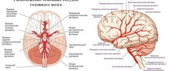
In anatomy, the olfactory brain is closely interconnected with the limbic system, which determines the joint regulation of many body functions, for example, the formation of emotions and the perception of odors. The olfactory system in the cortex is projected in the area of the anterior segment of the hippocampal gyrus. The olfactory analyzer perceives and analyzes odor. The system supports the following functions:
- Determining the suitability of food by smell.
- Regulation of eating behavior. The pleasant smell of food stimulates appetite.
- Stimulates the activity of the digestive tract. The smell of food, according to the principle of a conditioned reflex, triggers the mechanisms of digestion and food processing.
The structure of the olfactory brain involves close interaction between the departments - the amygdala within the limbic system, the hippocampus, the orbitofrontal and temporal (medial part) areas of the cortical layer. The vaulted gyrus (gyrus fornicatus) consists of parts - the cingulate, parahippocampal gyrus, and isthmus.
The central section within the olfactory brain includes the olfactory bulb, located in the area of the forebrain where the primary processing of information coming from the external environment occurs. The olfactory center within the brain is located in the lower segment of the temporal and frontal cortical layers covering the cerebral hemispheres.
The cortical sections perform secondary processing - they identify the smell, comparing it with stored data formed as a result of the accumulation of life experience. After identification, a general reaction of the body is formed that is adequate to the situation. As part of the central section of the combined structures - the olfactory brain and the limbic system, there are elements:
- Gyri – parahippocampal, cingulate, dentate, orbital.
- Hippocampal (seahorse) hook.
- Areas of the cortex in the frontal, parietal part, temporal pole, insular zone.
- Reticular formation.
- Basal ganglia (nuclei formed by gray matter inside white matter).
- Hypothalamus (a region of the intermediate part of the brain involved in the regulation of homeostasis - maintaining a constant environment in the body).
- Transparent septum (triangular, thin membrane separating the horns of the lateral ventricles).
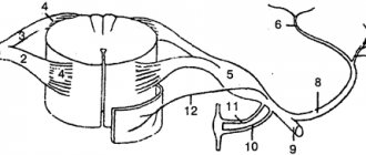
The peripheral section within the olfactory brain includes bulbs, triangles, and pathways. The peripheral section also includes the perforated substance (a section of the cerebral hemisphere pierced with holes for blood vessels, located on its lower surface posterior to the triangle) and olfactory stripes (branches of the olfactory sulcus). The part of the brain responsible for smell interacts with the analyzer, which is represented by the following departments:
- Receptor. Contains receptors located in the mucous membrane of the upper nasal passages. In total, humans have about 10 million receptors that perceive odors.
- Conductive. The olfactory nerve, consisting of 15-20 nerve filaments passing through the ethmoid bone in the direction of the anterior fossa of the cranium.
- Bulbs (bulbus olfactorius), pathways, triangles and other subcortical structures of the system.
- Cortical area. Located in the lower segment of the temporal lobe and uncus in the region of the parahippocampal gyrus.
The olfactory triangle is a part of the brain that regulates the function of smell, which is represented by the expansion of pathways that transmit signals from receptors to the cortical analyzer. The olfactory triangle (trigonum olfactorium) borders the perforated substance. The mechanism of odor perception is realized through the comparison of odorous molecules with receptors located in the villi of neurosensory cells.
Separate groups of receptors perceive certain aromatic molecules. Several groups of receptors are involved in the recognition of one odor, the total number of which can reach several thousand. Information entering the brain along the pathways is summarized and compared with data stored in memory.
The process of adapting information about an aromatic substance lasts several seconds or minutes. The duration of the process depends on the concentration of the aromatic substance in the air and the speed of air flow in the area of the olfactory epithelium. The sensitivity of human receptors is quite high. The receptor responds to a single molecule of aromatic substance.
Structure of the nose
In humans, the organ of smell is located in the nasal cavity. In fact, not all animals have a sense of smell connected to their nose. For example, in fish, the organ of smell (which also happens to be the organ of taste) is located directly on the surface of the body along its entire length. In humans, this works a little differently - we have a nose that performs several functions, one of which is smell.
Let's break down the basic anatomy of the nose. When we talk about the nose from an anatomical point of view, we mean two anatomical structures - the external nose (nasus externus) and the nasal cavity (cavitas nasi).
External nose
The external nose is represented by bone and cartilaginous parts. In this illustration from Sinelnikov’s atlas, the cartilaginous part of the nose is tinted blue:
The bony part of the external nose consists of two paired bones - the upper jaw, or more precisely, its frontal process (processus frontalis maxillae), and the nasal bone (os nasale). Many atlases also indicate that the frontal bone is involved in the formation of the external nose. In our illustration, I have highlighted the frontal process of the lower jaw in green and the nasal bone in red.
The cartilaginous part of the nose consists of several sections of cartilage tissue. These areas are quite large and have their own names. Thus, a large layer of cartilage that connects to the root of the nose is called lateral cartilage (cartilago nasi lateralis).
If we move a little to the top of the nose, we will see two large layers of cartilage tissue that are located right on the wings of the nose. These are large cartilages of the wings of the nose (cartilago alaris major). More lateral are the small cartilages of the wings of the nose (carttilagines alares minores), usually two or three paired sections of cartilaginous tissue.
Now we will dwell in more detail on the structure of the external nose as a whole. The external nose has a dorsum (dorsum nasi), an apex (apex nasi), wings (alae nasi) and a root (radix nasi). In this illustration I have marked with colors:
- Red - the top of the nose;
- Green - the back of the nose;
- Yellow - the root of the nose;
- Orange - the wing of the nose.
As you can see, the root of the nose is represented by a bone part, the back and wings are a cartilaginous part, and the tip of the nose consists of cartilage and a noticeable layer of subcutaneous fat.
Nasal cavity
When we talk about any body cavity, we, as anatomists, are interested, first of all, in its boundaries. The nasal cavity is no exception, so now we will examine its boundaries. First, let's look at the nasal cavity in an illustration from my favorite atlas:
This is what the upper and lower boundaries of the nasal cavity look like:
To figure out which bones form the boundaries of the nasal cavity, we need a slightly different drawing, a little less topographical and a little more bony:
The nasal cavity borders on top with the cranial cavity, more precisely, with the anterior cranial fossa. These two cavities are separated by the ethmoid bone, the frontal bone and the body of the sphenoid bone. In the picture above, the frontal bone is highlighted in light beige, the ethmoid bone is highlighted in green, and the sphenoid bone is highlighted in yellow. Pay attention to the cribriform plate of the ethmoid bone, it is fundamentally important for our topic today:
From below, the nasal cavity borders on the oral cavity. The nasal cavity and oral cavity are separated by the palatine process of the upper jaw and the horizontal plate of the palatine bone (these two bone formations are also called the hard palate). In our bone illustration, the upper jaw is highlighted in lilac and the palatine bone is highlighted in light blue.
At the front, the nasal cavity opens into the external environment through the openings of the nostrils, and at the back into the pharynx, more precisely, into its upper part, which is called the nasopharynx.
By the way, as you remember, the Eustachian tube also opens into the nasal pharynx, which connects it to the cavity of the middle ear. The pharynx communicates with three cavities at once - the mouth, nose and middle ear, and the mouth and nose communicate with the external environment. This creates quite convenient ways for the infection to spread in three directions at once. That is why one doctor, an otolaryngologist, treats diseases of the pharynx, ear and nose.
And we will return to the walls of the nasal cavity.
The lateral wall of the nasal cavity is formed by several bones. We are lucky because the bone specimen we are looking at shows the lateral wall perfectly.
So what bones form the lateral wall of the nose? First of all, this is, of course, the upper jaw, more precisely, its frontal process and the nasal surface. Next to it is the already familiar palatine bone with its perpendicular plate; it also forms the lateral wall of the nose.
If we go a little higher along the lateral wall and move towards the tip of the nose, we will see the nasal bone. Moving in the opposite direction, we will see the little lacrimal bone, it is indicated in gray. Two green round formations literally hang over it. These are parts of the ethmoid bone, which are called the nasal concha, superior and middle. The third rounded formation is the inferior nasal concha, which is an independent bone.
Finally, if we move towards the occiput, we see that the most posterior part of the lateral wall is formed by the medial pterygoid process of the sphenoid bone, this area is colored yellow.
Have you ever picked your nose? If you've ever done this, you may have noticed the thing that splits your nose into two parts. This is the nasal septum (septum nasi). The nasal septum is the medial wall of each half of the nasal cavity. In this illustration from Gray's atlas we see the nasal septum:
The nasal septum, like many things in our topic today, has bone (pars ossea septi nasi) and cartilage (cartilago septi nasi) parts. The cartilaginous part is located on the outside, it is elastic and mobile. The bony part is located internally, it is formed by the perpendicular plate of the ethmoid bone and the vomer. I note that all the structures of the ethmoid bone are very fragile; anatomists of the past called it paper bone because of this. I highlighted the boundaries of the vomer in yellow, and the perpendicular plate of the ethmoid bone in blue:
Nasal turbinates and nasal passages
This is a very easy question, which, for some reason, causes great difficulties for many students. As you remember, three large bony cells, called turbinates, are adjacent to the lateral wall of the nose.
The superior and middle turbinates are components of the ethmoid (paper) bone, and the inferior turbinate is an independent bone that motionlessly articulates with the upper jaw and the palatine bone. Let's use a naturalistic picture from Sinelnikov's atlas to look at the nasal turbinates and nasal passages.
There's nothing complicated about the turbinates, is there? Let's highlight the upper turbinate in blue, the middle one in yellow, and the lower one in orange.
The nasal conchas are not tightly adjacent to each other (otherwise we would not be able to breathe); there are quite large holes between them. These openings are called the nasal passages. The superior meatus is the opening between the superior and inferior turbinates, the middle meatus is the opening between the middle and inferior turbinates, and the inferior meatus is the opening between the inferior turbinate and the hard palate.
There is also a common meatus, which is the space between all the turbinates and the nasal septum.
Functions
Thanks to the ability to distinguish odors, a person receives information about the surrounding biochemical environment. Other functions of the olfactory brain are associated with the formation of instinctive reactions when interacting with the outside world. The sense of smell in the context of the formation of reflex behavioral patterns in response to external stimuli covers the nutritional, protective, sexual, and orientation spheres of the body’s life. The olfactory brain and limbic system are structures that are involved in the regulation of processes:
- Formation of emotions.
- Behavioral, motivational reactions.
- Sleep-wake mode.
- Learning, intuition and memory.
Odorants are volatile substances that have an odor. Once in the nasal passages, odorants reach the olfactory epithelium (Regio olfactorius), where they interact with chemoreceptors. The olfactory zone is located on the mucosal surface in the upper region of the nasal passages, covers an area of about 2.5 cm2, and contains about 50 million sensory neurons.
Axonal branches of nerve cells pass through the ethmoid bone towards the olfactory bulb within the brain. Here the processes form synaptic connections with the dendrites of second-level nerve cells and unite into glomeruli. Level 2 neurons include mitral (the largest cellular elements) and tufted cells. Their branches emerge from the bulb, which is a multilayer neural network.
The axons of nerve cells (mitral, fascicular) extending from the bulb transmit information to the primary zones of the cortex, where the stimulus is decoded, the information is interpreted and an adequate reflex response is formed. The signal is then transmitted to the center of smell, located in the cortical layer of the brain, which regulates the higher functions of the psyche, where a conscious sensation of aroma arises.
At the same time, the stimulus enters the limbic system, where emotions and conscious behavioral reactions are formed that are adequate to the perceived smell. Odorants are transmitted predominantly along unmyelinated (not covered with a protective layer of myelin) nerve fibers. Impaired smell function worsens a person's quality of life. For example, many people with such disorders have impaired digestive function, which is due to the close relationship between the smell and taste of food.
A little about smell
The sense of smell is a special sense that allows us to respond to aromas. Odorous substances act on the upper part of the nasal mucosa, where the olfactory nerve is located. Simply put, the sense of smell is the ability to sense smells. Each person perceives them differently, which is why experts distinguish three groups of people:
- Macromatics - have a keen sense of smell and are sensitive to aromas. They can distinguish all existing shades of odors.
- Micromatics - it will take them a little time to determine the intensity of the aroma. According to statistics, there are more such people.
- Anosmatics are people who cannot smell at all. Their number is small.
The sense of smell is gone
Sometimes it happens that a person’s sense of smell disappears or deteriorates. Why is this happening? Most often this is due to damage to the nasal mucosa or intracranial processes. Agree that the loss of smell, just like the loss of taste, is not a very pleasant situation for a person. What causes such a problem?
- Swelling of the mucous membrane of the nasal septum. This occurs due to the presence of diseases such as ARVI, rhinitis, sinusitis, as well as a deviated septum, allergies, and the presence of nasal polyps.
- Impaired secretion of the mucous membrane. At the same time, the cilia, thanks to which odors are captured, are immersed in the secretion.
- Disruption of the olfactory neuroepithelium. This occurs when inhaling toxic substances or acute infections.
- Traumatic brain injuries.
- Tumors.
- Taking neurotoxic drugs.
- Some congenital diseases.
- Neurosurgical intervention.
- Receptor dysfunction.
- Underdevelopment of the olfactory pathways.
- Smoking.
- Age-related changes.
How can you smell
The sense organs are not as complicated as it seems at first glance. The smell comes from the environment, irritates the receptors, then a signal is sent to the brain and its feedback determines how we smell odorous substances.
Research into the human sense of smell has been going on for several centuries. About 2000 years ago, the first theory was proposed about how and why a person perceives aromatic substances and which organs are responsible for this. If we turn to the theory of these studies and try to understand, then at the moment there are 4 assumptions about how we understand and hear the smelling world around us.
The classification includes the following methods of smell perception:
- Contact;
- Wave;
- Physical;
- Chemical.
Based on the research of scientists, the wave theory cannot fully apply to humans, but it can be successfully applied to mammals. For example, albatrosses can smell fish from an altitude of 3,000 meters, and bees can smell honey even in a sealed box.
The physical theory put forward by the scientist G. M. Deason indicates that the molecules of the odorous substance are capable of entering into a chemical reaction with the molecules of the human sense of smell and thereby transmit a signal to the brain through nerve endings.
Chemical theory says that in nature there are only 7 primary odors, which, when combined with each other, lead to the formation of various aromatic combinations. Scientist J. Eimour hypothesized that the molecules of all these combinations have their own shape. For example, the musk molecule is presented in the form of a disk, mint - in the form of a blade, ethers - in the form of a stick. Oddly enough, it was this theory that became established in human biology and anatomy.
Restoring the sense of smell
If the ability to recognize odors is lost, it should be returned. According to statistics, most often the lack of sense of smell occurs due to colds or the presence of polyps. In a word, when a mechanical obstacle appears that prevents you from enjoying the aromas. Based on the reason, a decision should be made on how to restore the sense of smell.
For diseases of the mucous membrane, doctors act as follows:
- Eliminate all factors that caused the loss of smell.
- Medications are prescribed on an individual basis.
- Physiotherapy is prescribed.
- If necessary, surgical treatment is used.
Taste
Taste sensations are contact sensations that arise when a sensory organ (tongue) comes into contact with the object itself. The sense of taste detects molecules dissolved in saliva.
There are four main qualities of taste stimuli: sour, sweet, bitter, salty. From the combinations of these four sensations, to which the movements of the tongue are added, a complex of taste sensations arises.
Initially, the sensory process occurs in the taste buds, and each of the papillae has from 50 to 150 receptor cells, which quickly wear out from contact with food and are then renewed. The sensory signals then travel along nerves to the hindbrain, thalamus, and gustatory cortex, which processes taste.
Taste sensations, like olfactory sensations, increase a person’s appetite. By analyzing the quality of food, taste also performs a protective function and is important for survival. When fasting, taste sensitivity increases, when saturated or satiated, it decreases.
What to do and what not to use
When the illness passes, the sense of smell is restored. If the patient uses nasal drops, which have the property of constricting blood vessels, then the sense of smell may not be restored after the swelling subsides. Also, their long-term use can lead to the human body getting used to them. Then they will not have a relieving effect, but, on the contrary, can lead to a worsening of the condition. Namely, due to long-term use of drops, swelling of the inner surface of the nose may appear. Further, a situation may arise that the recognition of odors will not return to a person, even when he is cured of the illness that bothers him.
To prevent your body from becoming accustomed to using the drops, you should wait a few days and not use them. To relieve nasal congestion and relieve swelling, it is recommended to use a salt solution of weak consistency. Pharmacies also sell special medications. They include sea salt and relieve swelling in the nose.