The dendrite of a neuron (dendrum - branch) is a process of the neuron body through which it receives a signal from other cells. The dendrite receives a signal from the axon of another neuron or a receptor protein that responds to the environment.
Answering the question of what dendrites are, we can say that traditionally dendrites are considered as antennas of a neuron. Information exchange occurs in one direction: from axon to dendrite. The more dendrites a neuron has, the more information channels, the more complex decisions the neuron makes.
Synaptic cleft
You may be interested in: Semantic barriers and ways to eliminate them
The signal from other cells reaches the body of the neuron along one of its dendrites. The dendrite in the human nervous system usually receives a chemical signal (neurotransmitter) from the axon. The junction of a dendrite and an axon is called a synapse.
Synapses allow precise messages to be passed from neuron to neuron. Thanks to synapses, there is neuroplasticity and the ability to fine-tune the functions and behavior of the body.
The dendrite contains receptors that receive the neurotransmitter. Receptors are specialized proteins that capture a neurotransmitter molecule and, depending on their type, trigger further reactions in the cell.
Differences from axons
The axon serves to transmit nerve impulses from the body of the nerve cell, which is otherwise called the soma, to the executive organs. The ending of the axon is an element of the synapse through which synaptic transmission of signals occurs between individual cells of the nervous tissue.
Dendritic cells have a branched structure. Dendritic processes branch throughout their entire length, in contrast to the axon, which branches only in the terminal segment, forming terminals. Unlike an axon, whose length can exceed 1 meter, a dendrite is a short process (about 700 µm). Other differences between dendrite and axon:
- Multidirectional conduction of impulses (dendrites - to the neuron body, axon - from the neuron body).
- Different thickness (the dendrite becomes thinner as it moves away from the body, the axon retains the same cross-sectional diameter along its entire length). The diameter of the axons of different cells is about 0.3-16 µm. The thicker the axon, the higher the speed of impulses propagating along it.
- The presence of a myelin sheath (dendrites of the central nervous system do not have a myelin sheath, but axons have a myelin sheath).
Dendritic transport involves the movement along the trunk of the process of protein substances and enzymes from the soma to the terminal segments. Unlike dendritic transport, axonal transport involves a continuous flow of axoplasm in both directions. The transport mechanism is supported by microtubules and proteins (kinesin - movement inside microtubules, dynein - movement along the surface of microtubules).
The movement of substances along the trunk is carried out through the expenditure of ATP. The size of dendritic spines correlates with neuronal activity. Stimuli coming from the external environment are converted into bioelectric signals. The nerve impulse is a wave of excitation spreading along the process. The process of generating the energy necessary to maintain dendritic transport occurs in mitochondria.
Dendritic spines
Small growths called spines form on the dendrites. The latter can take many forms, but the most persistent is that of a fungus.
The number of dendritic spines ranges from 20 to 50 per 10 µm of dendrite length. The spines are very variable in shape and volume.
There are 86 billion neurons in the brain. Axons, dendrites and cell bodies of neurons form huge neural networks.
Dendrites are responsible for learning and memory, and also control the balance of the system. When there is a local strengthening of connections between certain neurons, it is in the dendrites that the production of a protein increases, regulating the decrease in the activity of other synapses.
Dendrite processes are important receivers of nerve impulses
The dendritic tree, together with the neuron body (perikaryon), is the part of the nerve cell that receives electrical impulses. During the reception of this impulse, an important role is played by the change in the electrical potential of the membrane in the area of the synaptic connection. There is the concept of a threshold change in potential, and all impulses that are less than this threshold value will not be transmitted properly by the dendrite to the body of the neuron.
If the electrical impulse that comes from the axon is weak, then it will not be able to lead to a sufficient change in membrane potential, and the signal will fade. However, dendrites are very sensitive parts of a neuron because they possess so-called dendritic spines - small elongated areas of cytoplasm that lead to a significant increase in functional area. Thanks to this feature, the system of short processes is capable of collecting information about tens of thousands of weak impulses and transmitting them further through the neural network.
If during the growth and development of a nerve cell any failure occurs, as a result of which the dendrites of neurons do not develop their network of spines sufficiently, then a person may experience a perception deficit, that is, the function of receiving nerve impulses will be impaired.
Training and Spikes
Dendritic spines are responsible for the possibility of learning and memory formation. Thanks to the spines and their plasticity, a neuron can easily connect to one or another neighbor and quickly disconnect from them, controlling the possibility of receiving a signal.
It would be logical to assume that if synaptic connections are responsible for memories, then their plasticity is a problem for maintaining memories of the past. In 2009, Nature published a publication in which the authors examined how learning experience affects synaptic connections in mice.
The work showed that a large number of new spines formed from a new experience disappeared over time if the experience was not repeated periodically. But those that were preserved were most likely responsible for the acquired skills.
Moreover, if the training was repeated for a long time, spines were removed; apparently, the removed spines were responsible for incorrect actions. Learning and daily sensory experience leave permanent marks in the form of a small group of spines formed at different stages of learning.
What are dendrites if not a huge library of memories? But the main problem of dendritic spines is that they are very sensitive to any mechanical and chemical influences. Therefore, brain injuries, even if localized in one place, usually affect the entire neural network.
Definition of dendrites
Dendrites are projections of a neuron (nerve cell) that receive signals (information) from other neurons.
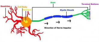
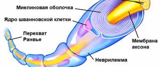
The transfer of information from one neuron to another is carried out using chemical signals and electrical impulses, that is, electrochemical signals. The transmission of information usually arrives at the dendrites through chemical signals, then it travels to the cell body (soma), continues along the neuron in the form of electrical impulses, and is finally transmitted to the next neuron at the synapse, which is the place where two neurons exchange information using chemical signals. . At a synapse, the end of one neuron and the beginning—dendrites—of another meet. This picture shows what a dendrite looks like in a neuron:
Sleep and learning
A 2014 study by ZG Yang showed how, after 24 hours of training and sleep, new dendritic spines appeared in mice and some of the existing ones disappeared. The authors note that the rate of formation of new spines in mice trained to learn a new behavior was significantly higher within 6 hours of training compared to untrained mice.
In addition, the authors showed that spines form much more slowly when mice are deprived of sleep. And neither new skill training nor late sleep can correct the situation.
Role in neural processes
A person is born with a genetically determined number of dendritic processes on each neuron. The gradual increase and complexity of brain structures and the construction of the nervous system, which occur during postnatal development, is realized through branching and an increase in the mass of dendrites.
According to numerous studies, at the peak of development of the nervous system, dendrites occupy about 60-75% of the total mass of nerve cells.
According to fundamental theories describing the principles of operation of the nervous system, dendrites have always been considered the part of the neuron that receives an impulse and conducts it to the body of the nerve cell.
However, modern research by neuroscientists using the latest technologies such as microelectrodes has revealed greater electrical activity of dendrites compared to the cell body.
These studies confirmed the fact that dendritic endings are capable of generating electrical impulses themselves - local action potentials.
Dendrite as an independent unit
What dendrites are is still being figured out. The fact is that it is difficult to study the behavior and functions of dendrites in living objects.
If the size of a neuron is about ten microns, then the length of a dendrite can reach up to a thousand. Dendrites are usually understood as not very active participants in the process.
In 2021, a study was published in the journal Science that reconsiders the classic view of dendrites. It turned out that dendrites generate signals several times more often than the neuron body does, which suggests that information is encoded at the dendritic level as well.
It was previously discovered that if during an experience the cell bodies of neurons were activated and the dendrites were silent, then long-term memory was not formed regarding this experience. It was suggested that the activity of neurons is associated more with real time, with current experiences, and dendrites - with what will remain in memory.
What are dendrites, given the new data? These are amazing structures that make up 90% of the nervous tissue and perhaps take on most of the work of storing and transforming experience.
General information
Neuron is the basic structural and functional unit of nervous tissue. A nerve cell is a formation with numerous processes measuring 4-130 microns. Several (less often one) dendrites and a single axon extend from a neuron. A dendrite in biology is a process that transmits excitation from peripheral receptors to the body of a neuron, which determines its leading role in the perception of external stimuli. Features of dendritic branches that are observed during microscopic studies:
- Microtubule system.
- The presence of spines extending from a common axis.
- Presence of branch nodes.
- The presence of endoplasmic reticulum (an intracellular organelle represented by a system of tubules, cavities, vesicles surrounded by a membrane).
The processes located next to the soma (body), thickened, form a large number of synaptic contacts. The membrane of the process, like the membrane of the neuron itself, consists of a large number of protein molecules that play the role of chemical receptors. Receptor formations are endowed with specific sensitivity to certain chemical compounds.
The designated chemical substances - neurotransmitters of inhibition and excitation, are actively involved in the process when impulses spread through the nervous tissue and arrive at the soma. The structure of the dendrite suggests the presence of spines that form synaptic contacts with terminals - the end sections of hundreds of thousands of nerve cells. A huge number of spines are located on the processes of nerve cells that form the cortex of the cerebral hemispheres.
The dendritic spine is formed from a body and a head. The size and shape of the structural components of the spine vary significantly. Thanks to the spines, the area of the postsynaptic membrane significantly increases. Spines in the dendritic structure are labile formations that, under the influence of external stimuli, change configuration, degenerate (destroy), and regenerate (appear again).
The number of synapses determines the quality of impulse transmission and the speed at which they propagate. Several dendritic branches form a single branch. The totality of all dendrites is a dendritic tree - a surface that perceives external stimuli. Research shows that dendritic trees make up 90% of the brain matter.