Diagnostics
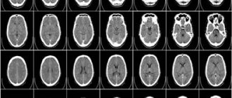
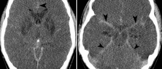
As we have already mentioned, leukoaraiosis is visible on CT and MRI neuroimages of the brain , with a decreased and increased frequency, respectively. MRI of the brain , including T2 and FLAIR series, is the most common technique for detecting leukoaraiosis. The Fazekas grading scale (Fig. 3) is most often used to determine the degree of white matter lesions on MRI images: – grade 0 : no lesions; – grade 1 : isolated lesions; – degree 2 : lesions are connected to each other; – grade 3 : lesions covering specific areas of the brain.
Fig. 3 Fazekas rating scale.
– According to the Filum System® method:
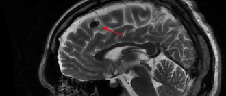
We often see images of leukoaraiosis in the white matter of the brain in horizontal MRI sections with a T2 signal, most often it is grade 1 and less often grade 2, in patients diagnosed with pathologies included in filum terminale disease or neuro-craniovertebral syndrome. Their number is about 45 % of our entire database and includes all age groups.
Degrees and symptoms
As a descriptive term, leukoaraiosis itself has no symptoms, but varying degrees of neuroimaging of these lesions are associated with corresponding symptoms. The main symptoms associated with some degrees of leukoaraiosis are ischemic stroke, cognitive impairment, gait disturbances, mood swings and sphincter disorders. There are the following degrees of leukoaraiosis from 0 to III: – 0 and I degrees are quite mild and without corresponding known specific symptoms. – II and especially III degrees are more severe, this type of leukoaraiosis affects a wider area of the white matter of the brain, damages both hemispheres, lesions are widely distributed throughout the brain, the disease becomes progressive. It is usually found in people with mental disorders that are associated with neurological damage or symptoms of dementia.
Causes
Currently, the pathogenesis of leukoaraiosis is controversial. Some authors recall that it has not yet been clarified to what extent the mechanisms that provoke small foci of ischemia coincide with those that cause extensive diffuse leukoaraiosis. In addition, it is unknown whether the pathological abnormalities associated with leukoaraiosis are the cause or consequence of lesions in the white matter. There are two main hypotheses: an abnormality in self-regulation of blood flow or a violation of the blood-brain barrier . The most recognized mechanism for the appearance of leukoaraiosis today is chronic ischemia due to damage to penetrating arteries. Hypertension and other pathologies affecting blood flow and oxygen supply to the brain appear to be closely associated with extensive leukoaraiosis. – According to the Filum System® method: Based on the observed association between leukoaraiosis and individual small foci of ischemia on images in patients diagnosed with filum terminale disease and neuro-craniovertebral syndrome , our center has developed a hypothesis for a possible pathogenesis: leukoaraiosis may also be associated with tension of the spinal cord due to too tight filum terminale. In this case, ischemia can appear in all tissues of the nervous system due to the collapse of small-diameter blood vessels.
Treatment
There are usually several effective treatments to delay the development of leukoaraiosis, which are recommended depending on the disease to which it occurs. The most appropriate treatment to delay the development of leukoaraiosis is to maintain a balanced diet, with foods rich in folic acid, folates and B vitamins. It is recommended to avoid toxic substances and an unhealthy lifestyle. – According to the Filum System® method : Given the fact that leukoaraiosis is common in patients with filum terminale disease, in our center patients who have areas of ischemia in the white matter on brain MRI are recommended to undergo a diagnosis using the Filum System® in order to determine whether there is disease of the filum terminale. If there is tension on the filum terminale, then dissection of the filum terminale is proposed with an exclusive minimally invasive technique. In several cases, a decrease in leukoaraiosis has been observed after cutting the filum terminale, further suggesting that its occurrence may be associated with abnormal tension in the nervous system.
Risk factors
On the one hand, recognized risk factors for cerebrovascular disease relate to hereditary or individual characteristics and lifestyle . On the other hand, the main risk factors for the appearance of leukoaraiosis are: age and hypertension , although it is also associated with diseases such as diabetes mellitus, heart disease or arterial stenosis, which affect the disturbance of the microcirculation of the brain, leading to demyelization of vascular origin, which can provoke the appearance of foci of ischemia. Leukoaraiosis is also associated with older age and senile dementia , although it is often found in relatively young people (approximately 40 years of age) who use drugs such as heroin, tobacco , or are in constant contact with toxic substances . Such people are prone to the appearance of leukoaraiosis of any degree. Arteriosclerosis also considered one of the factors in its appearance. – According to the Filum System® method : Patients with leukoaraiosis in the white matter of the brain and diagnoses of filum terminale disease and neuro-craniovertebral syndrome have a common risk factor: family predisposition . Tension in the nervous system caused by filum terminale (filum terminale disease) can cause disruption of the blood supply to the spinal cord and throughout the nervous system. This is a genetic disease that is inherited.