There are a number of anatomical features of the brain of each person. Sometimes such specificity is considered physiological; in other situations, deviations from the norm can give rise to manifestations of a pathological process. One such condition is asymmetry of the lateral ventricles of the brain. On the one hand, such cerebral specificity is not considered a separate nosological entity, and its clinical symptoms may be absent. However, often ventricular asymmetry may indicate the presence of a number of diseases.
Asymmetry of the lateral ventricles is a condition in which expansion of the lateral ventricular cavities occurs. However, their sizes do not correspond to each other. Most often, asymmetry is diagnosed in newborns and children in the first year of life, as a manifestation of perinatal pathology of the nervous system. However, cases of increased volume of the lateral ventricles in adults are not uncommon.
Asymmetry of the lateral ventricles is not an independent disease, but serves only as a symptom of a pathological condition.
Structure and functions of the ventricles of the brain
The brain has four chambers. They produce and circulate in the cerebrospinal fluid. Its main job is to transport nutrients to brain cells, remove waste products from the brain, and absorb head impacts.
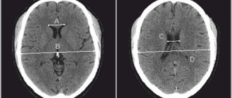
Two lateral ventricles (ventriculi lateralis) are located in the cerebral hemispheres - the first on the left, the second on the right. The normal depth of the sinuses is from 1 to 4 mm. An increase in the capacity of the organ to 5 mm leads to a noticeable change in shape: the lateral C-shaped curvature disappears, the defect acquires a rounded shape.
In this case we are talking about the initial stage of asymmetry of the lateral ventricles. Many people wonder what it is and how dangerous is this pathology?
Anatomy of the ventricular system of the brain
The ventricles of the brain are a cerebral system of cavities communicating with each other, the subarachnoid space and the spinal canal. Their inner surface is lined with ependyma. Under this layer are the choroid plexuses, which produce cerebrospinal fluid.
The lateral (or lateral) ventricles are the most voluminous. They are localized on both sides of the midline and have pairs of anterior, posterior and lower horns that are symmetrical relative to each other. The lateral ventricular cavities communicate with each other with the third ventricle, located between the thalami, through the foramen of Monroe. Between the cerebellum and the brain stem is the fourth ventricle. From it, the cerebrospinal fluid enters the subarachnoid space through the foramina of Luschka (paired) and Magendie (unpaired).
Causes of ventricular dilatation
An increase in the size of the lateral ventricles is formed as a result of impaired circulation of the cerebrospinal fluid. This picture emerges when:
- overproduction of cerebrospinal fluid;
- impaired adsorption of cerebrospinal fluid;
- difficulty in the outflow of cerebrospinal fluid.
Changes in liquor dynamics due to overproduction and slower reabsorption of cerebrospinal fluid occur as a result of irritation of the ventricular choroidal plexuses and the arachnoid membrane of the brain by a pathological process (most often as a result of neuroinfection).
Difficulty in the outflow of cerebrospinal fluid is caused by blockage of the cerebrospinal fluid pathways by neoplasms and cysts.
The main reasons for the expansion of the lateral ventricles are:
- neuroinfections (meningitis, meningoencephalitis);
- skull injuries;
- brain tumors;
- idiopathic hydrocephalus;
- formed hematomas;
- hemorrhagic stroke;
- thrombosis of cerebral vessels;
- atypical embryonic anlage of the ventricular system.
Clinical signs
Manifestations of asymmetry of the lateral ventricles in an adult are often absent. In this case, such a structure of cerebral structures is not considered a pathology; it is revealed as a finding during neuroimaging. Along with this, a pronounced disturbance of liquor dynamics leads to the following clinical symptoms:
- headaches;
- feeling of heaviness and fullness in the head;
- dizziness;
- nausea;
- vomiting that does not bring relief;
- anxiety-phobic syndrome;
- apathy.
In addition, the clinical picture of ventricular asymmetry is complemented by symptoms of the disease that forms the basis of this condition. Paresis, pathology of cranial nerves, sensitivity disorders, cognitive decline, and cerebellar disorders may occur.
Causes of asymmetry of the lateral ventricles
The expansion (expansion) of caverns is associated with the accumulation of liquors in them. There may be two reasons for this: excessive production of fluid or disruption of its outflow.
The following diseases can provoke an increase in the amount of cerebrospinal fluid:
- Hydrocephalus, in which the absorption of cerebral fluid is impaired.
- Changes in the central nervous system - schizophrenia, bipolar disorder.
Inadequate circulation of cerebrospinal fluid occurs when mechanical compression of the exit ducts occurs.
This phenomenon is most often caused by damaging factors:
- Traumatic brain injuries;
- Malformation of the Sylvian canal (a narrow canal connecting the third and fourth ventricles);
- Brain hemorrhages;
- Intracranial tumors, cysts, papillomas, hematomas;
- Meningitis;
- Thrombosis of cerebral veins.
Lateroventriculoasymmetry of the brain often occurs in newborns, more often in premature infants.
What it is
Ventriculoasymmetry is a pathological condition in which the lateral ventricles are asymmetrical in relation to each other. Normally, they have a symmetrical arrangement and the same volume, however, due to external or internal factors, their relative position changes.
Ventricular asymmetry is considered a pathology only if it leads to dysfunction of the nervous system, deterioration of the body’s functioning and the development of symptoms. If the violation of symmetry is minimal and does not lead to disorders, this is considered an anatomical feature of the brain.
In the medical classification there is no such nosological unit (independent disease) as ventricular asymmetry, however, their asymmetry can lead to disruption of the circulation of cerebrospinal fluid, which in turn determines the clinical picture.
Lateroventriculoasymmetry is often latent (hidden) and does not cause symptoms. As a rule, the pathology of this system is determined by chance, by neuroimaging on computer or magnetic resonance imaging during a routine examination or after a head injury.
The change in symmetry itself is not dangerous to humans. The consequences that asymmetry can lead to are dangerous. For example, impaired liquor dynamics (fluid outflow), compression of cranial nerves, increased intracranial pressure. All consequences trigger a chain that develops its clinical picture.
Asymmetry is mainly diagnosed at an early age (up to one year) and in children.
Interhemispheric asymmetry with asymmetry of the cavities is not disturbed.
Symptoms of enlarged ventricles
The mechanism of development of pathology has common features, regardless of the reasons. Fluid accumulates in the ventricles, which expand and contract brain tissue. The pressure in the cavities themselves also increases.
Patients may complain of sudden headaches, nausea and vomiting, a feeling of distraction in the eyeballs, hearing and vision problems. Performance gradually decreases, memory impairment, apathy, and drowsiness appear.
All these are symptoms of pathology of the spinal system. If the abnormality is not detected and treated early, it becomes a chronic condition.
Who needs it?
As already mentioned, it is advisable to do NSG for all infants at 1 month or at 3 months, if for some reason a medical examination was not carried out on the child at four weeks of age.
The advice of the Ministry of Health is, of course, advisory in nature, and therefore parents can refuse the examination, but this is not recommended, because if there are pathologies later, when the “fontanel” begins to close, diagnosis will be difficult.
However, there are categories of children for whom neurosonography is especially recommended. First of all, these are children who were born prematurely (up to 37 weeks of pregnancy inclusive). Premature babies are a special risk category, including the likelihood of developing pathologies in the brain and central nervous system. Experts also consider it obligatory to carry out NSG for children whose birth occurred surgically - if the woman had a cesarean section.
If a child exhibits the following symptoms during the first weeks of life, parents should also not refuse to undergo neurosonography:
- the child behaves strangely - in the absence of illnesses, he eats poorly, regurgitates frequently and profusely, he is inactive, does not show strong emotions, often cries, sleeps superficially, constantly wakes up, if the baby has pronounced limbs, chin, or strabismus;
- the baby often cries, throws back his head and arches his back (this may be an indirect sign of increased inflammation);
- the baby hears poorly or reacts poorly to visual stimuli, does not follow the toy with his eyes, and cannot focus his gaze on the mother’s face;
- low blood pressure in the baby, fainting, convulsions;
- severe coordination disorders (infant flapping and shuddering have nothing to do with it);
- the child has a birth injury or he fell, hit his head, or there was a sharp tilting of the head after birth.
If a child is scheduled for vascular or heart surgery in the near future, NSG is mandatory. An unscheduled neurosonographic examination will be carried out in the event of a fall, because the method allows one to establish signs of a concussion, bruise, or the formation of cerebral hematomas.
It is imperative to examine children who were born with a low weight (less than 2700 grams), as well as children who were born with asymmetry (who have one ear lower than the other, one eye larger than the other, etc.)
External anatomical malformations (presence of extra fingers and toes, absence of limbs, etc.) are also a good reason for a careful ultrasound examination of the baby’s brain.
Children born after pregnancy, which was accompanied by fetal hypoxia and Rh conflict, must undergo NSG, since the long-term consequences of these unfavorable intrauterine conditions can be quite severe.
Diagnostics
Lateral ventricular asymmetry is detected using the following procedures:
- Ultrasound of the brain is the most informative examination;
- Examination of the fundus, detection of swelling of the eye disc, spasms, hemorrhages;
- Neurosonography;
- Magnetic resonance imaging and computed tomography of the brain.
If the above methods give conflicting results, lumbar puncture is recommended. Analysis of the cerebrospinal fluid reveals the cause of the curvature of the lateral sinuses.
Normal and abnormalities on MRI of the brain
How do you know if there are signs of disease in the pictures? The most important thing is to remember what the brain of a healthy person looks like. The doctor, studying the photographs of patients, constantly compares them with normal photographs stored in his head. To understand how this happens, look at the pictures below:
Here are two pictures taken in the same mode. The photo below is normal. What disease, in this case, is in the top picture? To understand this, you need to compare these images. The difference is clearly visible - in the top image there is a tumor on the right side of the brain. The difference is even more noticeable if you compare the left and right sides of the same photo.
Let's mark it with a red circle. Visually, it is a node, heterogeneous in color and different from the gray and white matter of the brain. In such cases, in order to accurately determine the boundaries of the tumor and determine its type, the study is repeated with contrast. The introduction of a contrast agent into the blood through the cubital vein leads to the accumulation of the contrast agent in the tumor tissues - normal healthy tissues practically do not accumulate it. And we get the following picture, shown in the figure on the right. The bright color of the tumor corresponds to the accumulated contrast - now you can not only tell where the tumor is, but also roughly determine that it is a benign tumor, since it has clear boundaries (malignant tumors grow into surrounding tissues, which is why the boundaries will be blurred and not so clear ).
Thus, interpretation of the results of MRI of the brain is carried out by comparing the obtained images with the norm. If there are no differences, we can say that the patient whose images are examined by the doctor is most likely healthy. Everything is compared - shape, size of anatomical structures, localization, symmetry, amount of cerebrospinal fluid in the cavities of the brain, and many other parameters. Each disease, be it stroke or multiple sclerosis, has its own characteristic symptoms.
Symptoms of asymmetry
Clinical manifestations of lateral ventricular asymmetry may differ depending on the level of intracranial pressure. The main symptom in this case is pain, which accompanies many diseases, including brain pathologies.
Among the clinical manifestations characteristic of the condition are the following:
- headache;
- nausea and vomiting syndrome;
- increase in skull size;
- animated postural reflexes;
- constant anxiety syndrome;
- restlessness, tearfulness;
- weakened grasping and swallowing reflexes;
- divergence of the sagittal suture;
- increase and increase in fontanelle tension;
- decreased muscle tone;
- tremor of the upper extremities;
- swelling of the optic disc of the eye;
- insomnia;
- loss of appetite;
- anemia;
- the occurrence of hallucinations;
- the appearance of a veil, “flies” before the eyes.
The above symptoms are more typical for pathology occurring in newborns and young children. In adults, asymmetry of the lateral ventricles is rarely accompanied by severe symptoms and is more often diagnosed accidentally during an ultrasound examination to detect other diseases.
A child who has asymmetry of the lateral ventricles refuses the breast, becomes restless, and cries constantly.
How it manifests itself
The main function of the ventricles is to secrete cerebrospinal fluid, as well as ensure its normal circulation in the subarachnoid space. If the balance of exchange and production of cerebrospinal fluid is disturbed, then stagnation is formed and, as a result, the walls of the cavities are stretched. The same slight expansion of the lateral segments may be a normal variant, but their asymmetry and enlargement of individual parts (for example, only the horn) will be a sign of the development of pathology.
Enlarged ventricles of the brain in an infant can be diagnosed with a congenital disease such as ventriculomegaly. It varies in severity:
- Slight expansion of the ventricles of the brain up to 11-12 mm, with no significant symptoms. It manifests itself in the child’s behavior: he becomes more excitable and irritable.
- Increasing the depth of the ventricles up to 15 mm. Most often, the pathology is accompanied by asymmetry and impaired blood supply to the affected area, which entails the appearance of seizures, an increase in head size and a lag in mental and physical development.
- Ventricular dilatation up to 20 mm is characterized by irreversible changes in brain structures and is often accompanied by Down syndrome and cerebral palsy in infants.
In adulthood, an increase in ventricular volume is manifested by the following symptoms:
- Gait disturbance, with the child walking “on tiptoes” or vice versa, focusing only on the heels.
- The appearance of visual disorders, such as squint, insufficient focus of the gaze, as well as double images when trying to see small details.
- Tremor of arms and legs.
- Behavioral disorders that manifest themselves in excessive lethargy and drowsiness, while it is difficult to captivate the child with any activity.
- The appearance of headaches due to increased intracranial pressure, sometimes nausea and even vomiting may occur.
- Dizziness.
- Frequent regurgitation, loss of appetite. Some newborns are able to refuse breastfeeding.