What is the expansion of the subarachnoid space
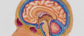
The human brain is surrounded by three protective membranes - hard, arachnoid and soft. The latter is directly adjacent to the medulla and provides its nutrition. The arachnoid membrane is connected to other membranes of the brain using connective tissue membranes. In areas where there are no membranes, there are cisterns.
With concomitant expansion of the subarachnoid space, arachnoiditis should be taken immediately.
The inflammatory process rapidly spreads to the brain tissue.The disease can lead to disability, serious visual impairment, hydrocephalus, and seizures.
Neurologist, reflexologist, hirudotherapist
Kislitsyna Ekaterina Nikolaevna
10 years of experience
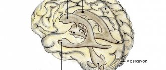
Cerebrospinal fluid circulates in the 4 ventricles of the brain (third, fourth and two lateral) and the subarachnoid space. Liquor performs the functions of feeding neurons, removing metabolic products, and mechanically protecting the brain.
The normal amount of cerebrospinal fluid in children is 80-120 ml, in adults 120-160 ml. It is updated 3-5 times per day. The rate of circulation of the cerebrospinal fluid is affected by the position of the body, the intensity of the heartbeat and breathing.
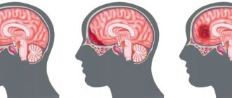
The expansion of the subarachnoid space occurs due to difficulty in the outflow of cerebrospinal fluid. Fluid accumulates in the subarachnoid spaces, dilating the ventricles of the brain.
The mechanisms of development of the disorder are different: the tumor mechanically blocks the paths for the flow of cerebrospinal fluid, inflammation provokes increased production of cerebrospinal fluid. Intracranial pressure may increase.
Signs
In almost all cases, when an enlarged gap is detected in a newborn, parents wonder how to treat it. However, if the widening of the gap is observed to a small extent, then no treatment is needed. Also, therapy is not necessary if the baby does not show other signs that bother him. If symptoms that accompany the clinical picture of a particular disease are identified, the necessary medications will be prescribed. For example, if a newborn shows signs of rickets, and he lives in a climate area where there is a lack of light, then the necessary vitamin complexes will be prescribed.
If symptoms are identified that indicate the development of intracranial pressure, the necessary diuretics will be prescribed. You also need to take potassium supplements in parallel, then this will prevent the development of hypokalemia and hypomagnesemia.
The specialist may also prescribe certain vascular drugs that are designed to improve cerebral circulation, and drugs with a calming effect before bed. All this is necessary only if there are alarming symptoms that indicate neurological problems.
Symptoms of the disease
The expansion of the subarachnoid space in an adult has characteristic signs:
- paralysis of the laryngeal muscles responsible for articulation and, as a result, speech impairment;
- loss of sonority ;
- difficulty swallowing;
- disturbances of visual and auditory perception;
- severe pain in the head, especially in the morning;
- nausea and vomiting accompanying headache;
- dizziness;
- drowsiness or sleep disturbances;
- dementia;
- impairment ;
- hallucinations;
- walking disorders - the patient is able to imitate walking movements in a lying position, but when standing up, walking is difficult, the gait is uncertain, shuffling;
- increased fatigue.
In young children, there is an abnormal enlargement of the skull as a result of developed hydrocephalus. Earlier symptoms are:
- increased sensitivity to sound and light stimuli;
- excessive regurgitation;
- unequal pupil size;
- strabismus;
- problems ;
- slowly overgrowing fontanel;
- swelling of the fontanel;
- restless behavior due to changing weather;
- tremor of the chin and limbs.
The intensity of symptoms depends on the causes of the disease and the severity. In the absence of adequate and timely treatment, expansion of the subarachnoid space in infants is dangerous due to the possible development of hydrocephalus.
Why can our articles be trusted?
We make health information clear, accessible and relevant.
- All articles are checked by practicing doctors.
- We take scientific literature and the latest research as a basis.
- We publish detailed articles that answer all questions.
This disease leads to a lag in motor development, seizures, paralysis, and intellectual impairment. The effects of hydrocephalus can last a lifetime.
Clinical symptoms of increased liquor spaces
Signs of dilation of the ventricles and subarachnoid cavities in newborns:
- Constant irritability;
- Negative reaction to strong noises, flashes of light;
- Restless sleep;
- Frequent regurgitation;
- Strabismus;
- Different pupil sizes;
- Moodiness when weather changes;
- Slow overgrowth of the fontanel;
- Twitching of the chin.
Deviations must be detected immediately after their occurrence. Correction of changes should be carried out immediately after detection.
Clinical symptoms of subarachnoid enlargement in adults:
- Dyspeptic disorders (vomiting and nausea);
- Headache;
- Drowsiness;
- Constant dizziness;
- Increased intracranial pressure;
- Dementia;
- Memory problems;
- Disorders of spatial orientation;
- Unsteady gait.
Determining clinical signs in a child and an adult requires contacting a neurologist. The specialist will detect the pathology and give a referral for an MRI of the convexital cerebrospinal fluid cavities.
At the initial stages there are no manifestations of pathology. Symptoms depend mainly on the severity of the deformity and the causes of the pathology. In small children, the disease is provoked by arachnoiditis, birth trauma, and meningitis. Tumors are a common etiological factor in the occurrence of pathology in adults. Compression of the cerebral parenchyma causes defects in the destruction of white and gray matter. The formed cavities are filled with liquor.
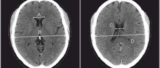
Analysis of brain tomograms in people with enlarged cerebrospinal fluid cavities: atrophic changes in the cortex of the frontal, occipital, and temporal regions. The number of convolutions is smoothed out, and blood accumulations are visible.
The presence of concomitant complications in a child is dangerous, so invasive diagnostic measures are postponed to the second year of life. A reliable diagnosis can be made after taking a biopsy of the enlarged cavities. Microscopic assessment of the properties of cerebrospinal fluid makes it possible to differentiate inflammatory, tumor, and ischemic changes.
Magnetic resonance imaging reveals accompanying pathological changes:
- Leukomalacia is a pathology of impulse signal transmission due to softening of the meninges;
- Inflammatory foci;
- Collections of blood.
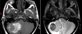
An MRI picture of a moderate expansion of the external cerebrospinal fluid spaces shows morphological changes, but a whole range of diagnostic methods is required to make a correct diagnosis.
Tomograms of hypertensive hydrocephalus
Reasons for expansion
In infants, the disorder occurs due to congenital malformations, birth injuries, brain tumors, and infectious diseases. In some cases, the subarachnoid space and ventricles return to normal by the age of two years. However, pathology requires constant monitoring by specialists who can determine the further prognosis of the disease and, if necessary, prescribe treatment.
In adults, expansion of the subarachnoid space occurs as a consequence of a stroke, vascular pathologies, head or spinal injuries, infectious and colds, and tumor cell degeneration. Meningitis, encephalitis, ventriculitis, tuberculosis, inflammation of the sinuses are diseases that affect the brain and can cause disruption of the circulation of cerebrospinal fluid.
The disease may indicate concomitant leptomeningitis or arachnoiditis - inflammatory processes in the tissues of the soft and arachnoid membranes of the brain. Long-term exposure to toxic substances, such as alcohol, arsenic, and lead, can cause inflammation.
The effectiveness of drug treatment
If the baby has signs of neurological disorders, the doctor may prescribe vascular drugs, the effect of which is aimed at improving cerebral circulation, as well as sedatives for a good night's sleep for the child. But before you start treating a child’s troubled sleep with drugs, you first need to analyze the general atmosphere in which he lives and the causes of the problems. The problem can be solved without the help of medications, by normalizing the child’s daily routine and eliminating irritants.
If the increase in the interhemispheric fissure of the infant occurs as a result of infection, then in this case the doctor prescribes a course of treatment with antibiotics. Thus, the conclusion about the expansion of the interhemispheric fissure in a child after undergoing an ultrasound is simply a statement of fact if the indicator is only slightly higher than the norm.
The consequences of widening the interhemispheric fissure in infants are quite different. In most cases, it leads to developmental delays. This is why it is so important to start treatment as soon as possible, if necessary. It is prescribed on the basis of a specific diagnosis made not only based on the results of neurosonography, but also on the basis of certain complaints and changes in the child’s behavior.
Diagnostics
An accurate diagnosis can be made after a comprehensive examination. To study the state of brain structures, the following methods are used:
- Computed tomography is a method for diagnosing pathologies of the brain and central nervous system using x-rays. It is highly informative and accurate. The disadvantages of the method are high cost and health risks from irradiation. Although the x-ray dose is much lower than permissible, the method is contraindicated for pregnant women.
- Magnetic resonance imaging is one of the most effective diagnostic methods; it allows you to obtain a layer-by-layer picture of each part of the brain. MRI with contrast is used to examine the condition of the vessels of the head and determine disturbances in the circulation of cerebrospinal fluid. Before the procedure begins, the patient is given a contrast agent intravenously. The disadvantages of the method are the high cost and the presence of contraindications: MRI is not performed if the patient has metal implants or a pacemaker.
- Cisternography is a type of X-ray diagnostic examination used in combination with CT to study the flow of cerebrospinal fluid. Requires injection of air or contrast agent.
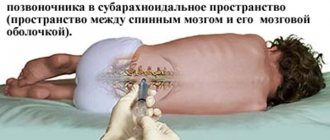
The patient is prescribed a general and biochemical blood test, urine and cerebrospinal fluid tests. To collect cerebrospinal fluid and measure intracranial pressure, a lumbar puncture is performed - a puncture of the spinal cord membranes at the lumbar level.
When examining cerebrospinal fluid, the concentration of protein and glucose, the number and structure of cells are determined, and a bacterioscopy of a Gram-stained fixed drop is performed. Additionally, an examination of the fundus is performed. If tumor cell degeneration is suspected, a biopsy is performed.
The neurologist also studies the patient’s medical history, conducts an examination and makes a conclusion based on all the data.
Features of brain examination in children
CT and MRI are considered the most effective modern diagnostic methods, however, their use is undesirable for young children. It is difficult for a child under one year of age to lie still for a long time. Therefore, in cases where there is still a need to resort to CT or MRI, children are given anesthesia.
To detect expansion of the subarachnoid space in children, the following is used:
- Neurosonography is an ultrasound examination of brain structures. To conduct the examination, the child must have a non-ossified fontanel. The procedure is safe, takes no more than 15 minutes, can be performed repeatedly, and does not require special preparation. A sonologist interprets the results.
- Echoencephalography is a method of examining the brain using ultrasound, used in children with already overgrown fontanelles and in adults. Can be carried out in one- and two-dimensional modes. Allows you to determine the damage to the brain and its displacement relative to the bones of the skull under the pressure of cerebrospinal fluid. Does not require preliminary preparation and is not harmful to health. The disadvantage of the method is its low accuracy.
To prevent brain damage due to birth injuries, hypoxia, and infectious diseases, neurosonography is performed in maternity hospitals. Pediatricians also monitor the growth of the skull in children - this simple procedure allows them to detect hydrocephalus.
What needs to be done in this situation and what will be the treatment?
Dr. Komarovsky says the following about the expansion of the interhemispheric fissure in infants: if the size of the expansion is insignificant, then treatment in this case is not necessary, since there is a high probability that it will disappear on its own with age. Also, therapy is not required if widening of the interhemispheric fissure is the only factor of concern. However, it will be necessary to have your doctor check you up from time to time.
Medicines are prescribed when additional symptoms appear that are characteristic of a particular disease. So, if a child lives in harsh climatic conditions with a lack of sunlight, or he has signs of rickets, he is prescribed vitamin D. If the symptoms indicate the development of intracranial pressure in the child, the doctor prescribes diuretics that cause the outflow of excess fluid from the brain. At the same time, medications containing potassium and magnesium are prescribed.
What can pathology mean?
The expansion of the subarachnoid convexital spaces indicates that there is an uneven distribution of fluid. This leads to cerebrospinal fluid pouring into the cavity and causing dropsy or hydrocephalus. Unevenly distributed cerebrospinal fluid can provoke intracranial pressure and dilate the ventricles of the brain. If during the course of this disease the ventricles are within normal limits, then there is a possibility that by the age of 2 the baby will be able to outgrow hydrocephalus, but in any case the doctor is obliged to prescribe treatment. Parents should not wait for the child’s skull to strengthen by the age of 2 and the disease to recede on its own; this may not happen, but you will still lose time.
Therapeutic measures in the fight against disease
The treatment of such pathologies in the brain is carried out by a neurologist, and it is he who should be consulted if you have reason to suspect hydrocephalus or arachnoiditis. Consultation with a doctor is necessary if there has been a head injury due to childbirth or mechanical impact. Even if the doctor does not diagnose anything, it is much better to play it safe and not worry in vain than to suffer from a serious illness and not even know it.
Treatment of an enlarged subarachnoid space in most cases involves eliminating the very cause that provoked this condition in the child. As a rule, the provoking factors of enlarged subarachnoid convexital spaces are sinusitis, otitis and increased intracranial pressure or infectious diseases. As a treatment for this deviation, a complex of antibacterial drugs and vitamins (especially group B) is prescribed. Treatment takes quite a long time and is prescribed individually, taking into account the patient’s age and the nature of the expansion of the subarachnoid spaces. Among the drugs that are used in the treatment of this disorder in children, the following can be noted:
- products that expel excess fluid (Diacarb, Veroshpiron, Asparkam);
- medications to improve brain trophism (Cavinton, Pantogam).
If we are talking about an adult or a child over 3 years old, then the treatment tactics will be slightly different. Treatment will include:
- diuretics;
- barbiturates;
- glucocorticosteroids;
- saluretics;
- vasoactive drugs;
- plasma expander solutions;
- painkillers.
In addition to drug therapy, the doctor can prescribe a number of physioneurological procedures that will eliminate the symptoms of the disease and restore normal metabolism of brain cells and tissues.
Speaking about treatment prognoses, they are favorable; the main thing is to start drug therapy in a timely manner.
If drug therapy does not provide the desired result, then surgical intervention is indicated.
Such diseases cannot be ignored and left to chance; at the first symptoms, qualified medical diagnosis is immediately required. If the disease is neglected, the person will experience dementia, gait disturbances, speech defects, problems with urination, developmental delays and a number of other unpleasant phenomena. Now you know what expansion of the subarachnoid space means. Being theoretically savvy in such medical issues, you will be able to cope with the disease and recognize its symptoms in a timely manner.
The subarachnoid space (SAS) exists in all people. It is located between the soft and arachnoid (arachnoid) membranes of the brain. Its purpose is the circulation of cerebrospinal fluid, that is, cerebrospinal fluid.
Expansion of SAP occurs when a lot of cerebrospinal fluid is formed, or its outflow is obstructed. Since the brain is placed in a limited cavity of the cranium, as the volume of fluid increases, its structures are subject to compression. There is a general (uniform) expansion and a local one .
Subarachnoid fissure - the layers of the meninges diverge above the recess of the gyrus and connect above its surface; these spaces are called fissures. The concept of SAP expansion is only a conclusion obtained from X-ray, ultrasound or tomographic diagnostics. Additional examinations are needed to determine.
Causes of pathology in uniform and local: increased formation of cerebrospinal fluid; malabsorption due to inflammation, swelling; obstructions to outflow - tumor, cyst, hemorrhage. They may occur during the period of intrauterine development . In this case, the baby is born with hydrocephalus (water on the brain). Provoking factors: developmental defects; anomalies in the structure of the skull, its connection with the spine; trauma during childbirth; maternal infectious diseases.
Diagnostic methods and goals
The disease can be diagnosed only after a comprehensive examination and laboratory tests. After receiving the results of magnetic resonance or computed tomography, blood biochemistry results, ultrasound examination of the cerebral hemispheres, assessment of the patient’s symptoms and behavior, the neurologist will establish the final diagnosis, the extent of the disease and prescribe medication.
Basic diagnostic methods:
- Neurosonography . It lasts no more than fifteen minutes and is carried out using an ultrasonic sensor through an open fontanel on the newborn’s head. The study can be carried out quite often, without negative consequences for the child. As a rule, neurosonography is performed on all newborns in the maternity hospital to identify pathologies in brain development at the initial stage. A neurologist or pediatrician interprets the examination data. Only by comparing symptoms and examination data can a doctor make a diagnosis.
- Computed tomography and magnetic resonance imaging are very expensive research methods and are carried out when serious abnormalities are detected. As a rule, for newborns it is enough to conduct neurosonography through the fontanel, but adults already need more serious diagnostic methods. Today these are the most reliable and accurate methods for studying the human body. MRI allows you to see a layer-by-layer image of the desired area of the brain. Examination of infants is very problematic, as it requires complete fixation and immobility, which is very problematic for small children. If a baby needs this type of examination, it is carried out under anesthesia.
- Cisternography is used to determine the direction of cerebrospinal fluid and clarify the type of hydrocephalus.
- Angiography is an examination method in which contrast is injected into the artery and abnormalities in the patency of blood vessels are detected.
- Neuropsychological examination - examination and questioning of the patient, collecting all tests and studies together to identify disorders in the functioning and functioning of the brain.