Human anatomy is undoubtedly a major core subject for study in medical schools. Despite the fact that normal human anatomy is a discipline that stood at the origins of the development of medicine, a large number of scientific works still appear that make adjustments to modern anatomical atlases.
It would seem that human anatomy cannot change so quickly with the course of evolution, but our understanding of it is constantly improving as new research methods appear - evidence of this is provided by ever new versions of the anatomy atlas.
Atlas of anatomy Sinelnikova R.D. in 4 volumes, this is perhaps the most authoritative and time-tested source of knowledge on this topic. It is constantly republished, delighting us with its visual illustrations and text accessible to everyone. Many students tried to download Sinelnikov’s atlas for study, but the links either did not work, or there was a virus in the folder... We solved this problem by making a website dedicated to this source.
The main goal of studying human anatomy is to create a fundamental knowledge base for students for further study of other medical disciplines. It is difficult to imagine mastering a curriculum in physiology, pathological physiology, pathological and topographic anatomy, operative surgery, and a whole range of clinical disciplines without a thorough study of normal human anatomy.
It is very important for a student to have a visual image of the material studied; for this it is necessary to study human anatomy in pictures. The main feature of this science. of course, is the structuring of its sections and subsections, as well as a clear systematization of the entire nomenclature.
Thus, we can distinguish the following directions that correspond to each system:
- osteology (section on the bones of the human skeleton). Studies the skeleton, both as a whole mechanism and as individual bones. There is also a study of age-related changes in bones.
- syndesmology (joints, ligaments). An extremely important section for future orthopedists and traumatologists.
- myology (muscular system). He studies not only the structure, but also the development and physiology.
- splanchnology (internal organs). Includes the anatomy of the endocrine, digestive, respiratory, excretory and genitourinary systems.
- angiology (vessels and their derivatives). Information is presented on the structure of blood and lymphatic vessels.
- neurology (central and peripheral nervous system). An extremely important section for successful diagnosis of diseases and perhaps the most difficult.
- aesthesiology (the science of the sense organs). Everything about vision and hearing. And also about taste, olfactory and tactile sensitivity. Closely related to neurology.
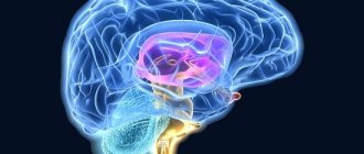
Anatomy of the ventricles of the brain
The ventricles of the brain are cavities in the brain filled with cerebrospinal fluid. The ventricles of the brain include: Lateral ventricles - ventriculi laterales (telencephalon); The lateral ventricles of the brain (lat. ventriculi laterales) are cavities in the brain containing cerebrospinal fluid, the largest in the ventricular system of the brain.
The left lateral ventricle is considered the first, the right - the second. The lateral ventricles communicate with the third ventricle through the interventricular (Monroy) foramina. They are located below the corpus callosum, symmetrically on the sides of the midline. In each lateral ventricle, there is an anterior (frontal) horn, a body (central part), a posterior (occipital) and an inferior (temporal) horn.
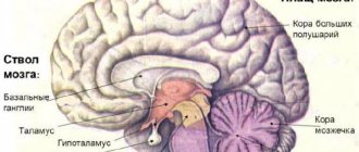
Third ventricle - ventriculus tertius (diencephalon); The third ventricle of the brain - ventriculus tertius - is located between the visual hillocks, has a ring-shaped shape, since the intermediate mass of the visual hillocks - massa intermedia thalami - grows into it. In the walls of the ventricle there is a central gray medulla - substantia grisea centralis; subcortical autonomic centers are located in it. The third ventricle communicates with the cerebral aqueduct of the midbrain, and behind the nasal commissure of the brain - comissura nasalis - with the lateral ventricles of the brain through the interventricular foramen - foramen interventriculare.
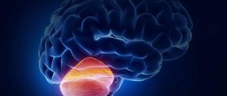
The fourth ventricle is ventriculus quartus (rhombencephalon). Placed between the cerebellum and the dorsal surface of the pons and medulla oblongata. Its arch is the worm and brain sails, and its bottom is the medulla oblongata and the bridge. It is a remnant of the cavity of the hindbrain and therefore is a common cavity for all parts of the hindbrain that make up the rhombencephalon, rhombencephalon (medulla oblongata, cerebellum, pons and isthmus). The IV ventricle resembles a tent, in which a bottom and a roof are distinguished. The bottom, or base, of the ventricle has the shape of a rhombus, as if pressed into the posterior surface of the medulla oblongata and the pons. Therefore it is called the rhomboid fossa, fossa rhomboidea. The central canal of the spinal cord opens into the posteroinferior corner of the rhomboid fossa, and in the anterosuperior corner the fourth ventricle communicates with the aqueduct. The lateral angles end blindly in the form of two pockets, recessus laterales ventriculi quarti, curving ventrally around the inferior cerebellar peduncles.
The two lateral ventricles are relatively large, C-shaped, and unevenly wrap around the dorsal portions of the basal ganglia. The ventricles of the brain synthesize cerebrospinal fluid (CSF), which then enters the subarachnoid space. Violation of the outflow of cerebrospinal fluid from the ventricles is manifested by hydrocephalus.
Source
Lateral ventricles // Encyclopedic Dictionary of Brockhaus and Efron: in 86 volumes (82 volumes and 4 additional). - St. Petersburg, 1890-1907. Wikipedia
Lateral ventricles
The lateral ventricles are the cavities of the cerebral hemispheres (see Fig. 3.33). They are symmetrical slits in the thickness of the white matter containing cerebrospinal fluid. They have four parts corresponding to each lobe of the hemispheres: the central part - in the parietal lobe; anterior (frontal) horn - in the frontal lobe; posterior (occipital) horn – in the occipital lobe; the lower (temporal) horn is in the temporal lobe.
central part
looks like a horizontal slit. The upper wall (roof) of the central part is formed by the corpus callosum. At the bottom are the body of the caudate nucleus, partly the dorsal surface of the thalamus and the posterior leg of the fornix. In the central part of the lateral ventricles there is a developed choroid plexus of the lateral ventricle. It has the shape of a dark brown strip 4–5 mm wide. Posteriorly and downwardly it is directed into the cavity of the lower horn. The roof and bottom in the central part converge with each other at a very acute angle, i.e. There are no lateral walls near the central part of the lateral ventricles.
Front horn
is a continuation of the central part and is directed forward and laterally. On the medial side it is limited by the plate of the septum pellucidum, on the lateral side by the head of the caudate nucleus. The remaining walls (anterior, superior and inferior) form the fibers of the forceps minor of the corpus callosum. The anterior horn has the widest lumen compared to other parts of the lateral ventricles.
Posterior horn
has a pointed posterior shape with a convexity facing the lateral side. Its upper and lateral walls are formed by the fibers of the large forceps of the corpus callosum, and the remaining walls are represented by the white matter of the occipital lobe. There are two protrusions on the medial wall of the posterior horn: the upper one, called the dorsal horn bulb, corresponds to the parieto-occipital groove of the medial surface of the hemisphere, and the lower one, called the bird's spur, corresponds to the calcarine groove. The lower wall of the posterior horn has a triangular shape, slightly protruding into the cavity of the ventricle. Due to the fact that this triangular elevation corresponds to the collateral groove, it is called the “collateral triangle”.
Lower horn
located in the temporal lobe and directed downward, forward and medially. Its lateral and superior walls are formed by the white matter of the temporal lobe of the hemisphere. The medial wall and partly the lower wall is occupied by the hippocampus. This elevation corresponds to the parahippocampal sulcus. Along the medial edge of the hippocampus stretches a plate of white matter - the hippocampal fimbria, which is a continuation of the posterior leg of the fornix. On the lower wall (bottom) of the lower horn there is a collateral elevation, which is a continuation of the collateral triangle from the area of the posterior horn.
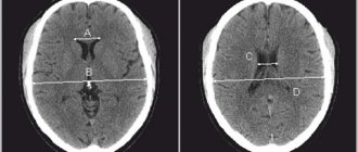
The lateral ventricles communicate with the third ventricle through the interventricular foramen (foramen of Monro). Through this opening, the choroid plexus penetrates from the cavity of the third ventricle into each lateral ventricle, which extends into the central part, the cavity of the posterior and inferior horns. The choroid plexuses of the ventricles of the brain produce cerebrospinal fluid. The shape and relationships of the ventricles of the brain are shown in Fig. 3.35.
Rice. 3.35.
Ventricles of the brain:
a – lateral ventricles: 1 – anterior horn; 2 – corpus callosum; 3 – central part; 4 – posterior horn; 5 – lower horn; b – cast of the ventricular system of the brain: 1 – interventricular foramina; 2 – anterior horn; 3 – lower horn; 4 – third ventricle; 5 – cerebral aqueduct; 6 – fourth ventricle; 7 – posterior horn; 8 – central channel; 9 – median opening of the fourth ventricle; 10 – lateral openings of the fourth ventricle
General system structure and some important terms
Cavities containing cerebrospinal fluid communicate with a number of organs. In particular, with the spinal cord canal, subarachnoid space. The structure of the system is as follows:
- 2 lateral ventricles;
- third and fourth ventricles;
- choroid plexuses;
- choroid ependymocytes;
- tanycytes;
- blood-cerebrospinal fluid barrier;
- liquor fluid.
Contrary to their name, the ventricles are not bags filled with cerebrospinal fluid, but hollow spaces, or cavities, located in the brain. The produced liquor performs a huge number of functions. The common cavity, formed from the ventricles of the brain with canals, overlaps with the subarachnoid space and the median canal of the spinal part of the central nervous system.
Most of the total cerebrospinal fluid is produced in the area of the choroid plexuses located above the 3rd and 4th ventricular cavities. A little substance is located in the wall areas. Soft membranes emerge into the lumen of the cavities, from which plexuses of blood vessels are created. Ependymal cells (choroid ependymocytes) play a huge role and are quite functional in stimulating nerve impulses. An important criterion is the promotion of cerebrospinal fluid with the help of special cilia. Tanycytes provide connections between blood cells and spinal cord fluid in the ventricular lumens; they have become a specialized variety of ependymal cells. The blood-cerebrospinal fluid barrier is a highly selective filter. It performs the function of selectivity in the flow of nutrients into the brain. It also displays metabolic products. Its main purpose is to maintain the homeostasis of the human brain and the multifunctionality of its activities.
Consequences
The consequences of increased intracranial pressure are manifested in the following symptoms:
- The baby becomes lethargic and loses appetite.
- A pronounced venous network appears on the forehead due to obstruction of outflow from the cranial cavity.
- Muscle tone in the limbs changes, tendon reflexes, which normally should be moderate, are revived.
- Changes occur in the fundus in the form of congestion.
- Sometimes tremor appears in the limbs.
- Both grasping and swallowing and sucking reflexes are reduced.
- The baby spits up often. This is caused by the fact that increased cerebrospinal fluid pressure irritates the vomiting centers at the bottom of the rhomboid fossa (in the fourth ventricle). The adult equivalent is cerebral vomiting without nausea.
- Tension and bulging of the fontanelles appear, and an increase in their linear dimensions occurs.
- The baby has a disproportionately large head.
Of course, the symptoms described above are not necessarily associated with ventriculomegaly. As mentioned above, a slight increase in size, and even asymmetry of structures, in the absence of a clinical picture, changes in reflexes and the fundus of the eye, should not bother parents. They should simply observe the baby and do neurosonography on him regularly.