Damage to any of the arteries supplying the brain leads to disruption of the function of parts of the brain in the basin of this artery. Lack of blood supply causes temporary dysfunction, and its cessation leads to the death of a part of the brain and a stroke. With the development of circulatory failure in the carotid arteries, paralysis of half the body and impaired speech functions occur. The basin of the vertebral arteries is responsible for the area of the brain that maintains balance and provides auditory perception. This area is supplied by the vertebrobasilar system and therefore one of the most important causes of such symptoms is a violation of blood flow in the vertebrobasilar system.
In 70% of cases, cerebral circulatory disorders are associated with atherosclerosis or tortuosity of the arteries in the neck - carotid and vertebral. Therefore, any microstroke or vertebrobasilar insufficiency should be a reason to study these vessels.
Vascular surgeons of the Innovative Vascular Center have significant experience in unique operations on the carotid and vertebral arteries. The advantage of our approach is the use of local anesthesia for operations on the carotid and vertebral arteries. Local anesthesia provides contact with the patient during arterial clamping and minimizes the risk of stroke during surgery.
Cardioneurology: integration and specialization
Stroke is one of the most painful problems of modern medicine [1]. Increasing morbidity, high mortality and severe consequences for survivors are bringing the problem of cerebrovascular accidents to the national level. The pressure of the economic, social and medical components of the problem on the public health care system, society and family not only persists, but also increases. The costs of treating patients with acute cerebrovascular accidents have increased many times over the past decades, but this, so far, has only led to a decrease in mortality in the acute period of the disease. It is becoming increasingly clear that the strategy to combat stroke has not only technological, but also ideological gaps [2]. In the fight against stroke, new interdisciplinary solutions are needed in the field of prevention and treatment of a wide range of diseases of the cardiovascular system, the course of which is complicated by cerebrovascular accidents. Stroke is the outcome of various diseases of the heart, blood vessels, and blood; its development is caused by disorders of carbohydrate and fat metabolism [2]. But the pathogenesis of stroke is most closely associated with heart pathology. Back in the seventies of the last century, in the works of N.K. Bogolepov and his colleagues, the basic mechanisms of cardio-cerebral relationships in vascular diseases of the brain were studied. More than 20 years ago, N.V. Vereshchagin identified the most pressing clinical problems of a new integral direction of medical science and practice - cardioneurology [3]. The commonality of the etiology and pathogenesis of cardiovascular diseases has led to the formation of generally accepted ideas about “coronary heart and brain disease” (Gusev E.I., 1992). Modern cardioneurology is not a new specialty, but a current integrative scientific and practical direction in medicine, the main goal of which is the study of the heart in various forms of vascular lesions of the brain and the study of the brain in heart diseases, hemodynamic disorders associated with cardiac surgery [1]. Cardioneurology solves a number of pressing clinical problems that require the consolidation of efforts of cardiologists, neurologists, cardiac surgeons, specialists in interventional medicine, representatives of radiation, functional and laboratory diagnostics. The high level of specialization of modern medicine, reflecting the need for in-depth knowledge in the context of an increasing volume of information, does not contradict the modern trend of comprehensive study of the most pressing problems. The effectiveness of this approach was demonstrated by the 1st National Congress “Cardioneurology” held in December 2008. The field of interest of cardioneurology is very wide and goes far beyond clinical neurology. It involves studying the cardiac aspects of ischemic stroke, the influence of heart pathology on the course of the post-stroke period and the progression of chronic cerebrovascular insufficiency [1-5]. Cardioneurology opens up new opportunities in the field of preventing cerebrovascular accidents. The study of the mechanisms of brain damage in diseases of the cardiovascular system and the development of methods for their compensation complements the traditional ideas about risk factors that underlie the modern system of stroke prevention. Thus, already within the framework of cardioneurology, such independent directions as cardioneurology of emergency conditions, cardioneurology of the post-stroke period, cardioneurology in cardiovascular surgery, and preventive cardioneurology are being developed. All of them require the participation of practitioners and researchers of various specialties. The development of cardioneurology has been facilitated by the latest advances in the field of functional diagnostics, imaging of the brain, blood vessels and heart, and cardiovascular surgery. For many decades, the subtle mechanisms of cerebral ischemia were inaccessible for in-depth study. Ultrasound methods for studying blood vessels and the heart, long-term recording of blood pressure and ECG, contrast angiography, and MRI, stress tests, modern laboratory tests - these and many other diagnostic methods, which have become widespread in clinical practice, have created the necessary information base for the formation of new ideas about the nature of the stroke. The concept of stroke heterogeneity determined the transition of vascular neurology to a new level of development, at which heart diseases began to play a major role and the term “cardioneurology” began to replace the usual “angioneurology”. Since it became obvious that the division of strokes into hemorrhagic and ischemic is not exhaustive, a rapid and extremely fruitful study of the pathogenetic subtypes of ischemic strokes began. It turned out that in most clinical observations the doctor is faced with the need to assess the state of the cardiovascular system as a whole (cardioembolic, hemodynamic stroke), and the role of the neurologist in the diagnostic process cannot be limited only to a topical diagnosis. The course of the disease and its outcomes are largely determined by the state of central hemodynamics and heart function. A significant part of cerebrovascular disorders is pathogenetically closely related to changes in peripheral blood (stroke of the hemorheological microocclusion type). Often severe brain damage occurs due to gross pathology of extracranial arteries (atherothrombotic stroke). Within a short period of time, a refined pathogenetic diagnosis of ischemic stroke became part of clinical practice. It became clear that ideas about the nature of stroke go far beyond the competence of a neurologist. The addition of a cardiologist to the staff of the neurovascular departments became the logical organizational completion of the structure of a modern hospital designed to provide care to patients with acute cerebrovascular accidents. It would be a mistake to consider cardioneurology as a simple sum of knowledge accumulated in the field of cardiology and vascular neurology. Cardioneurology is a field of medical science and practice that is limited to the participation of the brain in pathological processes associated with the circulatory system. Within the framework of cardioneurology, a number of pressing clinical problems are currently being solved, which include [1,2,4]: • Development of effective areas for stroke prevention; • Study of pathogenetic subtypes of ischemic stroke; • Prevention and treatment of acute cerebrovascular accidents in patients with arrhythmias and heart failure; • Prevention and treatment of acute cerebrovascular accidents in patients with stenosis and occlusion of the main arteries; • Prevention and treatment of acute cerebrovascular accidents in patients with coagulopathies; • Study of the influence of cardiac pathology on the course of the post-stroke period and the progression of chronic cerebrovascular insufficiency; • Prevention of fatal cardiac events in the immediate and late post-stroke periods; • Study of the cerebrovascular effects of antihypertensive therapy and the prevention of associated hypoperfusion cerebral complications; • Prevention of cerebral complications during and after open heart surgery (coronary artery bypass surgery, valve replacement).
Causes of stroke
The etiology of stroke is based on any diseases and factors that reduce or completely stop the supply of arterial blood to the brain and its various parts.
- Hypertension, coronary heart disease, atherosclerosis, transient ischemic attack (ministroke), hyperlipidemia (high blood fat), thrombosis, diabetes. All these diseases affect the properties of blood supply to the blood vessels of the brain and the rheological (viscosity, fluidity) properties of blood.
- Smoking, alcoholism, drug addiction, and taking oral contraceptives are also risk factors for stroke.
- Changes in blood vessels and vascular wall: dysplasia, vasculitis, arthritis, etc.
- Blood pathologies: coagulopathy (clotting disorder), damage to central and cerebral hemodynamics, leukemia, polycythemia (increased number of red blood cells).
- Heart diseases: myocardial infarction, rheumatism, cardiomyopathies, etc.
Cardioneurological examination
The development and implementation of algorithms for cardioneurological examination of patients at high risk of stroke and patients undergoing stroke is one of the important tasks of cardioneurology [5]. At present, the optimal scope of such an examination has been determined, which allows identifying the most significant mechanisms of cerebral circulatory disorders and suggesting the development of stroke of one or another pathogenetic subtype (Table 1). Diagnostic methods and pathogenetic subtypes of stroke
| Instrumental and laboratory studies | Pathogenetic subtype of ischemic stroke |
| Ultrasound Doppler diagnostic technologies: DS, TC USDG*, ultrasound transcranial monitoring of cerebral blood flow with embolodetection; | Hemodynamic Cardioembolic |
| Cardiological diagnostic techniques: echocardiography, ECG, 24-hour ECG and blood pressure monitoring; | Hemodynamic Cardioembolic Lacunar |
| X-ray contrast cerebral and coronary angiography; | Atherothrombotic Hemodynamic |
| X-ray or magnetic resonance imaging using special angioneuroimaging modes; | Atherothrombotic Hemodynamic Lacunar |
| Detailed laboratory study of the hemostasis system and rheological properties of blood; | Stroke type of hemorheological microocclusion Atherothrombotic |
| Laboratory study of biochemical parameters that determine the state of lipid and carbohydrate metabolism, reflecting the participation of immune mechanisms of atherogenesis; | Stroke by type of hemorheological microocclusion Atherothrombotic Hemodynamic |
* - duplex scanning and transcranial Doppler ultrasound Determining the required volume and nature of diagnostic studies seems to be the most important part of the diagnostic process in the context of a significant increase in the cost of treatment and the overcrowding of clinical practice with numerous, often uninformative, techniques. Many of these research methods are becoming a thing of the past (REG, EEG, EchoEG, fundus examination, lumbar puncture with cerebrospinal fluid analysis). This does not mean that for the purposes of differential diagnosis in clinical practice these and other techniques cannot be used that can provide additional information in cases requiring a detailed assessment, but in the process of studying the state of the cardiovascular system they have ceased to play an important role. It seems obvious that the main place in terms of examining patients with vascular diseases of the brain should be occupied by non-invasive techniques that carry information about the structure and function of the heart, blood vessels and brain. The main ones here are ultrasound research methods: duplex and triplex scanning, transcranial Dopplerography, echocardiography [2,3]. The addition of ultrasound methods, MRI and CT of the brain, and modern laboratory tests, allows, in most cases, to obtain sufficient information about the pathogenetic triangle “heart-vessels-blood”. However, in some cases, to clarify the state of the vascular bed, it is necessary to use angiography, high-speed spiral CT with contrast of the coronary and cerebral arteries, stress tests, transesophageal echocardiography and other more complex diagnostic methods. The result of a cardioneurological examination should be a clinical diagnosis, reflecting not only the picture of brain damage, but also the state of the cardiovascular system, hemostasis and blood rheology.
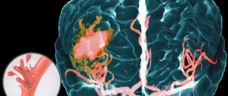
Mechanism of development of ischemic cerebral stroke
A stroke is always a condition resulting from stroke (acute cerebrovascular accident). With stroke, the supply of oxygen and nutrients to the brain is disrupted. This leads to a stop in metabolic processes and the development of hypoxia - both of these processes are called ischemia (hence the name “ischemic stroke”). It is known that any ischemic process leads to necrosis (death) of cells, the duration of which depends on the type of ischemic tissue. Many studies have found that only 8 minutes of complete ischemia are required for necrosis of neurons in the cerebral cortex to occur, and 15 minutes for neurons in subcortical formations.
Ideas about the pathogenesis of stroke from the perspective of cardioneurology
Cardioembolic stroke
is one of the most common pathogenetic subtypes of ischemic cerebrovascular accidents.
According to modern research, cardiogenic embolism causes 20-30% of all ischemic strokes, and this number may increase as cardiological diagnostic methods are introduced into clinical practice. To date, more than 20 cardiac disorders associated with embolic complications have been described [4]. These are non-rheumatic atrial fibrillation, myocardial infarction, thrombosis of the left ventricle of the heart, mitral stenosis, endocarditis and other, rarer heart diseases. Not all of these pathological conditions can be detected by physical examination. Latent paroxysms of atrial fibrillation, mitral valve prolapse with myxomatous degeneration of the leaflets, aneurysm of the interatrial septum, atheroma of the aortic arch, myxoma of the left atrium, patent foramen ovale, thread-like fibers of the mitral valve and other heart pathologies do not manifest characteristic, specific symptoms. It can be difficult to detect these violations even with targeted research. Diagnosis and correct clinical assessment of heart diseases that can lead to cerebral complications are also important because cardiocerebral embolism is a pathological process and not a completed event. Transcranial Doppler monitoring of the middle cerebral arteries allows us to detect microembolic signals in more than half of patients who have suffered a stroke due to the mechanism of cardiogenic embolism. In approximately 40% of patients, such signals are detected long-term (months and years) after the stroke [2,4]. A feature of cardioembolic stroke should also be considered the heterogeneity of the embolic substrate. Fibrin-erythrocyte thrombi, platelet aggregates, tumor particles, myxomatous elements, calcifications and atheromatous particles, fragments of valve vegetations can act as emboli. The morphological diversity of the substrate influences the treatment tactics of patients, which involves the use of both conservative and surgical methods. But conservative tactics, depending on the results of the examination, become very differentiated. It may include not only the prescription of anticoagulants and antiplatelet agents, but antibacterial therapy (endocarditis), the use of statins, antiarrhythmic drugs (paroxysmal atrial fibrillation). Surgical treatment brings encouraging results when detecting a cardiac tumor and patent foramen ovale [3,4,5]. In some cases, there is no alternative to heart valve replacement in severe endocarditis. Endovascular occlusion of the left atrial appendage is promising in order to prevent cardiogenic embolism in atrial fibrillation. It is obvious that with the expansion of knowledge about the nature of cardioembolic stroke, the possibilities of preventing and treating brain damage will increase. Hemodynamic stroke
accounts, according to various authors, from 8 to 53% of all ischemic strokes.
Such significant differences in the frequency of this pathogenetic subtype of ischemic stroke reflect the magnitude of unused diagnostic capabilities in determining the mechanisms of cerebral ischemia. A hemodynamic stroke is based on a discrepancy between the blood supply needs of the brain and the capabilities of the cardiovascular system. The formation of clinical symptoms of stroke occurs against the background of a long-term deficiency in the blood supply to the brain, caused by changes in its vascular system. A decrease in autoregulation of cerebral circulation, adaptation to the action of meteorological factors, physical and emotional stress, leads to a direct dependence of cerebral hemodynamics on the efficiency of the heart. Under these conditions, any pathological conditions that lead to a decrease in cardiac output (coronary insufficiency, arrhythmia, increased load on the myocardium) are inevitably accompanied by cerebral ischemia. Hemodynamic stroke is always the result of circulatory decompensation, in which the brain suffers as one of the main consumers of cardiac output [2,3]. Data obtained at the Scientific Center of Neurology of the Academy of Medical Sciences indicate the priority role of cardiac disorders in hemodynamic stroke [4]. As a result of cardiac examination, a variety of heart pathologies are detected in 70% of patients with hemodynamic stroke. Stratification of cardiogenic causes of hemodynamic stroke demonstrates that the most common causes of cerebral ischemia are silent myocardial ischemia and persistent atrial fibrillation. Next in frequency: sick sinus syndrome, paroxysmal atrial fibrillation and acute myocardial infarction. Less commonly, hemodynamic stroke is caused by: frequent ventricular extrasystole, transient atrioventricular block, ventricular fibrillation and pacemaker failure. All of these disorders can cause a reduction in cardiac output. In most cases, transient, asymptomatic cardiac disorders, revealed during cardioneurological examination, are associated with the hemodynamic mechanism. It should be noted that not only cardiac pathology underlies hemodynamic stroke. In some cases, decompensation of cerebral hemodynamics is associated with disturbances in the autonomic regulation of vascular tone and heart function. An example is hypotensive crises in patients suffering from parkinsonism. The hemodynamic mechanism of brain damage is also present in cases of stroke associated with arterial hypertension. The pathological basis of such damage is pathological processes in the arteries of the muscular type, occurring with their sclerosis, thickening and deformation. Hypertensive angioencephalopathy is the result of restructuring of the arterial system of the brain due to long-term hypertension. In hypertension, the entire cardiovascular system, and above all, the heart, is subject to remodeling. Left ventricular hypertrophy, dilation of the cavities of the heart, relative insufficiency of the valve apparatus, cardiosclerosis - lead to the formation of systolic and diastolic dysfunction. The result of remodeling is a decrease in hemodynamic reserve - the ability to maintain sufficient cardiac output under changing loads. Under these conditions, the immediate cause of stroke is most often a hypertensive crisis. An increase in the load on the myocardium leads to a decrease in minute blood volume, which in conditions of hypertensive angiopathy results in a hemodynamic ischemic stroke. Hemorheological stroke
is a special form of acute cerebral ischemia, which is based on altered blood properties.
Cerebral ischemia during hemorheological stroke is directly related to the blockade of microcirculation. Numerous factors lead to an increase in blood viscosity, among which the most important are: erythremia, hypercoagulation, hyperfibrinogenemia, hyperglycemia, dyslipidemia. Timely assessment and correction of these factors is absolutely necessary in both preventive and treatment programs aimed at compensating cerebral hemodynamics. Atherothrombotic stroke
is the most common and, as a rule, severe pathogenetic variant of stroke. Atherothrombosis usually affects large main arteries that have significant atherosclerotic changes. The absolute annual risk of stroke with stenosis of one carotid artery reaches 12–16% [5]. Large atheromas of unstable structure create favorable conditions for thrombus formation, which leads to artery obstruction. Timely diagnosis of atherosclerotic changes in the large arteries of the brain is one of the most important areas of cardioneurology. Assessing the degree of risk, feasibility and method of surgical intervention is possible only with a comprehensive examination of the state of the cardiovascular system.
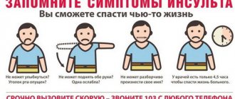
Main clinical symptoms of stroke and its consequences
The clinical picture of stroke is usually divided into focal and cerebral neurological lesions. It should also be noted that clinical strokes are divided according to the location of the lesion and the reason that caused them. So, for ischemic stroke, the classification will look like this:
- By etiology: Thrombic stroke (caused by thrombosis)
- Embolic stroke (caused by an embolism)
- Stroke in the carotid region: patients complain of darkening or lightening of the visual field of one eye (one-sided blindness), aphasia - impaired ability to pronounce speech.
Ischemic strokes in the vertebrobasilar region also include: lacunar stroke (ischemia in the system of small arteries of the brain, such strokes begin suddenly, without precursor symptoms) and subarachnoid hemorrhage - this condition is characterized by a sudden onset and severe headache before the attack. The symptoms of subarachnoid hemorrhage, classic for ischemia of the vertebrobasilar region, are sometimes supplemented by photophobia.
The consequences and complications of a stroke are no less pressing problem than the disease itself. Stroke affects many brain functions, including motor, neurological and psycho-emotional. After a stroke, patients are unable to move and are confined to bed for a long time. This is where the first complication arises - bedsores. Bedsores are essentially areas of necrosis on the skin. The process occurs due to a disruption of metabolic processes in the subcutaneous fat and dermis “pressed” under body weight.
Another, no less terrible, complication of a stroke is pneumonia. This condition occurs in cases where a stroke has affected the areas of the brain responsible for expectoration and coughing (these are natural reflexes of the body). As a result, a large amount of mucus accumulates in the lungs, which gradually becomes infected (because there is no outflow), leading to infectious pneumonia.
Numbness, loss of sensation and paralysis are also consequences of a brain stroke. These pathologies are quite common. The mechanism of their occurrence is also simple - a violation of the neurofunctional abilities of the brain due to ischemia and necrosis. Psycho-emotional and cognitive disorders are conditions in which patients lose the ability to normally perceive and reproduce psycho-emotional activity, together with depression of thinking, memory and speech and other cognitive functions.
Heart diseases and the course of the post-stroke period
The role of heart pathology in the pathogenesis of ischemic stroke is not limited to the acute period of the disease. These diseases and syndromes have a significant impact on the post-stroke period. Decompensation of cardiac disorders, including anginal attacks, heart failure and cardiac arrhythmias, can aggravate and slow down the rehabilitation process, leading to serious complications. . Cardiac dysfunction is very common in patients who have had a stroke. Upon careful examination, they are found in 70-75% of patients [4]. Most often, ischemic stroke is diagnosed with various forms of coronary artery disease, including acute myocardial infarction, post-infarction cardiosclerosis, unstable and stable angina, cardiac arrhythmias and chronic heart failure. Heart damage due to rheumatism and endocarditis, the presence of prosthetic valves are not uncommon among this category of patients. It has been established that in patients with post-infarction changes in the heart muscle and a permanent form of atrial fibrillation, ischemic brain damage is more extensive, and the neurological deficit is more pronounced than in patients who do not have such disorders. In patients with severe strokes, relatively low rates of left ventricular contractility are recorded and episodes of silent myocardial ischemia are more often detected. The post-stroke recovery period against the background of chronic cardiac pathology is associated with latent hemodynamic disorders, which can cause circulatory decompensation during the period of active rehabilitation. The mortality rate of stroke patients over the next 5-year period significantly exceeds the expected mortality rate in the general population of persons of the same age and sex [4]. It is noteworthy that one of the main causes of mortality is cardiac pathology: acute and chronic heart failure, myocardial infarction and cardiac arrest. Active Holter monitoring in the post-stroke period makes it possible to detect life-threatening ventricular arrhythmias in 22% of patients, including paroxysms of ventricular tachycardia, and in 16% - silent myocardial ischemia. Patients with negative cardiac factors are at increased risk of serious complications, including sudden cardiac death, even in the absence of subjective or objective cardiac disorders at the time of examination. Another important factor associated with a high likelihood of sudden cardiac death is dysregulation of cardiac activity caused by focal brain damage. Neurogenic hypertension during brainstem ischemia, neurogenic arrhythmias, coronary syndrome are widely known to clinicians. Damage to the insular lobe of the brain is associated with increased mortality in the long-term post-stroke period. Autonomic regulation of cardiac activity suffers with multiple focal brain lesions in the periventricular zones.
Common myths about stroke and its consequences
Let's look at the most common myths about stroke.
- Stroke is transmitted genetically. The myth exists only for 10-12% of patients, because in fact, the patients’ history contains a risk factor - a stroke in a family member in one or two generations. But also from the anamnesis of such patients, data on many other risk factors (lifestyle, etiologically important diseases, etc.) is revealed. Therefore, genetic predisposition to stroke cannot be considered as the main cause of the disease.
- Pumpkin and legume plants are excellent for preventing stroke. This myth has no basis in existence. Of course, pumpkin and legumes have a beneficial effect on some blood properties, but they cannot be considered as the main preventive methods for preventing stroke.
- A stroke only happens once. In fact, one in three stroke patients suffers another ischemic attack within five years.
- Stroke occurs only in older people. This statement has no evidence base. According to many statistical studies, ischemic stroke occurs in people aged 30-45 years in 10-12% of cases. It should also be noted: unfortunately, childhood and infant strokes are common.
- Men are more likely to suffer from strokes. This myth also has no scientific basis. According to recent monitoring data, it was found that women suffer more often from stroke (55/47%).
Prevention of cerebral ischemia during antihypertensive therapy
Currently, there is no doubt that adequate antihypertensive therapy can prevent the development of cerebrovascular complications, including recurrent stroke [1,4,5]. However, in patients with cerebrovascular pathology there is a risk of neurological complications caused by a significant decrease in blood pressure when its level falls below the lower limit of autoregulation of cerebral circulation. There are a number of factors that determine the safe tactics of antihypertensive therapy in the post-stroke period. It has been established that in the acute period of stroke, the increase in blood pressure is compensatory in nature and is aimed at maintaining adequate perfusion pressure in the peri-infarction zone. Therefore, the optimal level of systolic blood pressure in the acute phase of ischemic stroke is 160-180 mmHg. Art. and diastolic blood pressure 95-105 mm Hg. Art., which is associated with a lower incidence of early and delayed cardiovascular and neurological complications and less pronounced residual neurological deficit. In patients who have suffered a hemorrhagic stroke, the risk of recurrent hemorrhages is directly dependent on the level of blood pressure: the minimum risk is found at 120/70 mm Hg. Art., lower values were not achieved in studies. On the contrary, after an ischemic stroke, blood pressure levels must be maintained slightly higher, and optimal blood pressure values depend on the condition of the great arteries. In the presence of occlusive atherosclerotic lesions of the carotid artery, the lowest incidence of complications and the best outcomes are recorded with a systolic blood pressure of 130-140 mm Hg. Art., at higher or lower blood pressure levels, the risk begins to increase. With bilateral stenosis of the carotid arteries of more than 70% of the lumen of the vessels, there is an inverse relationship between the level of systolic blood pressure and the risk of stroke. Due to the danger of iatrogenic cerebral ischemia, blood pressure is maintained at 160-170 mm Hg. Art. – this tactic brings the best results. In patients with long-term and high hypertension, cerebral circulatory failure can occur at relatively normal blood pressure levels, so it is very important to plan a safe range of its reduction during long-term antihypertensive therapy. Studies by the Scientific Center for Neurology of the Russian Academy of Medical Sciences have shown that with the relative preservation of the cerebrovascular adaptive reserve and the absence of pronounced damage to the main arteries of the head, it is permissible to reduce systolic blood pressure by 20%, and diastolic blood pressure by 15% from the initial drug-free level. During treatment, as a rule, blood pressure approaches the so-called “target” levels - 120 - 130 mmHg. However, for patients with clinical manifestations of cerebral ischemia, gross atherosclerotic changes in the main arteries, hypertensive angiopathy, this requires a longer time.
Diagnosis of stroke
When a patient enters a medical institution, a doctor can make a diagnosis of “ischemic stroke”, based on basic and additional research methods, as well as when collecting an anamnesis. When treating such patients, it is necessary to carry out a clear differential diagnosis with other strokes, and to understand what kind of stroke (hemorrhagic or ischemic) the patient is suffering from. Let's consider the most informative modern methods of instrumental research.
- MRI (magnetic resonance imaging). MRI is based on scanning the area under study using radio waves and magnetic fields. The method is very informative, especially when studying late-stage stroke (or when studying the consequences of a stroke). There are, of course, contraindications: the patient has electronic implants, a pacemaker, and early pregnancy.
- Ultrasound examination (phonography). A method for visualizing sound waves reflected from body tissues. Ultrasound waves are most strongly reflected from the aqueous media of the body, so ultrasound is effective in studying the blood supply of blood vessels, in various hemorrhages, etc. It should be noted that, with a relatively inexpensive cost, ultrasound is an absolutely harmless research method that gives a good diagnostic result.
If you have any questions, ask our specialist! Ask a Question
Prevention of complications during and after open heart and large vessel surgeries
One of the most pressing tasks of modern cardioneurology is the prevention of neurological complications during open-heart surgery, which is due to the steady growth of surgical interventions of this kind. Cerebral complications of cardiac surgery can be divided into two types [4]. Complications of the first type: death due to stroke and hypertensive encephalopathy, non-fatal stroke, transient cerebrovascular accident. Complications of the second type: deterioration of intellectual functions, confusion, memory impairment, seizures. The incidence of stroke in the postoperative period is about 2%; with diabetes mellitus and atherosclerosis of the carotid arteries, the risk of stroke reaches 8%. Risk factors for complications of the first type are atherosclerosis of the aorta, previous stroke, the use of intra-aortic balloon counterpulsation, diabetes mellitus, postoperative atrial fibrillation, arterial hypertension, old age, left ventricular thrombosis, perioperative hypotension. The immediate causes of intra- and postoperative cerebral damage are embolism, decreased cerebral blood flow, contact activation of blood cells during artificial circulation and metabolic disorders. The most common source of embolism is atheromatous masses of the ascending aorta during the insertion of cannulas for the heart-lung machine and manipulation of vessels during the creation of anastomoses with shunts. A significant point should be considered a decrease in cerebral perfusion caused by atherosclerosis of the cerebral arteries and relatively low average blood pressure, non-pulsatile blood flow during certain types of artificial circulation. Prevention of neurological complications of open heart surgery is based on accurate knowledge of their causes. Since aortic atherosclerosis is one of the main predictors of neurological complications, it is necessary to accurately assess the condition of the ascending aortic arch. Identification of atherosclerotic plaques protruding into the lumen of the aorta, mobile, heterogeneous in structure, is an indication for the use of alternative surgical approaches (prosthetics of the affected area of the aortic arch in conditions of hypothermic circulatory arrest). New atrial fibrillation in the early postoperative period requires emergency treatment with anticoagulants and restoration of sinus rhythm. Patients who have recently suffered a myocardial infarction need to be examined using echocardiography to identify mural thrombi in the cavities of the heart. Timely preventive anticoagulant therapy can delay surgery for some time, but prevent severe complications. Delayed surgery in patients with recent stroke significantly reduces the risk of perioperative complications. The danger of cerebrovascular complications has a significant impact on the management of patients with combined damage to the blood vessels of the heart and brain. In most large vascular centers, reconstruction of the main arteries of the head is performed before myocardial revascularization, with the exception of cases of coronary artery bypass grafting for emergency indications.
Rehabilitation after a stroke at the MART clinic
Rehabilitation of patients after a stroke in our clinic is carried out with the aim of eliminating the consequences and complications of this disease. It should be aimed at restoring normal body functions.
The main types of measures to prevent the consequences of stroke.
- Therapeutic physical education (physical therapy). Conducting exercise therapy classes for stroke patients is an integral part of rehabilitation therapy. A specialist (trainer) creates a group of exercises individually for each patient. Exercise therapy helps to cope with stroke complications such as loss of sensation, numbness, and partial paralysis.
- Therapeutic massage. The massage is performed by a certified specialist, on the basis of a medical institution. Massage helps to cope with bedsores and numbness. Suitable for both able and incapacitated patients. Excellent complement to exercise therapy.
- Physiotherapeutic treatment. The benefits of physiotherapeutic treatment methods have been known for a long time. They are mainly used as rehabilitation and preventive measures: Transcutaneous electrical neurostimulation (TENS). Based on the use of weak currents. Helps cope with the consequences of a stroke, such as pain. Effectively relieves severe and moderate pain. The method can also be used to facilitate the delivery of drugs through the skin.
- Ultraphonophoresis, ultraphonotherapy. It has a relaxing, soothing, analgesic effect through ultrasound. Just like TENS, it can be used for accelerated administration of medications.
- Magnetic therapy (and magnetophoresis). Like TENS and ultraphonotherapy, it has an analgesic and relaxing effect. But the main difference from previous methods is the use of a magnetic field of different frequencies.
- Laser therapy (and laser phoresis). In addition to analgesic and relaxing effects, laser beams have anti-inflammatory and warming effects. Under the influence of the laser, blood supply and metabolism improves, which is very important in the treatment of the consequences of a stroke.
- Reflexology. It is based on stimulating certain active points of the body using various methods. These methods include: TENS, acupuncture (use of the finest needles), pharmacopuncture (injection of medicinal substances into active points).
- Kinesio taping (kinesiotherapy). When treating with this method, it is possible to influence muscle groups using special adhesive tapes (tapes).
Nowadays, stroke is one of the most common pathologies from the stroke group. The problem of stroke is scary not only because of its high mortality rate, but also because of the large percentage of post-stroke consequences.
Most people who have suffered the disease remain disabled and require constant care and therapeutic measures aimed at eliminating the consequences of a stroke. We must remember about stroke prevention measures, which include a normal diet, moderate exercise and a healthy lifestyle.
The article was reviewed by Doctor of Medical Sciences, Professor Grigory Isaakovich Shvartsman, Northwestern Medical University. I.I. Mechnikov.
Sign up at the MART medical center in St. Petersburg (see map) by calling 8 or leave a request on the website.
Literature
1. Suslina Z.A. Vascular diseases of the brain in Russia: achievements and unresolved issues. Cardioneurology. Proceedings of the 1st National Congress “Cardioneurology”. Edited by M.A. Piradov, A.V. Fonyakin. –M.: 2008. P. 7-10. 2. Simonenko V.B., Shirokov E.A. Preventive cardioneurology. – St. Petersburg: FOLIANT Publishing House LLC, 2008. 224 p. 3. Simonenko V.B., Shirokov E.A. Fundamentals of cardioneurology: A guide for doctors. – 2nd edition, revised and expanded. – M.: Medicine, 2001. – 240 p. 4. Fonyakin A.V. Modern concept of cardioneurology. –2006.