Key Facts
Muscular dystonia is a syndrome characterized by slow or repeated fast movements causing rotation, flexion or extension of the trunk and limbs with the formation of pathological postures.
A characteristic feature of dystonic hyperkinesis is the occurrence or intensification of voluntary movements. Dystonic posture initially occurs only with a certain movement, but gradually becomes permanent, remaining at rest. Another feature of dystonia is the dynamism of hyperkinesis: improvement after sleep, the influence of certain “corrective gestures” and changes in posture, the influence of the emotional state.
Diagnosis of muscular dystonia syndrome
Diagnostic criteria for diagnosing muscular dystonia syndrome are as follows:
- determining the presence of dystonic postures or movements;
- assessment of maternal labor and early development of a child suspected of having dystonia;
- determining the absence of diseases or taking medications that can cause dystonia;
- determination of the absence of paralysis and epilepsy;
- determination of the results of instrumental and laboratory tests (for example, copper metabolism studies, electroencephalography, computed tomography and magnetic resonance imaging).
Story
The term “dystonia” was introduced by H. Oppenheim in 1911, who used “dystonia musculorum deformans” to define generalized dystonia with onset in childhood. Today, almost 100 years later, many types of dystonia have been identified, including primary (idiopathic), as well as dystonia and secondary to other diseases (symptomatic).
The prevalence of dystonia is difficult to determine. According to various data, the prevalence of primary dystonia ranges from 11.1 to 300 per 100,000.
Classification of muscular-tonic syndromes
There are several types of muscle-tonic pain syndromes. They are classified depending on location:
- compression syndrome of the anterior scalene muscle: covers the region of the III-VI cervical vertebrae and the upper rib. Muscle spasm compresses the subclavian artery and brachial nerve plexus
- piriformis syndrome: compression of the sciatic nerve and inferior gluteal artery
- facet syndrome: local pain and stiffness in the area of the facet joints of the spine. Accompanied by subluxations of the vertebral segments and rupture of the joint capsules. Depending on the location of the affected facet joint, pain may radiate to the head, shoulders, chest, lumbar, sacral, thigh or buttock
In addition to the above, the trapezius, latissimus pectoralis, quadratus lumborum, and levator scapulae muscles often undergo pathological changes. Pain syndrome can be primary (covering only spasmodic tissues) and secondary, localized outside the damaged area.
Species and types
According to the prevalence of hyperkinesis, there are:
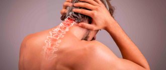
- focal dystonia, involving a small part of the body: head and facial muscles (cranial dystonia), neck (cervical dystonia), vocal cords (laryngeal dystonia), arm or leg (limb dystonia), torso (trunk dystonia);
- segmental dystonia involving two or more adjacent parts of the body, such as the head (face) and neck or vocal cords, neck and arm, or neck and torso;
- multifocal dystonia involving two or more non-contiguous parts of the body (for example, the face and leg);
- hemidystonia involving the limbs on one side;
- generalized dystonia involving both legs (or one leg and trunk) and at least one other part of the body.
The prevalence of generalized dystonia in the world is 0.3-3 per 1 million; focal dystonia: in the USA - 40 cases per 100 thousand population, in Europe - 15-16 per 100 thousand; idiopathic blepharospasm - 3.6, spasmodic torticollis - 5.7, oromandibular dystonia (OMD) - 0.09 per 100 thousand population.
Muscular dystonia is one of the most common movement disorders, ranking third in frequency after essential tremor and Parkinson's disease.
The term “dystonia” was first proposed by B. Oppenheimer in 1911; one of the forms of primary muscular dystonia bears his name. Recent decades have become significant for the modern understanding of the etiology, pathophysiological basis and effective methods of treating dystonia.
The modern definition characterizes muscular dystonia as non-rhythmic violent rotational movements in various parts of the body, with fanciful changes in muscle tone and the formation of pathological postures [2, 44]. Recently, clinical criteria, along with dystonic posture and dystonic movements, began to include corrective gestures (sensory tricks), mirror dystonia and motor overflow [38]. Corrective gestures can be varied. For example, touching the eyelid or eyebrow with a finger, wearing sunglasses, chewing gum or candy for blepharospasm; wearing a Shants collar, touching the back of your head to a wall or headrest, touching your cheek with a finger or squeezing your earlobe for cervical dystonia; cooling a limb before writing with writer's cramp, singing, sucking a lollipop with oromandibular dystonia, rhythmic foot tapping with hyperkinesis of the lower extremities and much more. Patients with dystonia use such corrective gestures to reduce hyperkinesis.
Motor excess and mirror dystonia have been well studied using the example of writer's cramp. Mirror dystonias are dystonic hyperkinesis or postures that occur during specific actions performed by the contralateral limb [11]. Motor excess is untargeted muscle contractions that accompany, but differ from, dystonic movements [17]. O Sitburana and J. Jankovic identified three patterns of abnormal muscle activity in writer's cramp. In addition to mirror dystonias, they also included ipsilateral redundancy (involuntary contraction of muscles adjacent to those involved in focal dystonia) and contralateral redundancy (involuntary muscle contractions and dystonic postures in a healthy, contralateral limb, occurring during dystonia in the primary involved limb). The detection of these phenomena indicates a lack of supraspinal inhibition of ipsilateral excessive movements and suppression of the function of transcollosal fibers during contralateral motor activity in dystonia [27].
The results of studies on the prevalence of dystonia are contradictory and depend on the methodology used. Thus, in the USA (Minnesota), generalized primary dystonia occurs with a frequency of 3.4 per 100,000 population, focal - 29.5 per 100,000 [28].
In Europe, the prevalence of primary dystonia is 15.2 per 100,000, of which 11.7 are focal forms [21, 33]. Blepharospasm in the general population is diagnosed in 5 people per 100,000, cervical dystonia - in 1.2-5.7 people. A generalized study found that early-onset dystonias are recorded in 2-50 cases per 1 million and late-onset dystonias in 30-7320 cases [19].
There is no exact information about the epidemiology of secondary dystonia and dystonia-plus. It is known that dystonia occurs in 40% of patients with early-onset Parkinson's disease, 100% of patients with progressive supranuclear palsy and corticobasal degeneration, 60% of patients with multiple system atrophy, 30% of patients with post-stroke paresis, 5-15% of patients with cerebral palsy , 20-60% of patients who have had Japanese encephalitis; tardive dystonia develops in 2-20% of patients treated with antipsychotics [3, 20, 29, 35, 40, 41].
Epidemiological studies are complicated by the fact that in 50% of patients the diagnosis of dystonia is established only 1 year after the onset of symptoms, and in 24% - 5 years after the onset of the first symptoms. In 36%, dystonia is regarded as a disease of a “psychogenic” nature [18].
Dystonic syndromes include a large group of diseases classified according to various principles. Classic categories include distribution by age, anatomical distribution, temporal pattern, association with other movement disorders, etiology, and genetic defect.
Classification by age
Based on the age of onset, dystonia is classified into infant (up to 2 years), childhood (3-12 years), adolescent (13-20 years), early adult (21-40 years) and older adult (over 40 years) dystonia. Classification by age of onset of the disease has important diagnostic and prognostic significance. Earlier manifestation of clinical manifestations of primary dystonia characterizes more rapid progression and generalization. For example, the onset of dystonias with autosomal dominant inheritance is DYT1, DYT5 corresponds to childhood or adolescence, and DYT7 corresponds to the onset in an older age period [5, 32]. Most primary dystonias that occur in adulthood are focal or segmental in nature. Primary cervical dystonia develops in early adulthood, and blepharospasm occurs in older adults [2, 9].
Infantile onset and aggressive progression characterize most X-linked neurodegenerations, multiple inheritance patterns, and mitochondrial diseases [37]. During the same period, manifestations of the hyperkinetic form of cerebral palsy are formed, which requires careful differentiation with neurodegenerations.
Classification by anatomical distribution
According to the anatomical distribution, focal, segmental, multifocal, generalized and hemidystonia are distinguished. Focal dystonia affects a small part of the body: the head and facial muscles, neck, vocal cords, arm or leg, torso. Segmental dystonias involve two or more adjacent parts of the body, such as the head and neck, neck and torso, and neck and arm. Multifocal dystonia involves two or more non-contiguous body parts (eg, face and leg, arm and leg). Hemidystonia is localized according to the hemitype with or without involvement of the face and neck. Generalized dystonia involves both legs (or one leg and trunk) and at least one other part of the body [44].
The distribution of dystonia along the body often indicates the etiology and prognosis of the disease. Dystonia with initial localization in the lower extremities in children is characterized by an unfavorable prognosis [5]. Hemidystonia indicates its secondary nature.
The secondary localization of dystonia is also evidenced by the primary localization of hyperkinesis in the facial area in children and in the lower extremities in adults [44].
Classification according to pathoanatomical principle
A new aspect of the classification is the pathological principle [18]. In accordance with it, dystonias without degeneration, with “static” damage and with degeneration are distinguished. In primary dystonia, no signs of degeneration of any brain structures have yet been identified. At the same time, functional neuroimaging methods have revealed a number of neurophysiological processes: decreased cortical inhibition, increased plasticity, dysfunction of sensory processes, disruption of metabolic processes in the cerebello-thalamocortical pathway [32].
Secondary persistent irreversible dystonia is most often the result of “static” damage to the basal ganglia, thalamus or brain stem [1, 24, 31].
The main pathogenetic substrate of hereditary or sporadic neurodegenerative diseases is a violation of the synthesis, metabolism or catabolism of neurotransmitters, which leads to degeneration of not only the basal ganglia or other areas of the brain, but also somatic organs and systems [37].
Etiological principle of classification
The basis of the modern etiological classification was laid by S. Fahn, who proposed to distinguish 4 groups: primary dystonia, dystonia-plus, neurodegenerative and secondary dystonia (see table)
[22].
In primary dystonia, it is the only symptom of the disease.
Primary dystonia can be hereditary and with an unknown hereditary factor, local and generalized. The dystonia-plus group includes dystonic syndromes with additional symptoms, but without signs of degeneration. Additional symptoms may include manifestations of parkinsonism or myoclonus [12]. Neurodegenerative diseases can be hereditary (for example, Wilson's disease) or sporadic in nature (for example, progressive supranuclear palsy), in which dystonia is the leading, but not the only symptom of a progressive degenerative process [37]. And finally, secondary dystonias are non-degenerative, sudden onset conditions with a known cause. Stable posture in one or more parts of the body may be a symptom of a “non-dystonic” disease that mimics dystonia. These include orthopedic (rotational atlantooccipital subluxation, spinal deformities, Arnold-Chiari malformation, etc.) and neurological diseases (pseudoathetosis with damage to the posterior columns of the spinal cord, tumors of the posterior cranial fossa, vestibular torticollis, dystonic tic, etc.).
Genetic classification
The genetic classification includes a large group of diseases with a known gene or gene locus of autosomal dominant, autosomal recessive or X-linked inheritance patterns, which are referred to as DYT [30]. The list contains at least 21 monogenic diseases, 7 of which - DYT1, DYT2, DYT4, DYT7, DYT13, DYT17, DYT21 - belong to primary dystonias; 5 - to dystonia-plus - DYT5 and DYT14 (dopa-sensitive), DYT11 and DYT15 (myoclonus-dystonia), DYT12 (sudden onset dystonia). DYT3 (X-linked dystonia-parkinsonism) and DYT16 (dystonia-parkinsonism PRKRA) are neurodegenerative diseases. DYT8, DYT9, DYT10, DYT18, DYT19, DYT20 are included in a separate group of various paroxysmal dyskinesias. With the exception of a few rare variants (DYT2, DYT3, DYT5b, DYT16, DYT17), all other dystonias are autosomal dominant [32].
Most of the genetically determined dystonias are associated with a very specific phenotype, although sometimes they have atypical clinical features. Many of the dystonia genes show low penetrance and variable expressivity [15, 39].
Secondary dystonia
Dystonia can be a symptom of various neurological diseases. The diagnosis of “secondary dystonia” is valid when the history, clinical assessment and laboratory data indicate the cause of the disease. Symptomatic dystonia is more often formed with striato-pallidal or thalamic lesions [24]. Striato-pallidal lesions are characterized by the appearance of pure dystonia, while lesions of the thalamus are characterized by myoclonus dystonia [25]. The cause of hemidystonia or focal dystonia in the arm or leg is damage to the contralateral putamen [31]. Manganese poisoning causes foot dystonia with the formation of “cock walk” [36]. Unlike most secondary dystonias, acute drug-induced dystonias are completely reversible and disappear after discontinuation of the dopamine-blocking drug. Late (tardive) drug-induced extrapyramidal disorders are partly reversible, 70% of them have a stationary course [3].
In general, secondary dystonias are characterized by: sudden onset; fixed and painful dystonic postures or resting dystonia; early onset of dysarthria; hemidystonia; the presence of a relevant factor (central or peripheral trauma, encephalitis, perinatal hypoxia, dopamine blockers, stroke, etc.); the presence of other, non-dystonic neurological symptoms (mild paresis, cerebellar symptoms, cognitive impairment, dysarthria, etc.); pathological changes according to laboratory studies and neuroimaging [44].
Treatment of dystonia
Treatment of dystonia still remains symptomatic, although significant progress has been made in this area over the past decades. New oral medications, the advent of botulinum toxins and modern neurosurgical technologies have significantly improved the quality of life of patients with dystonia. The main methods of treating muscular dystonia include drug therapy (anticholinergics, muscle relaxants, benzodiazepines); chemical denervation (botulinum toxin injections); intrathecal administration of baclofen; chronic brain stimulation (GPi).
Botulinum therapy has become the main treatment method for local and segmental forms of muscular dystonia, regardless of etiology [4, 8, 9]. Intrathecal administration of baclofen is used when previous therapy is ineffective in patients with severe generalized dystonia, mainly of a secondary nature (hyperkinetic form of cerebral palsy) [32]. Chronic stimulation of the internal part of the globus pallidus is indicated for primary generalized dystonia and in cases of unsatisfactory results from repeated administrations of botulinum toxins in local forms [4, 10, 43]. Unfortunately, neurosurgical methods are expensive and are not widely used in Russia.
The effectiveness of anticholinergic drugs for the treatment of primary generalized dystonia was established in a double-blind prospective study in 1986 [16]. Today, anticholinergics, baclofen, benzodiazepines and levodopa (for Segawa disease) remain the main methods of drug therapy [2, 13].
One of the anticholinergic drugs is biperidene hydrochloride (akineton). According to its biochemical and pharmacodynamic properties, biperiden hydrochloride is a centrally acting M-anticholinergic blocker with a slight peripheral anticholinergic effect, which allows avoiding undesirable effects in the form of accommodation disturbances, cardiac arrhythmias, arterial hypotension, urinary retention, etc. Its advantage should be considered relative selectivity in blocking M1-cholinergic receptors, widely represented in the basal ganglia. The symptomatic effect of akineton in the treatment of dystonia is associated with the blocking of cholinergic striatal interneurons. Biperiden is able to block NMDA glutamate receptors, which is of pathogenetic importance in the treatment of progressive neurodegenerative diseases associated with dystonia.
Anticholinergics are effective against local and generalized forms of dystonia of a primary, secondary, neurodegenerative nature and in dystonia-plus syndromes with both early and late onset [2, 6, 13, 42]. Their effectiveness has been proven in the treatment of primary torsion dystonia (DYT1) [34], parkinsonism-dystonia (DYT16), X-linked dystonia-parkinsonism [6, 23], dystonic form of cerebral palsy [14], acute secondary dystonia [3, 42], with tardive dyskinesias [7, 45].
The undoubted advantage of Akineton is the availability of both tablet and injection forms. Under standard conditions, biperiden is administered orally; The initial dose is 1 mg 2 times a day with a daily increase of 1-2 mg until the therapeutic dose is reached. The maximum daily dose is 16 mg.
In severe dystonic conditions, emergency assistance is required. Injection treatment with Akineton is the leading method of emergency treatment. Such emergency conditions include dystonic stridor with respiratory dysfunction, decompensated generalized dystonia, including “dystonic storm”, acute poisoning with amphetamines, drug surrogates and other toxic conditions, complications associated with taking neuroleptics, including neuroleptic malignant syndrome [3, 7, 26 , 36]. Parenterally (intramuscularly or intravenously), 2.5-5 mg of Akineton (0.5-1 ml of 0.5% solution) is administered. If necessary, the administration is repeated after 30 minutes, but no more than 4 times a day. Discontinuation of the drug should be done gradually.
When prescribing anticholinergics, it is necessary to remember possible peripheral side effects. They should be prescribed with caution for angle-closure glaucoma, prostate adenoma, and elderly patients. Cholinergic receptor blockers enhance the effects of antihistamines, anticonvulsants and antiparkinsonian drugs and are incompatible with alcohol. The cumulative side effects of various groups of drugs used in the treatment of dystonia can be avoided, without reaching maximum doses, by combining biperiden with muscle relaxants and benzodiazepines. Biperiden is combined with all other methods of treating muscular dystonia.
The knowledge accumulated in the process of studying pathophysiological, morphofunctional, and genetic mechanisms formed the basis for modern classifications of dystonia and its symptomatic treatment. Further research with the aim of implementing pathogenetic approaches to therapy seems promising.
Focal forms
Focal forms are approximately 10 times more common than generalized forms. Common focal variants include cranial dystonia, including blepharospasm and oromandibular (orofacial) dystonia, and cervical dystonia. The combination of orofacial dystonia with hyperkinesis of other facial muscles, including blepharospasm and dystonia of the neck muscles (segmental craniocervical dystonia), is referred to as Meige syndrome.
The most common focal dystonias are:
- Cervical (cervical dystonia, spasmodic torticollis), which affects the muscles of the neck and is manifested by forced turning of the head to the side, bending forward or straightening backward.
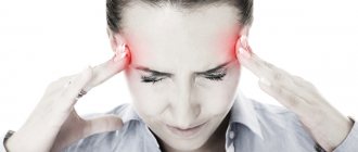
- Blepharospasm. Damage to the muscles surrounding the eyes. Manifested by rapid, forced closing of the eyes.
- Oromandibular dystonia. It is characterized by violent contractions of the jaw and tongue, which makes it difficult to open and close the mouth and often leads to chewing and speech disorders.
- Spasmodic dysphonia. Focal dystonia affects the vocal muscles of the larynx, resulting in speech impairment. The voice may become strained, hoarse, strangled, breathy, or whisper.
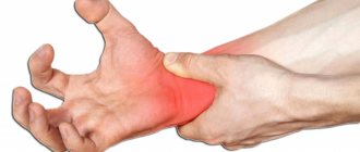
- Writer's cramp. Task-related hand dystonia affecting the fingers, hands, and forearm. Occurs when trying to perform any actions with a brush, for example: while writing or playing a musical instrument.
In 80% of cases, muscular dystonia is a genetic disease (primary dystonia), although it often begins to manifest itself in adulthood. Why a genetic disease does not begin to manifest itself at birth remains unclear. It is assumed that the pathological gene begins to work only under certain conditions, under the influence of external factors.
Although the disease is genetic, dystonia rarely affects more than one member of the same family.
Muscular dystonia can also be a consequence of other reasons: taking certain drugs, toxic effects, stroke, encephalitis, etc. Muscular dystonia can also be one of the many symptoms of a progressive degenerative disease.
The diagnosis of “primary muscular dystonia” does not require additional methods of confirmation. A visual examination, history taking and clinical testing are sufficient.
Read also
Blepharospasm
Blepharospasm is a chronic disease characterized by involuntary squinting of the eyelids due to increased tone of the orbicularis oculi muscles.
Most often, the causes of blepharospasm are extremely difficult to establish or… Read more
Torsion dystonia
This is a chronic nervous disease that manifests itself in the form of uncontrolled tonic muscle contractions, which can result in pathological postures and hyperkinesis. Torsion dystonia…
More details
Hyperhidrosis
Hyperhidrosis is a condition that is manifested by excessive, inadequate sweating due to increased activity of the sweat glands. It happens that a person sweats so profusely that his clothes get wet very quickly, from his hands...
More details
Facial hemispasm
Facial hemispasm is a forced contraction of the facial muscles of one half of the face. Most often, the orbicularis oculi muscle is first involved in the process, then the muscles of the lower half of the face join.…
More details
Treatment of spasticity
Spasticity is an excessive increase in muscle tone, the cause of this condition can be a stroke, traumatic brain injury, spinal injury, spinal cord problems, neuroinfection, multiple…
More details
Treatment
The vast majority of patients with both primary and secondary dystonia require treatment, most often it is aimed at the symptoms rather than at the causes of the disease.
The most common treatment for focal dystonia in the world is repeated local injections of botulinum toxin (botulinum toxin). Treatment of focal dystonias with botulinum toxin should be carried out by a neurologist who specializes and has experience in the treatment of movement disorders, therefore, throughout the world, this specialized medical care is usually assigned to outpatient clinical centers for movement disorders and botulinum therapy.
Also, various groups of drugs are used to treat dystonia, especially those spread throughout the body. Treatment is aimed at relaxing the muscles.
There are also surgical treatment methods that include stereotactic surgery and stimulation of deep brain structures - highly effective methods for treating generalized muscular dystonia and other movement disorders. Previously, before modern neurosurgical methods, peripheral operations were used (cervical radicotomy, decompression of the accessory nerve, selective denervation and rhizotomy, myectomy). However, the therapeutic effect of these peripheral influences is often temporary and is also associated with a high risk of functional impairment. Typically, surgical treatment is used in cases of persistent therapeutic resistance to other treatment methods.
Symptoms and manifestations
Children have different symptoms than adults. Thus, at an early age, parents may notice unilateral or completely asymmetrical symptoms. For example, a child turns over only on one side and completely ignores the other. Or turns his head only to one side.
In adults, the main symptom is an unnatural gait, abnormal, atypical body postures and limb positions. The disorder manifests itself mainly in skeletal muscle symptoms. Sometimes the poses are a little unusual, and sometimes they seem unnatural even at first glance.
Other symptoms:
- Impaired motor function, loss of motor skills.
- Children have delayed motor development. The child does not crawl or sit at the required age.
- With a progressive disease in adults, loss of ability to work occurs quite quickly.
- Activity and well-being largely depend on muscle tone.
In adults, dystonia can lead to disability and complete disability, especially if the cause is cerebrovascular accident.
Therefore, it is important to consult a doctor promptly. The only way to maintain your ability to work is to start treatment on time.
The benefits of massage as a method of combating dystonia
The main advantages of massage procedures are:
- the painlessness of all the specialist’s manipulations, which means there is no risk of causing physical or mental trauma to the baby during the sessions;
- positive attitude of children towards massage;
- complete safety for the child’s health, compared to drug treatment, and maximum benefit.
In addition to the beneficial effect on the main problem, children's massage in Moscow can improve the overall well-being of the baby in the form of normalizing sleep, increasing the baby's appetite and mood.
When performing the procedure, blood circulation improves, the body's defenses increase, and the functioning of internal organs and systems - the heart, gastrointestinal tract, lungs and central nervous system - is normalized.
In addition, massage is an excellent way to prevent colds in young children.