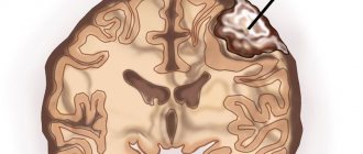
The spinal cord is a section of the central nervous system that transmits nerve impulses from the brain to other organs and systems, and in the reverse order. It is also responsible for the normal functioning of the skeletal muscles and takes part in the activities of the following systems:
- respiratory;
- cardiovascular;
- urogenital;
- digestive.
Tumors in the spinal cord or those located nearby can cause irreparable harm to the entire body. Despite the fact that neoplasms in the spinal cord are rarely diagnosed, it is important to have some useful information for timely detection of pathology. The disease may not make itself felt for years, and in the lost time the tumor can affect neighboring organs.
Classification of the disease
Formations in the spinal cord are classified according to various parameters that are important for the diagnosis and further treatment of the tumor. First of all, pathology is divided into the following types:
— Primary
. Neoplasm cells consist of nerve or brain cells.
— Secondary
. It is metastasis of a tumor located elsewhere.
Depending on the anatomical location of the tumor, they are divided into:
Intramedullary
. Intracerebral formations consist of spinal cells and occur in 18-20% of cases of pathologies in the spinal cord. The location of the tumors is inside the spinal cord, they affect its tissues. Intramedullary tumors, in turn, are divided into:
- ependymomas;
- astrocytomas;
- hemangioblastoma;
- oligodendrogliomas;
Extramedullary.
Neoplasms are located outside the brain and make up 80-82% of the total number of tumors. Localized near the spinal cord, their ingrowth is possible. Extramedullary formations are also classified into:
- subdural (intradural): affect the dura mater of the spinal cord and nerve roots;
- epi- or extradural (vertebral): located on both sides of the meninges, they are metastases, non-malignant and malignant tumors.
In relation to the spinal canal, neoplasms are:
- Intravertebral
. Localization - inside the spinal canal. - Extravertebral
. They are characterized by growth from outside the spinal canal. - Extraintravertebral
. Tumors of this type resemble an hourglass. One part is located inside the spinal canal, the other is located outside.
According to the type of spread, types of neoplasms are distinguished:
- craniospinal;
- cervical, thoracic or lumbosacral regions;
- coccygeal and lower sacral segments;
- horse tail.
Based on the type of histology, tumors are divided into the following:
- meningiomas;
- schwanommas;
- neuromas;
- angiomas;
- hemangiomas;
- hemangiopericytomas;
- ependyomas;
- sarcomas;
- oligodendrogliomas;
- medulloblastomas;
- astrocytomas;
- lipomas;
- cholesteatomas;
- demrmoids;
- epidermoids;
- teratomas;
- chondromas;
- chordomas;
- metastases.
Meningiomas are the most common. All types of spinal cord tumors (SCT) are pathological neoplasms - benign or malignant.
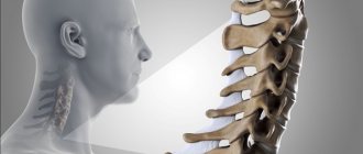
Cervical lesion
The cervical region is the most dangerous place, because here the spinal cord passes into the brain, and there is also a large neurovascular line in the neck. And any pathology concentrated in this department poses a great threat not only to the spinal structures, but also to the components of the higher part of the central nervous system, namely the brain. In general, cancer in any other segment - thoracic and lumbosacral - is associated with its own risks and complications. Let's consider what symptoms can be observed during tumor processes that attack one or another department.
MRI of the cervical spine.
Symptoms of a cervical tumor
With neoplasms of the cervical spine, you may be concerned about:
- constant or recurrent neck pain;
- radiating pain to the arm;
- stiffness of the neck muscles;
- painful sensations between the right shoulder blade and the spine, in the jugular fossa;
- dizziness, loss of consciousness;
- nausea, lack of appetite;
- problems swallowing;
- lack of coordination;
- impairment of vision and/or hearing;
- headaches, discomfort in the head;
- memory impairment, low concentration;
- partial or complete failure of motor functions of the hand;
- paralysis of the respiratory muscles is possible, up to respiratory arrest.
Common signs of a lumbar spine tumor:
- regular pain in the lumbar region, often radiating to the buttock or thigh;
- loss of leg strength, problems with movement, the skin on the leg may acquire a marbled tint (if treatment is not started in a timely manner, the limbs may completely fail);
- spontaneous urination or uncontrolled bowel movements;
- impotence, infertility, menstrual irregularities;
- atrophy of the quadriceps (thigh muscle);
- paresis of the lower legs, especially in the area of the ankle and ankle (in severe cases, paralysis of the legs is possible);
- local tingling, numbness and other unpleasant sensations in the area of the sciatic nerve and in the legs.
Clinical manifestations in the thoracic region are expressed:
- pain in the back, chest, ribs, upper abdomen;
- disturbances in the functioning of the cardiovascular system (blood pressure surges, arrhythmia, tachycardia, etc.);
- failure of the digestive system;
- rapid fatigue during physical activity;
- breathing problems, shortness of breath, shortness of breath;
- an increase in pain when trying to take a deep breath, when coughing (if untreated, suffocation may occur; artificial ventilation is sometimes urgently needed);
- complete or partial paralysis of those parts of the body that are located below the resulting lesion.
Causes of spinal cord tumors
The appearance of neoplasms of this type may be preceded by the following risk factors:
- the effects of radiation on the body;
- ultraviolet irradiation;
- poor environmental situation;
- frequent use of a mobile phone;
- being near high-voltage lines;
- exposure to microwave and electromagnetic radiation from household appliances;
- exposure to chemicals or nuclear waste;
- smoking;
- genetic abnormalities;
- consumption of products with nitrates and carcinogens;
- family history of skin, bladder, breast and pancreatic cancer.
The likelihood of developing the disease also increases with age. In addition, doctors identify specific causes of ACM:
- hereditary neurofibromatosis;
- Hippel-Lindau disease.
The appearance of spinal cord tumors, like other types of cancer, is also due to constant stress and lack of emotional balance.
Spinal cord tumor symptoms
The complex of symptoms depends on the etiology and severity of the pathogenesis of the disease. At the initial stage of the disease, symptoms are mild. Mostly the patient is unaware of the presence of a dangerous process in the body. Treatment of a spinal cord tumor is most effective if you seek medical help in a timely manner. Therefore, if the following signs appear, it is better to see a specialist:
- pain in the back or neck, usually spreading to other parts of the body;
- loss of sensation in the limbs;
- dysfunction of the musculoskeletal system;
- impaired coordination of movements;
- numbness, both at the site of the tumor and in the extremities, in the head, chest and abdomen;
- paresis;
- disruption of the genitourinary system;
- intestinal dysfunction;
- decreased potency;
- infertility;
- increased intracranial or blood pressure;
- headache;
- increased body temperature;
- loss of body weight.
Even with minor discomfort in the back area, you should consult a doctor. The clinical picture of ASM often resembles osteochondrosis or intervertebral hernia.
In addition, spinal tumors include the following syndromes:
1. Root. Intense pain is observed, which intensifies with coughing and sneezing, bending the head and physical activity.
2. Brown-Sequard. The clinical picture is expressed by sensory disorders, including a decrease in muscle-joint sensitivity and a decrease in temperature and pain perception.
3. Complete transverse lesion. In this case, there are disorders of the pelvic functions and the development of bedsores and paresis.
The size, type and stage of development of cancer pathology play a significant role in the manifestation of certain symptoms. One of the most important signs that should not be ignored is constant back pain, independent of physical activity. Taking painkillers for ASM does not have a long-term effect.
Causes of spinal tumors at an early stage
There is still no clear answer to why spinal column cancer occurs. However, a number of observations name the most rationally justified reasons. Thus, the heredity factor largely determines the occurrence of cancer. Also, injuries to the back and spine often lead to dire consequences. Deterioration of nutrition or the pursuit of fashionable diet programs can also lead to the formation of this serious disease. If you eat food that contains carcinogens, you can “count on” cancer of the spine or other organs. For prevention purposes, it is worth building a daily routine. Don't forget to get enough sleep, healthy food and physical activity. This triad will help avoid many diseases. A healthy lifestyle is always on guard of your health.
Diagnosis of spinal cord tumors
The presence of a modern diagnostic complex in the neurosurgical department of the city clinical hospital named after A.K. Yeramishantsev allows for high-quality tests. Diagnostics helps to identify oncogenes in the patient’s blood with a hereditary predisposition. Progressive technologies and techniques provide the opportunity to:
- prescribe preventive anti-cancer measures;
- detect the first signs of illness;
- promptly begin effective treatment of the tumor.
In our clinic, after examination by a neurologist, the following methods of further monitoring are used to determine the location and spread of tumor formation and its histological type, and to identify disturbances in the patency of the cerebrospinal fluid:
- Magnetic resonance imaging;
- Ultrasound;
- CT scan;
- radionuclide diagnostics;
- spinal puncture and further analysis of the cerebrospinal fluid.
MRI of the spine today is considered the safest and most informative research method. To make the most accurate diagnosis, it is necessary to study samples of tumor tissue.
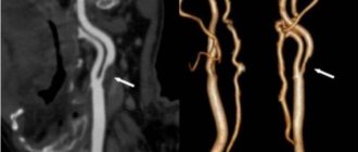
Spinal epidural hemorrhage and hematomyelia
In the case of hemorrhage in the spinal cord (hematomyelia) in the subarachnoid and epidural space, the patient may develop the clinical picture of acute transverse myelopathy within a few minutes or hours. Hemorrhages in the spinal cord (hematomyelia) are accompanied by severe back pain. The source of hemorrhage in hematomyelia can be:
- arteriovenous malformation (AVM, angioma, hemangioma) of the spinal cord
- hemorrhage into the tumor
- anticoagulant therapy with warfarin for thrombosis of the cerebral venous sinuses
- spontaneous hemorrhage in the spinal cord (occurs most often in patients)
Epidural hemorrhages of the spinal cord can develop as a result of:
- minor spinal injuries
- lumbar puncture
- anticoagulant therapy with warfarin
- secondary to blood diseases
Patients' complaints of back pain and radicular pain due to hemorrhages in the spinal cord (hematomyelia) often precede the onset of weakness by several minutes or hours. Weakness and pain can be significant, forcing patients to assume antalgic positions when moving. An epidural hematoma at the level of the lumbar segments of the spinal cord is accompanied by loss of the knee and Achilles reflexes, while with retroperitoneal hematomas only the knee reflexes are usually lost.
Extradural hematoma on MRI of the spine that occurred in a patient after surgery to remove a hemangioma.
When diagnosing spinal hemorrhage in a patient, a volumetric process is determined by myelography. A computed tomography (CT) scan of the spine sometimes does not detect this hematoma, since the blood clot cannot be distinguished from the adjacent bone tissue.
Spinal cord hematomas can form as a result of spontaneous bleeding. Blood clots can be caused by the same factors as epidural hemorrhages, giving particularly severe pain in the subdural and subarachnoid spaces. With an epidural hemorrhage, the cerebrospinal fluid (CSF) is usually clear or contains a small number of red blood cells. With subarachnoid hemorrhage, the cerebrospinal fluid is initially bloody, and later acquires a pronounced yellow-brown tint (xanthochromia) due to the presence of blood pigments. In addition, pleocytosis and a decrease in glucose concentration can be detected in the cerebrospinal fluid. This creates a false picture similar to bacterial meningitis.
Intraspinal (intramedullary) hematoma can be caused by spontaneous rupture of an internal vascular malformation, such as telangiectasia in the gray matter. More often it is the result of injury. If bleeding begins in the central region, it usually spreads up and down the axis of the spinal cord into several segments and is referred to as hematomyelia. Clinically, an acute syndrome develops that may closely resemble the chronic syndrome characteristic of syringomyelia.
Spinal epidural bleeding is rare. It is usually caused not by trauma, but by rupture of a vascular malformation, most often a small-vessel hemangioma (AVM, angioma) in or near the epidural space in the bones of the spine. X-rays reveal vertical trabeculae in the spongy substance of the spinal bone, characteristic of angioma. Blood does not always collect in the area of the angioma. The hematoma usually develops over the dorsal portion of the midthoracic spinal cord. It can cause acute radicular pain at the level of bleeding. Then the syndrome of transverse myelopathy develops with paresthesias, followed by sensory loss. Motor paresis begins in the fingers and feet and rises to the level of compression of the spinal cord. In such cases, immediate consultation with a neurosurgeon is indicated.
Methods of treating the disease
The main treatment for a spinal cord tumor is surgical removal. Operations are prescribed after a complete examination of the patient and an accurate diagnosis. Today, the capabilities of neurosurgeons are significantly increased by the latest medical equipment:
— endoscopy technology provides the opportunity to gain access to tumors in hard-to-reach places;
— thanks to microsurgical instruments, the risk of complications during and after the procedure is reduced.
Our neurosurgery department has modern operating microscopes made in Germany with the highest degree of resolution. Their use allows surgeons to avoid damaging healthy areas.
The recovery period after removal of benign tumors ranges from several months to two years. In 70% of cases, complete recovery is achieved. In the case of intramedullary and low-quality formations, the prospect is not so comforting. Metastatic tumors are usually inoperable.
Further treatment for a spinal cord tumor after surgery includes:
- chemotherapy;
- radiation therapy.
To rehabilitate the patient, physiotherapeutic methods, fixation of the spine from the outside, and learning to walk again are prescribed. It is advisable to carry out social and psychological rehabilitation.
You can make an appointment using the contact number. We will provide a free consultation about the treatment of a spinal cord tumor, approximate prices and answer other questions.
The cost of surgical removal depends on the type of tumor and the nature of the surgical procedure. We guarantee operations at the highest level and at reasonable prices.
Treatment and rehabilitation of spinal tumors at an early stage
The main tasks of the doctor are to recognize the type of cancer and its stage. After this, treatment is prescribed. If we talk about its surgical option, then it is worth mentioning the increased risks. The slightest inaccuracy, the slightest damage, and the spinal cord can be damaged. Therefore, the surgeon is required to have the highest degree of skill and professionalism. Chemotherapy slows tumor growth. This fact has been scientifically proven. However, it gives many side effects. Radiation therapy is effective: it kills tumor cells. But it also has side effects.
It is worth remembering an important fact: the earlier treatment begins, the greater the chances of a successful outcome of all procedures. In addition, a team of doctors must act. Each of them is responsible for certain areas of medicine. So, the oncologist draws up an individual treatment plan, and the radiologist adds the nuances. In general, specialists should assemble a consultation. At this meeting, all aspects of treatment and rehabilitation are decided and discussed through joint efforts. Only this approach allows us to judge that a medical error will be excluded.
Rehabilitation should take place in a special center staffed by doctors and nurses. After all, at first you may need medical help at any time. The hospital conditions are suitable. It is important to enlist the support of family and friends. After all, the psychological factor sometimes plays a key role in matters of treatment and rehabilitation.
Surgery to remove spinal cord tumors
Neoplasms in the human spinal cord can be benign or malignant. Predominantly, surgery is prescribed for treatment. Surgical intervention is carried out after a complete examination of the patient, using magnetic resonance and computed tomography, as well as ultrasound. You will also need a spinal puncture to analyze the liquor fluid and conduct radionuclide diagnostics. Modern research methods make it possible to accurately determine the size, location and type of tumor in the spinal cord.
Diagnostics
Diagnostics is a step-by-step process that allows you to determine the size and type of tumor and draw up a plan for its treatment. First of all, the doctor is interested in the patient’s complaints, symptoms, and medical history. It is important when symptoms developed and how they changed over time.
A full neurological examination is also carried out - determination of muscle tone, reflexes, muscle strength, spinal mobility, sensitivity.
To refine the pathology you need:
- MRI with contrast - this method helps to determine the condition of the entire spinal cord, its structures and determine the tumor;
- CT myelography is a technique for assessing the tumor border;
- diffusion-weighted and diffusion tensor MRI;
- scintigraphy;
- direct angiography.
After clarifying the type, location and size of the tumor, a treatment plan is drawn up.
Indications for surgery
If the tumor develops rapidly and grows to a large size, surgical intervention is indicated. Metastatic formations, in most cases, are inoperable. In addition, the patient’s age, the presence of chronic diseases and the location of the tumor are taken into account. After preliminary studies, examination by a neurologist and neurosurgeon, surgical procedures are prescribed on an individual basis. The latest endoscopy instruments and microsurgical devices allow us to perform operations in our clinic, even in hard-to-reach areas and with a reduced risk of complications for the patient.