The brain is a complex and poorly understood human organ that is difficult to diagnose. However, the brain is the control center responsible for the operation of all systems in the body. Therefore, any pathological processes in it affect the physical and mental health of a person.
MRI is one of the methods for studying the brain to determine negative conditions in it. Diagnostics is prescribed for adults and children and is a safe examination method.
What it is
The method refers to a non-invasive examination using radio waves and a magnetic field. During the scanning process, the doctor receives an image showing parts of the brain. Diagnosis is carried out without radiography. The study reveals disturbances in the functioning of the nervous and vascular systems, aneurysms, and tumors.
Also, based on the results of the examination, the level of activity of the cerebral cortex is determined. When using a contrast agent, tissues are better visualized, which helps to more accurately identify pathologies.
Advantages of the method
Advantages of brain tomography:
- the patient does not experience pain during diagnosis;
- During the examination, no foreign substances are introduced into the human body;
- no ionizing radiation;
- clear images help to accurately recognize pathology and prescribe treatment;
- as prescribed by a specialist, an examination of the head and cervical/upper spine is carried out to assess the functional activity of the brain - based on the results, a safe method of surgical intervention is prescribed;
- areas of the brain hidden by the bones of the skull are examined - other methods do not diagnose these areas;
- MRI helps to obtain a complete picture of the brain and vascular system even without the use of a contrast agent;
- tumors and other pathologies are detected in the early stages of development.
Make an appointment now!
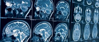
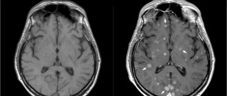
Normal brain MRI
Normal state of brain tissue on an MRI image of the head (sagittal projection)
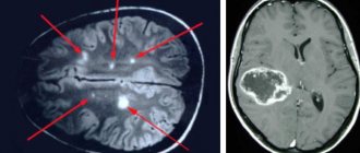
Forming a conclusion when deciphering an MRI of the brain is a complex multi-stage process. The study of the picture begins with the hemispheres. The contours should be clear and round in shape. Normally, there are no gross indentations or protrusions on the images of the hemispheres. Next, the structure of the brain substance is examined. In the absence of pathologies, focal and diffuse changes are not observed in the photo. The third stage of studying the images is the assessment of the ventricular system. Any decrease or increase in the size of the latter or its individual parts is considered a deviation. During enhanced MRI in a healthy person, the contrast agent quickly leaves the vascular bed and concentrates in the area of pathological changes.
Why is an examination prescribed?
MRI diagnostics helps to recognize pathological changes in the soft tissues of the lining of the brain, inflammatory reactions, and disorders of the central nervous system. Also, with the help of an examination, the conditions of the parts of the head are studied: the visual and occipital lobes, the pituitary gland, the cerebellum, areas of thinking and memory, and the ventricles of the brain.
Before the procedure, the patient undergoes tests, the results of which determine the type of diagnosis. For example, tests showed that the patient had an excess level of the hormone prolactin. The doctor writes a referral for examination of the cerebellum.
Examination of the brain in a magnetic field helps determine the presence of:
- Multiple sclerosis - lesions of the myelin sheath of nerve fibers. The diagnosis determines the stage of the disease, the level of damage, after which the doctor prescribes therapy.
- Benign and malignant tumors. Brain scanning is used to identify the affected area, monitor the development of the tumor, as well as the treatment process.
- Disorders in the cerebral cortex - Parkinson's disease, Alzheimer's disease. MRI determines the density of white and gray matter, the presence of pathologies in the subcortex and cortex of the head.
- Ischemic strokes, heart attacks. The images determine the area and density of ischemic lesions, the presence of cortical necrosis, the appearance of edema, and the stage of development of the disease.
- Brain injuries are damage to blood vessels and tissues due to trauma. Diagnostics reveals the first signs of VSD.
- Mental disorders of endogenous and exogenous types. The method helps to identify hereditary pathologies, changes after traumatic brain injury, viral infection, and poisoning with toxic substances. MRI diagnostics determines schizophrenia.
For children, head tomography is prescribed for the following pathologies:
- head injury, concussion after injury, intrauterine infectious processes;
- hypoxia, ischemia and other disruptions in brain development;
- epileptic seizures, cerebral hemorrhage;
- the first stages of multiple sclerosis;
- dangerous diseases such as pituitary adenoma, brain tumors, cysts and suspicions of them;
- a sharp deterioration in vision and hearing, malfunction of the inner ear;
- high intracranial pressure.
Diagnostics helps to study the state of the brain and its parts, identify the causes of headaches in children, and prevent the development of autism.
Differences between MRI and CT scan of the brain
Magnetic resonance imaging of the head differs from other diagnostic methods in the following:
- the examination is performed in 3-4 projections, which provides more information for identifying diseases and choosing treatment methods;
- pathologies are detected in the early stages of development - ischemic changes are detected 2-3 hours after a stroke;
- even minimal changes in the brain that cause multiple sclerosis are determined;
- parts of the brain that are not diagnosed by computed tomography (CT) are studied - the brain stem, the cerebellum.
How to overcome fear of the procedure
There is no need to worry before an MRI - the examination is absolutely painless and safe. The radiologist remains in contact with the patient throughout the procedure and, if necessary, will answer any questions that arise.
If the patient experiences severe anxiety, then before the examination, you can take sedatives.
MRI is a modern type of diagnostics that makes it possible to accurately identify any pathological changes in the brain, even at the initial stages of their occurrence. Magnetic resonance imaging has virtually no contraindications and is a painless and safe technique used to diagnose a wide range of diseases.
| What does an MRI show? |
| How is an MRI of the brain done with contrast? |
| What does an MRI of the pituitary gland show? |
| MRI of the brain with contrast |
| MRI of the sella turcica and pituitary gland in Rostov-on-Don |
| MRI of the cerebellum of the brain |
When is a diagnosis prescribed?
A brain examination is prescribed for the following ailments:
- developmental anomalies and diseases of cerebral vessels;
- head injuries and bruises, cerebral hemorrhages;
- sharp deterioration of vision and hearing;
- tumor development;
- infectious reactions in the central nervous system - meningitis, HIV infections, abscesses;
- pituitary adenoma, epilepsy;
- pathologies of the base of the skull;
- multiple sclerosis, sinusitis, neurodegenerative diseases;
- aneurysms, thrombosis and other anomalies in the vessels of the head.
Diagnosis is also carried out after surgery. Prescribed for frequent migraines and fainting, tinnitus, sudden deterioration of memory and attention, impaired coordination of movements, and mental disorders.
Indications
MRI of the brain is prescribed in the following cases:
- For head injuries, when there is a suspicion of traumatic brain injury, for the diagnosis of diffuse axonal damage, subdural and epidural hematoma, as well as intracerebral hemorrhage.
- Suspicion of benign and malignant neoplasms of the brain: glioma, hemangioma, sarcoma, neuroblastoma, astrocytoma, ependymoma, as well as tumors of the meninges: meningioma, cranial nerves and individual structures (epiphysis, pituitary gland, medulla oblongata, cerebellum), as well as cysts of various origins and localization.
- If you have a persistent headache, the cause of which cannot be identified by other diagnostic methods.
- To exclude or confirm cerebral ischemia (ischemic stroke) or hemorrhage (hemorrhagic stroke) in acute cerebrovascular accident, as well as for the differential diagnosis of these pathological conditions.
- If you have a history of seizures or an established diagnosis of epilepsy.
- If a demyelinating process of the brain is suspected or a demyelinating disease has been diagnosed, as well as to assess the degree of progression of the process and the effectiveness of treatment for this disease.
- For the diagnosis of dyscirculatory encephalopathy.
- If congenital defects or abnormalities of brain development are suspected.
MRI of the brain is prescribed after surgical interventions to assess the effectiveness of treatment. In this case, the study is performed before and after surgery, then the doctor compares the results and draws appropriate conclusions.
Contraindications
Head tomography is not prescribed if the patient has absolute contraindications, but is allowed at the discretion of the doctor in case of relative contraindications. Diagnostics is prohibited if the following conditions exist:
- the patient has prosthetic heart valves, pacemakers or neurostimulators;
- presence of insulin pumps, inner and middle ear prostheses, cochlear implant;
- the patient has an Ilizarov apparatus;
- the presence of metal implants, ferromagnetic particles, fragments in the body.
Relative indications include:
- tremor, the patient’s inability to hold his breath for a long time during examination;
- presence of dental braces, dentures, stents, vena cava filters;
- heart failure, clip instead of gallbladder;
- pregnancy;
- claustrophobia;
- severe pain while standing still;
- coronary artery bypass grafting.
Indications for examination
The description of the tomography procedure highlights its unique capabilities. MRI of the head helps to find out the reasons if the patient has frequent and severe headaches, inflammation, or impaired blood supply to the nervous system. There are several modes for conducting a medical examination. Don't know where to get a head tomography? The MRI CENTER clinic will help you. The duration of the procedure is 15–40 minutes.
Prices for head MRI in Moscow depend on the specifics of the process. The procedure must be done when:
- Brain tumors and various disorders in the functioning of its blood vessels.
- Diseases of the eyes and inner ear.
- Pituitary gland diseases.
- Stroke.
- As prescribed after the patient has suffered injuries.
- Headaches.
- Diseases of the nervous system.
- Detection of pathologies associated with dementia.
This method allows you to get a holistic picture of the patient’s condition and detect various organ diseases. This diagnostic method allows you to comprehensively study the patient’s condition and detect various diseases of organs and systems. However, there are also contraindications. For example, the examination is not recommended if you have a pacemaker, steel implants, or claustrophobia.
Preparatory stage
The doctor, based on the test results, determines whether to perform a tomography with or without the introduction of a contrast agent. If the first option is chosen, then the patient does not take food or liquid 5 hours before the diagnosis. Before the procedure, you must remove all jewelry and accessories, watches and objects with metal elements.
During a consultation with a doctor, the patient reports the presence of chronic diseases, allergies to medications, claustrophobia, and pregnancy.
When preparing a child for a head tomography, it is not recommended to give him anything to drink or eat 3 hours before the diagnosis. If an MRI is performed with the administration of contrast, the examination is performed on an empty stomach. Before the procedure, the child should be shown to an allergist to check for an allergic reaction to the injected substance.
Advantages of performing MRI at the Central Clinical Hospital of the Russian Academy of Sciences
When choosing a clinic where to undergo an examination, remember that the accuracy of the examination results largely depends on the quality of the equipment, as well as on the experience and professionalism of the specialist responsible for interpreting the images. The Central Clinical Hospital of the Russian Academy of Sciences uses modern equipment that meets international quality standards to perform magnetic resonance imaging. Diagnosticians are highly qualified and have extensive experience. With us, you can do an MRI of the head on the day of your visit, or by appointment. All examinations are performed with maximum convenience for patients. The price of MRI diagnostics depends on the duration and complexity of the procedure.
You can sign up for magnetic resonance imaging through the form on the website or by phone
Diagnostic features
If a brain tomography is performed with the introduction of a contrast agent, the examination takes 1-1.5 hours.
Stages:
- the patient removes objects and clothing with metal elements;
- lies on the tomograph table on his back;
- the doctor administers a contrast agent intravenously;
- if there is tremors or inability to lie still, the patient takes sedatives;
- legs and arms are secured with belts, bolsters are placed under the neck;
- the table is pushed into the tomograph capsule - the doctor monitors the procedure from a special room with equipment;
- during the diagnosis, the patient hears slight noises of the tomograph operating - the procedure is safe and painless, a slight sensation is felt in the area of the injection of the substance;
- head diagnostics lasts 15 minutes - the patient is recommended to remain in a stationary position to obtain accurate scan results.
Features of examination of children
Brain tomography in children is carried out under the drug n8a8p00ko70z0o-7m9, -6v5 Propofol is administered. Anesthesia helps keep the child still during diagnosis. Children over 5 years old are given a sedative and given psychological adjustment and support before the tomography.
During the examination, the baby is shown cartoons and toys. Some clinics use open tomographs, when only the child's head is placed in the capsule, and the parents stand nearby and hold his hand.
Before diagnosis, it is recommended to take the child to the toilet, remove electronic items and jewelry with metal inserts from clothes and body. Next, the child is dressed in special clothes, “introduced” to the tomograph and allowed to listen to how it works. This helps to calm the baby before the procedure and obtain his consent to the examination.
Side effects
After tomography, the patient may experience nausea, dizziness, vomiting, weakness, and disorientation in space. This is a normal reaction. It occurs in people with increased sensitivity, when there are metal objects on the patient’s body, and when the rules for preparing for an MRI are not followed. Unpleasant sensations go away on their own. However, if side effects do not disappear for a long time, it is recommended to consult a doctor.
If the rules are followed, the procedure takes place without pain or side effects. The images help doctors accurately determine the cause of the disease and prescribe effective treatment.