Anatomy of the Human Cerebral Vessels - information:
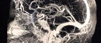
The arteries of the cerebrum originate from the branches of a. carotis interna and a. basilaris, forming the circulus arteriosus cerebri at the base of the brain. The anterior, middle and posterior cerebral arteries branch on the surface of each hemisphere.
A. cerebri anterior supplies blood to the medial surface of the hemisphere up to the sulcus parietooccipitalis, on its outer surface to the superior frontal gyrus and the upper edge of the parietal lobe, and on the lower surface of the hemisphere to the gyrus rectus of the frontal lobe.
A. cerebri media supplies blood to the insula, both central gyri, the inferior frontal gyrus and most of the middle frontal gyrus, the parietal lobe and the superior and middle temporal gyri.
A. cerebri posterior branches on the medial, inferior and lateral surfaces of the temporal and occipital lobes, with the exception of the superior and middle temporal gyri.
The listed arteries, with their branches in the pia mater, form an arterial network, from which they penetrate vertically into the thickness of the medulla:
- cortical arteries - small branches that branch only in the cerebral cortex, and
- medullary arteries, which, after passing through the cortex, go into the white matter.
The central arteries enter from the base of the brain. Cortical, medullary and central arteries anastomose with each other, forming a single vascular network. The cerebellum receives blood from three arteries on each side.
Two a. cerebelli inferior anterior (branch of a. basilaris) and a. cerebelli inferior posterior (branch of a. vertebralis), branch on the lower surface of the cerebellum, the third branch, a. cerebelli superior (branch of a. basilaris), goes to its upper surface. From a. cerebelli superior is also supplied to the lower colliculi of the roof of the midbrain, and the superior colliculi receive their branches from a. cerebri posterior. The arteries of the remaining parts of the brain, related to the pons and medulla oblongata, originate from a. vertebralis, a. basilaris and their branches. In addition to the described arterial vessels, there are also special arteries of the choroid plexus, four in number on each side.
The cerebral veins are divided into superficial and deep. The superficial veins mostly collect blood from the cerebral cortex and flow partly into the sinus sagittalis superior (superior veins), partly (lower veins) into the sinus transversus and the sinuses of the base of the skull. The veins lack valves and are distinguished by their numerous anastomoses. The deep veins collect blood from the central gray nuclei and ventricles of the brain and merge into one large v. cerebri magna, flowing into the sinus rectus. The veins of the cerebellum form groups: the upper ones pour blood into the sinus rectus and v. cerebri magna, lower - in sinus transversus, sigmoideus, petrosus inferior.
Brain blood supply device
As you know, in order for the brain to work and all its cells to function correctly, a continuous supply of a certain amount of oxygen and nutrients to its structures is required, regardless of the physiological state of a person (sleep - wakefulness). Scientists have calculated that the needs of the central nervous system consume about 20% of the oxygen consumed, while its mass in relation to the rest of the body is only 2%.
The brain is nourished through the blood supply to the organs of the head and neck through the arteries that form the arteries of the circle of Willis on the brain and penetrate through it. Structurally, this organ has the most extensive network of arterioles in the body - its length in 1 mm3 of the cerebral cortex is approximately 100 cm, in a similar volume of white matter about 22 cm.
The largest amount is located in the gray matter of the hypothalamus. And this is not surprising, because it is responsible for maintaining the constancy of the internal environment of the body through coordinated reactions, or in other words, it is the internal “steering wheel” of all vital systems.
The internal structure of the blood supply to arterial vessels in the white and gray matter of the brain is also different. For example, the arterioles of the gray matter have thinner walls and are elongated compared to similar structures of the white matter. This allows for the most efficient gas exchange between blood components and brain cells; for this reason, insufficient blood supply primarily affects its performance.
Anatomically, the blood supply system of the large arteries of the head and neck is not closed, and its components are interconnected through anastomoses - special connections that allow blood vessels to communicate without forming a network of arterioles. In the human body, the largest number of anastomoses is formed by the main artery of the brain - the internal carotid. This organization of blood supply allows you to maintain constant movement of blood through the circulatory system of the brain.
Structurally, the arteries of the neck and head are different from the arteries in other parts of the body. First of all, they do not have an external elastic shell and longitudinal fibers. This feature increases their stability during surges in blood pressure and reduces the force of blood pulsation shocks.
The human brain works in such a way that at the level of physiological processes it regulates the intensity of blood supply to the structures of the nervous system. In this way, the body’s defense mechanism is triggered - protecting the brain from surges in blood pressure and oxygen starvation. The main role in this is played by the sinocartoid zone, the aortic depressor and the cardiovascular center, which is associated with the hypothalamic-mesancephalic and vasomotor centers.
Anatomically, the largest vessels bringing blood to the brain are the following arteries of the head and neck:
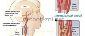
- Carotid artery. It is a paired blood vessel that originates in the chest from the brachiocephalic trunk and the aortic arch, respectively. At the level of the thyroid gland, it, in turn, is divided into an internal and external artery: the first delivers blood to the medulla, and the other leads to the facial organs. The main branches of the internal carotid artery form the carotid basin. The physiological significance of the carotid artery lies in supplying microelements to the brain; about 70-85% of the total blood flow to the organ flows through it.
- Vertebral arteries. The vertebrobasilar basin is formed in the cranium, which provides blood supply to the posterior sections. They begin in the chest and follow the bony canal of the spinal part of the central nervous system to the brain, where they unite into the basilar artery. According to estimates, the blood supply to the organ through the vertebral arteries supplies about 15-20% of the blood.
The supply of microelements to the nervous tissue is ensured by the blood vessels of the Circle of Willis, which is formed from the branches of the main blood arteries in the lower part of the skull:
- two forebrains;
- two midbrain;
- pairs of hindbrain;
- anterior connecting;
- pairs of rear connecting ones.
The main function of the circle of Willis is to ensure stable blood supply during blockage of the leading vessels of the brain.
Also in the circulatory system of the head, experts identify the Zakharchenko circle. Anatomically, it is located on the periphery of the medulla oblongata and is formed by the union of the collateral branches of the vertebral and spinal arteries.
The presence of separate closed systems of blood vessels, which include the Circle of Willis and the Zakharchenko Circle, makes it possible to maintain the supply of the optimal amount of microelements to the brain tissue when blood flow in the main channel is disrupted.
The intensity of blood supply to the brain of the head is controlled using reflex mechanisms, the functioning of which is controlled by nerve pressoreceptors located in the main nodes of the circulatory system. For example, at the site of the branch of the carotid artery, there are receptors that, when excited, can give a signal to the body that it needs to slow down the heart rate, relax the walls of the arteries and lower blood pressure.
Venous system
Along with arteries, the veins of the head and neck participate in the blood supply to the brain. The task of these vessels is to remove metabolic products from nervous tissue and control blood pressure. The venous system of the brain is much longer than the arterial system, which is why its second name is capacitive.
In anatomy, all veins of the brain are divided into superficial and deep. It is assumed that the first type of vessels serves as a drainage of the decay products of the white and gray matter of the terminal section, and the second type removes metabolic products from the structures of the trunk.
A cluster of superficial veins is located not only in the meninges, but also extends into the thickness of the white matter up to the ventricles, where it unites with the deep veins of the basal ganglia. Moreover, the latter entangle not only the nerve ganglia of the trunk - they are also sent to the white matter of the brain, where they interact with external vessels through anastomoses. Thus, it turns out that the venous system of the brain is not closed.
The superficial ascending veins include the following blood vessels:
- The frontal veins receive blood from the upper part of the terminal section and send it to the longitudinal sinus.
- Veins of the central sulci. They are located on the periphery of the Rolandic gyri and run parallel to them. Their functional purpose is to collect blood from the middle and anterior cerebral arteries.
- Veins of the parieto-occipital region. They differ in branching in relation to similar structures of the brain and are formed from a large number of branches. They supply the blood to the posterior part of the terminal section.
The veins draining blood in a descending direction will unite into the transverse sinus, the superior petrosal sinus and the vein of Galen. This group of vessels includes the temporal vein and the posterior temporal vein - they send blood from the same parts of the cortex.
In this case, blood from the lower occipital zones of the terminal section enters the inferior occipital vein, which then flows into the vein of Galen. From the lower part of the frontal lobe, the veins run to the inferior longitudinal or cavernous sinus.
Also, the middle cerebral vein, which is neither an ascending nor a descending blood vessel, plays an important role in collecting blood from brain structures. Physiologically, its course is parallel to the line of the Sylvian fissure. At the same time, it forms a large number of anastomoses with branches of the ascending and descending veins.
Internal connection through anastomosis of deep and external veins allows the removal of cell metabolic products in a roundabout way when one of the leading vessels is insufficiently functioning, that is, in a different way. For example, venous blood from the superior Rolandic fissures in a healthy person drains into the superior longitudinal sinus, and from the lower part of the same convolutions into the middle cerebral vein.
The outflow of venous blood from the subcortical structures of the brain goes through the large vein of Galen; in addition, venous blood from the corpus callosum and cerebellum collects in it. The blood vessels then carry it to the sinuses. They are a kind of collectors located between the structures of the dura mater. Through them it is directed to the internal jugular (jugular) veins and through the reserve venous outlets to the surface of the skull.
Despite the fact that the sinuses are a continuation of the veins, they differ from them in their anatomical structure: their walls are formed from a thick layer of connective tissue with a small amount of elastic fibers, which is why the lumen remains inelastic. This structural feature of the blood supply to the brain promotes the free movement of blood between the meninges.
Impaired blood supply
The arteries and veins of the head and neck have a special structure that allows the body to control blood supply and ensure its consistency in the structures of the brain. Anatomically, they are designed so that in a healthy person, with an increase in physical activity and, accordingly, an increase in blood movement, the pressure inside the vessels of the brain remains unchanged.
The process of redistribution of blood supply between the structures of the central nervous system is carried out by the middle section. For example, with increased physical activity, blood supply to motor centers increases, while in others it decreases.
Due to the fact that neurons are sensitive to a lack of nutrients, and especially oxygen, disruption of blood flow to the brain leads to a malfunction of certain parts of the brain and, accordingly, a deterioration in a person’s well-being.
For most people, a decrease in the intensity of blood supply causes the following signs and manifestations of hypoxia: headache, dizziness, cardiac arrhythmia, decreased mental and physical activity, drowsiness and sometimes even depression.
Disruption of cerebral blood supply can be chronic and acute:
- A chronic condition is characterized by insufficient provision of brain cells with nutrients for a certain amount of time, with a smooth course of the underlying disease. For example, this pathology may be a consequence of hypertension or vascular atherosclerosis. This may subsequently cause gradual destruction of gray matter or ischemia.
- An acute circulatory disorder or stroke, unlike the previous type of pathology, occurs suddenly with sharp manifestations of symptoms of poor blood supply to the brain. Usually this condition lasts no more than a day. This pathology is a consequence of hemorrhagic or ischemic damage to the brain substance.
Arteries of the brain[edit]
Carotid arteries[edit]
The carotid arteries form the carotid basin. They originate in the chest cavity: the right one from the brachiocephalic trunk truncus brachiocephalicus, the left one from the aortic arch arcus aortae. The carotid arteries provide about 70-85% of blood flow to the brain.
Vertebro-basilar system[edit]
The vertebral arteries form the vertebrobasilar basin. They supply blood to the posterior parts of the brain (medulla oblongata, cervical spinal cord and cerebellum). The vertebral arteries originate in the thoracic cavity and pass to the brain in the bone canal formed by the transverse processes of the cervical vertebrae. According to various sources, the vertebral arteries provide about 15-30% of blood flow to the brain.
As a result of the fusion, the vertebral arteries form the main artery (basilar artery, a. basilaris) - an unpaired vessel, which is located in the basilar groove of the bridge.
Circle of Willis[edit]
Near the base of the skull, the main arteries form the Circle of Willis, from which arteries branch that supply blood to the brain tissue. The following arteries participate in the formation of the Circle of Willis:
- right and left anterior cerebral arteries (A1 segments)
- right and left middle cerebral arteries (M1 segments)
- right and left posterior cerebral arteries (P1 segments)
- anterior communicating artery
- right and left posterior communicating arteries
If all of the above arteries are visible, the circle of Willis is considered closed
.
If any of the connecting arteries, or one of the segments of the cerebral arteries is not visualized, the circle of Willis is considered not closed
.
Zakharchenko circle[edit]
Located on the ventral surface of the medulla oblongata. Formed by two vertebral arteries and two anterior spinal arteries.
