What is duplex (triplex) scanning of blood vessels?
Duplex or triplex scanning - synonyms, duplex of vessels and triplex of vessels, ultrasound duplex angioscanning or simply UZDAS .
This is a non-invasive ultrasound research method that allows you to obtain a two-dimensional image of blood vessels with the ability to assess the condition of the vascular wall, the nature and speed of blood flow through them. Simply put, duplex scanning allows the diagnostician to see the vessels being examined, evaluate the places of their narrowings, blockages or, on the contrary, dilated areas, and also determine the presence of blood clots, atherosclerotic plaques, and blood flow disorders. The traditional duplex (triplex) scanning technique uses high-frequency sound waves that are not perceptible to the human ear. The computer receives information from the ultrasonic sensor and converts it into a two-dimensional dynamic image on the monitor screen.
Duplex (triplex) has great advantages over its outdated predecessor - Doppler or Dopplerography of blood vessels, a popular technique in the past that allows one to evaluate only the direction and speed of blood flow, but does not evaluate structural changes in the wall and lumen of blood vessels.
Ultrasound of head and neck vessels
This diagnostic method allows you to obtain detailed information about the condition of the most important vascular branches in the head and neck area.
The phrase “ultrasound of the vessels of the head and neck” also often means:
- Dopplerography of the vessels of the head or neck
- Duplex or triplex ultrasound scanning of the head or neck
- Transcranial Dopplerography of the vessels of the head (TCDG)
- Study of extracranial or intracranial vessels
- Dopplerography of bracheocephalic arteries (BCA)
- Dopplerography of the main arteries of the head (MAG)
If your doctor's prescription contains one of these wordings, ok - you should definitely come to us!
Our prices will pleasantly surprise you, and the quality and thoroughness of the services will surprise your attending physician!
Phone number for registration:
8-495-011-00-30
8-499-390-18-59
Why is duplex scanning of the vessels of the head and neck necessary?
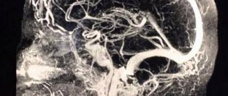
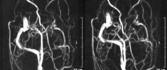
Duplex ultrasound examination of the brachiocephalic vessels (BCA) includes a study of the structural features and speed of blood flow in the carotid and vertebral arteries of the neck. Using this method, it is possible to simultaneously determine the diameter of the vessels, tortuosity, changes in their wall and the speed of blood flow. Usually, in parallel with performing a transcranial ultrasound of the brain with an assessment of the condition of the main arteries of the head to study intracerebral circulation, an ultrasound of the brachiocephalic vessels and vice versa. These ultrasound examinations complement each other.
Indications for ultrasound of head and neck vessels (transcranial and extracranial vessels):
- Dizziness and tinnitus;
- Transient visual disturbances, the appearance of fog in both eyes;
- Diseases of the neck organs;
- Injuries, congenital malformations and acquired diseases of the cervical spine;
- Head injuries;
- Constriction of the pupil and drooping of the upper eyelid on one side;
- Headache.
Another indication is the impact of factors that increase the risk of stroke - smoking, diabetes, heredity, excess body weight.
For any of the listed conditions or suspicion of them, it is recommended to do an ultrasound of the vessels of the brain and neck . A neurologist, surgeon, therapist or ophthalmologist, functional diagnostics specialist (based on the results of an EEG-Electroencephalogram) can prescribe a study. This type of ultrasound diagnostics is absolutely safe, so you can undergo it without a doctor’s prescription, on your own initiative for preventive purposes.
What diseases does the method help identify?
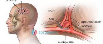
Duplex (triplex) scanning of the vessels of the head and neck makes it possible to determine congenital or acquired disorders of the course and diameter of blood vessels. Their increased tortuosity, aneurysms, the presence of atherosclerosis, neoplasms and developmental anomalies (anomas, congenital hypoplasia, etc.) are detected. Transcranial ultrasound allows you to see signs of cerebral circulatory disorders, spasm and atherosclerosis of blood vessels, and evaluate the pathways of reserve circulation in case of impaired blood flow.
In addition, duplex/triplex ultrasound of the brachiocephalic arteries and main arteries of the head makes it possible to observe the problem over time in order to monitor the result of the treatment. Thanks to the method, you can accurately determine the cause of cerebrovascular accident and choose the most appropriate treatment method.
Advantages of ultrasound of head and neck vessels
The method is absolutely painless, does not require surgical interventions, violation of the integrity of the skin and other unpleasant manipulations associated with risks and complications. To undergo an ultrasound scan of cerebral vessels, no prior preparation is required. This method is completely safe and does not involve x-ray exposure or the need to use contrast fluids. At the same time, MAG ultrasound allows you to quickly obtain detailed information important for making a diagnosis and determining the cause of the disease.
Where to do duplex ultrasound of the vessels of the head and neck?
Our clinic took special care in the selection of diagnostic equipment, as well as in the training of specialists conducting the study. You can easily make an appointment with us by phone or online, saving time, and no longer worry about the quality of the examination. Duplex examination of the brachiocephalic arteries (BCA) and the great arteries of the head (MAG) at the Medford clinic can be done for both adults and children. If you have the results of past studies and analyses, our specialists will help you evaluate changes in dynamics and advise the further course of action.
Our clinic is located in the South-Eastern Administrative District on Aviamotornaya street, not far from the Aviamotornaya metro station, and we can also be easily reached from the Elektrozavodskaya, Baumanskaya and Semyonovskaya metro stations. The Medford Clinic will help you get the maximum health benefits from modern diagnostic ultrasound techniques.
| Ultrasound of cerebral vessels | RUB 1,900 |
| Ultrasound of neck vessels | RUB 1,900 |
| Ultrasound of the vessels of the head and neck (simultaneously) | RUB 3,600 |
| Visit of an ultrasound specialist to your home, within the Moscow Ring Road (not including the cost of the examination). The cost of the visit depends on the number of studies. The more research, the lower the cost of the trip itself. | from 4,500 rub. |
| Visit of an ultrasound specialist within Lefortovo | from 2,000 rub. |
Where can duplex be used?
A doctor may recommend vascular duplex for the following diseases and conditions:
- Atherosclerosis, endarteritis and diabetic angiopathy of the vessels of the lower extremities
- Atherosclerosis of the visceral branches of the abdominal aorta (vessels supplying the gastrointestinal tract, liver, spleen and kidneys)
- Aneurysm of the abdominal aorta and other vessels
- Varicose veins of the lower extremities
- Vasculitis (inflammatory vascular disease)
- Vascular diseases of the brain and neck
- Control of performed surgical intervention on blood vessels
- Postthrombophlebitic disease
- External vessel compression syndrome
- Screening examination (a study to identify asymptomatic forms of the disease)
- Thrombophlebitis and phlebothrombosis of the veins of the extremities
- Thrombosis of intestinal vessels
- Vascular trauma and its consequences
Triplex scanning MAG in Moscow
A person who is scheduled for diagnosis seeks to find a clinic in close proximity to his home or place of work. If triplex scanning of the main arteries of the head is required, then the Nagornaya metro station is one of the most accessible and convenient for travel.
Near the Nagornaya metro station there is the Yusupov Hospital, which has several clinics, including a neurology clinic. Experienced doctors treat patients with various diseases of the nervous system. To diagnose violations, specialists use modern high-precision equipment. In the neurology clinic, the main arteries of the head are examined using ultrasound machines.
Patients of the clinic can undergo various tests at affordable prices. For the convenience of clients, various options for making an appointment are provided; in addition, you can visit a specialist at a convenient time without a long wait in line. In order to make an appointment with a neurologist and undergo an ultrasound scan of the main arteries of the head, please contact the clinic staff by phone.
Which technique to choose - duplex or triplex scanning?
There is no big difference in information content between duplex and triplex scanning. The only advantage of triplex scanning (triplex) is the ability to assess the condition of the vessel in three different ultrasound modes simultaneously, while duplex scanning allows this to be done using only two modes. Thus, the quality of the study performed is primarily determined by the quality of the ultrasound equipment and the experience of the diagnostician, and not by the chosen technique.
Who is prescribed such a study?
The study is prescribed to identify the causes of headaches, with elevated cholesterol levels in tests, for early diagnosis of atherosclerosis, with pathologies of the spine, suspected tortuosity of the vessels supplying the brain, and frequent dizziness. To make a full diagnosis, an ultrasound examination of the vessels is performed both at the level of the neck and inside the skull.
Symptoms for which duplex scanning is indicated:
- pain in the head that appears with alarming and noticeable frequency;
- memory loss for a short period of time;
- dizziness;
- bleeding from the nose for no apparent reason;
- high cholesterol;
- mechanical damage in the neck area;
- noise in ears;
- the presence of certain diseases - stroke, heart attack, TIA, diseases caused by circulatory disorders, weakened blood flow, diabetes.
When assessing the results of a duplex study, a specialist considers a lot of characteristics and data, comparing them with normal indicators. What is important for the doctor is the thickness of the vessel wall (normally 0.12 cm), the location of the arteries relative to each other, surrounding tissues and organs, the speed of blood in different branches, the nature and presence of branches in certain areas, etc.
Duplex scanning not only reveals pathological processes when symptoms have already appeared, but also shows vascular pathologies at an early stage of their development. This allows you to prevent the disease in time and quickly begin treatment.
If the wall is thickened, it can be assumed that fat accumulates on it and connective tissue grows. If, with a thickened wall, the vascular layers are poorly distinguishable, this indicates vasculitis. When observing characteristic plaques in the vessels, we can talk about atherosclerosis (data on the location and size of plaques are of great importance for treatment).
How should I prepare before duplex scanning?
Most often, specialists prescribe one of the following studies - duplex scanning of the arteries or veins of the lower extremities, as well as duplex scanning of the brachiocephalic arteries (arteries of the head and neck). Before these types of examinations, no special preparation is required from the patient.
However, in the case of a duplex examination of the vessels of the abdominal cavity and small pelvis (for example, duplex of the visceral branches of the abdominal aorta), especially in obese people, a 3-day preparation in the form of a diet is prescribed (meat, milk, black bread, cabbage and other vegetable foods rich in fiber) and taking medications that prevent gas formation in the intestines (Espumizan capsules, etc.).
Doppler ultrasound of the main arteries of the head: indications for performing
An ultrasound examination is prescribed by a neurologist for patients who have factors contributing to the development of diseases of the cardiovascular system, as well as during the treatment phase. Ultrasound scanning of the main arteries of the head, the price of which in the neurology clinic is available for patients with various financial capabilities, is performed in the following cases:
- for early diagnosis of thrombophlebitis;
- for thrombosis;
- for varicose veins, diagnosis is especially important in the severe stage of the disease;
- when preparing the patient for surgery to determine the location of the lesion;
- for pathologies of deep veins;
- when a patient is diagnosed with atherosclerosis of the main arteries of the head.
Neurologists at the Yusupov Hospital are attentive to the condition of each patient, therefore, before making a diagnosis, a comprehensive diagnosis and collection of information are carried out. When referring a patient for examination, the specialist explains what a triplex scan of the main arteries of the head shows and what results can be expected.
What happens during the study?
The procedure for duplex (triplex) vessels is absolutely painless and does not require the use of local or general anesthesia. Depending on the area of the body being examined, diagnostics can be carried out while lying down, sitting or standing. After applying a special gel to the skin, the doctor places the sensor in the projection of the vessels being examined. The gel improves the conduction of ultrasound signals between the transducer and the patient's body. During the procedure, the researcher may ask the patient to help him by changing his body position, briefly holding his breath, and other simple actions. Duplex scanning lasts about 30-40 minutes on average and, as a rule, does not cause any discomfort to the patient.
General description of the study
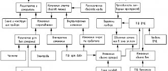
The essence of the duplex scanning method is to combine the capabilities of ultrasonic waves, which visualize the structure of the vascular walls, and the Doppler effect, which reflects the nature of blood movement using multi-color coding (for clarity).
The software combines the two images and displays the data in a clear form for medical analysis.
Thus, scanning is carried out in 2 modes:
- two-dimensional (B-mode) - vessels and adjacent tissues are viewed, complete data on blood supply is not obtained in this mode;
- duplex (Dopplerography) - a two-dimensional color drawing is constructed, from which an expanded description of the condition of the vessels can be made.
The Doppler effect reflects the change in wavelength for an observer when a signal is reflected from a moving object, which is different types of blood cells. Ultrasound is reflected from them, helping to assess the direction of blood flow and calculate its speed and dynamics. In this case, pathologies will be indicated by turbulence, retrograde movement, and slowdown.
There are 3 main areas of research:
- transcranial ultrasound Doppler ultrasound - examines the blood vessels of the brain;
- Doppler ultrasound of the brachiocephalic vessels - the area of study lies in the neck area;
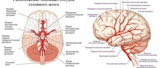
- Doppler ultrasound MAG is an examination of the main arteries of the head, which include two vertebral and two carotid, forming a circle at the base of the brain (their changes affect the entire body).
Characteristics of the procedure
MAG ultrasound is a combined procedure in which standard ultrasound is used, and the Doppler effect is also used. Thanks to these measures, you can assess the condition of the vascular system of the head, as well as observe the movement of blood in the skull. Ultrasound examination is carried out using sound waves that penetrate the human body . The patient will not feel anything, so there is no need to be afraid of discomfort.
A special sensor for MAG ultrasound will capture the waves, and all information will be displayed on the computer in a two-dimensional image. Doppler will begin to display red blood cells moving through the vessel. This research method allows you to diagnose congenital anomalies of the arteries that supply blood to the brain. An aneurysm can be detected, as well as disorders of the circle of Willis vessels can be studied.
When performing a duplex MAG scan, it will be possible to identify chronic diseases, as well as determine whether treatment affects blood circulation in the cranial cavity. The procedure is safe, so it is often prescribed to patients to identify head pathologies.
How the research is carried out
The patient is asked to lie on his back, turn and tilt his head back slightly. The direction of rotation does not matter. The course of the procedure depends on the chosen diagnostic method:
- two-dimensional mode;
- duplex color scanning.
In the first case, the condition of the carotid arteries (internal, common, external), jugular vein, as well as small branches of these vessels is analyzed. Extracranial scanning makes it possible to examine the vessels within the skull and neck.
For children and adults
Due to its safety, MAG ultrasound is prescribed regardless of age. The subject should not be stationary for a long time, which simplifies the diagnosis of children. With young patients, psychological preparation plays an important role: the child must be explained the principle of future manipulations, their painlessness and the specifics of their implementation. Video research materials posted on the Internet can be useful in this regard.
Duplex and triplex modes
Modern ultrasound diagnostic equipment allows examination in duplex and triplex modes. Two-dimensional (or B-mode) makes it possible to find a vessel, and through color duplex scanning, the state of blood flow can be assessed. To obtain the flow spectrum, it is necessary to set the scanning depth in the artery lumen. The combination of any two modes is called duplex, and research in three modes at once (spectrum, color, B) is called triplex.
Preparation for the procedure
In order to get the most accurate result, it is recommended to properly prepare for the procedure. There is no need to take any special actions, but some important points need to be taken into account so as not to distort the clinical picture. You will need to give up foods and substances that can affect vascular tone . It is better not to use them on the day of MAG ultrasound.
First of all, you should not smoke shortly before the examination, and you should also not drink tea or coffee. Energy drinks are contraindicated, especially those containing guarana. You should not eat chocolate, because it can also affect the results of the study.
It is recommended to consult with a neurologist about whether you should stop taking the medications. This point is relevant for those people who regularly use medications. Ultrasound of the great vessels of the head will be useful only when it is possible to obtain an accurate picture of the condition of the vessels without the influence of side factors.
What can be detected with Doppler ultrasound examination of blood vessels?
This diagnostic procedure makes it possible to detect the formation of blood clots not only in the vascular system of the cervical spine and head, but also in the upper and lower extremities. Using ultrasound, you can determine:
- causes of headaches;
- narrowing of the arteries;
- stage of diseases, the development of which was provoked by atherosclerosis or thrombosis;
- the presence of vascular aneurysms;
- the speed of blood flow in the main arteries and its disturbances;
- condition of the spinal vessels.
Changes identified during a diagnostic examination may indicate the development of:
- vasculitis – the echogenicity of the lumen of the vessel, the thickness of its walls and differentiation into layers changes;
- atherosclerosis – the thickness of the diameter of the vascular walls increases, an uneven type of change in echogenicity appears;
- cholesterol plaques in the arteries - hypo-echoic formations with a thin rim are detected.
Conditions in which there is a narrowing of the lumen of the arteries of more than 50% require immediate treatment.
MAG ultrasound can detect deformation of the vascular walls of the neck - a harbinger of coronary heart disease