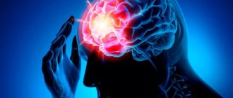
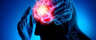
All infections are dangerous to varying degrees, and even more so are neuroinfections. The most dangerous neuroinfections are those that affect the brain. There can be no “frivolous” diseases here. Every pathogen that can cross the blood-brain barrier poses a huge danger to human health and life.
Types of Brain Infections
The variety of neuroinfections affecting the brain can be divided into five groups:
- bacterial;
- parasitic;
- viral;
- prion;
- fungal.
Bacterial infections
A huge number of pathogens related to bacterial infections can affect the brain. Diseases such as meningitis, encephalitis or brain abscess may well be caused by such “ordinary” pathogens as pneumococcus, staphylococcus, and enterobacteria. But this can only happen if:
- damage to the bones of the skull, with disruption of the integrity of the membranes of the brain;
- introduction of pathogens during neurosurgical operations;
- the presence of a purulent focus in the body and weakened immunity.
However, with other pathogens the situation is different.
Meningococcal infection is a traditional neuroinfection that affects the brain. The peak incidence is observed in the autumn-winter period, when the immune system is reduced due to frequent hypothermia and lack of vitamins. If the immune system is normal, then you will limit yourself to ordinary nasopharyngitis; otherwise, the likelihood of getting meningitis or meningoencephalitis increases.
Symptoms of meningococcal infection
- fever,
- increase in body temperature to 39-40° C.
- chills,
- headache
- weakness
- neck muscle tension
- nausea,
- vomit,
specific symptoms
- A red-violet rash protruding above the surface of the skin, the elements of which resemble a star in shape
- The disease begins very acutely (often you can specify a specific time (hour) when the person fell ill)
- Treatment must begin within 24 hours while the person is conscious, otherwise he may fall into a coma.
Mycobacterium tuberculosis, among other things, can also affect the brain. Children, elderly people and people suffering from immunodeficiency are more often affected.
The symptoms of the onset of the disease are not clearly expressed, most often it is general weakness, malaise, lack of appetite, headache and irritability, body temperature is subfebrile (the temperature rises over a long period of time within 37.1 - 38°C.). Subsequently, “usual” meningeal symptoms appear.
Afterwards, neurological disorders appear - paresis and paralysis of the facial nerve, oculomotor muscles, dizziness. Mental disorders occur against the background of neurological disorders.
Neurosyphilis, now almost never occurs, but before the discovery of penicillin it formed the basis of the work of neurologists. Neurosyphilis comes in several types:
- Asymptomatic, occurs without any special symptoms, the disease can only be detected by testing.
- Meningitis - often appears during the first year of the disease, manifested by disturbances in the functioning of cranial nerves and increased intracranial pressure (ICP).
- Cerebrovascular - occurs mainly in the 2-5th year of the disease and can lead to a stroke or transform into tabes dorsalis or progressive paralysis.
- Progressive paralysis is a disease that was also called “crazy paralysis.” Occurs 15-20 years after infection and first manifests itself with mental symptoms, then muscle paralysis occurs and progresses, which ultimately leads to death.
- Congenital, which, strictly speaking, affects the entire body and is characterized by multiple defects in the development of the child.
- Gumma of the brain - manifests itself as a space-occupying formation. Symptoms include increased ICP and focal symptoms, depending on the location of the gumma.
An unpleasant feature of the disease is its difficult diagnosis.
CHAPTER 2. MAIN NEUROPSYCHOLOGICAL SYNDROMES IN
Findings of recent years related to the study of the selective influence of various drugs on brain structures and, accordingly, on various components of mental processes (factors) are increasingly being implemented in the clinic of brain dysfunctions. Correct use of these drugs requires not only knowledge of which structural and functional areas of the brain they can have a therapeutic effect on. Targeted pharmacological action in this case is possible with an understanding of the specifics of the mental defect, neuropsychological syndrome and the disturbance of the factors underlying it. Only in this case can one predict not only the direct effect of the drug, but also the changes in the mental system that may occur.
Thus, the tasks of a clinical neuropsychological examination of a patient can be combined into two interconnected classes: 1) differential topical diagnosis and 2) description of the structure of mental function disorders based on the syndrome-forming component in the form of an indication of a violation of the factor(s) underlying their deficiency and functional restructuring. The possibility of solving the second class of problems provides the prospect of neuropsychological clinical diagnostics going beyond the limits of local brain pathology into a wide range of diseases, the consequences of which are disorders of mental activity that require therapeutic intervention, correctional and rehabilitation measures. Generalization and comprehension of the data obtained in this way creates new prerequisites for the further development of ideas about the connection of mental processes with the brain substrate, i.e. for the development of theoretical neuropsychology in its dialectical unity with practice.
The main neuropsychological syndromes, their structure, the release of a syndrome-forming radical associated with the elimination of certain structural and functional units of the brain from normal functioning and the loss of one (or several) factors from the functional system are quite well developed and described in relation to lesions of the left hemisphere of the brain (A.R. Luria, 1969; 1973). At the same time, syndromes of damage to the structures of the right hemisphere of the brain, subcortical nuclei, limbic-reticular complex and commissural formations that ensure joint integrative activity of the right and left hemispheres of the brain require further development. To date, clinically verified data have already been obtained on neuropsychological symptoms associated with damage to these particular brain areas, and allowing for neuropsychological diagnostics aimed at differentiating the side of the brain lesion, the lateral or medial location of the pathological focus, as well as the level of its localization in the systemic system. structural vertical structure of the central nervous system. The interpretation of these symptoms in theoretical terms and in the context of understanding brain mechanisms and factors has not yet been fully and systematically understood. It requires the development of ideas about the features of the structural and functional organization of the listed brain zones and a new approach to the analysis of those factors in the structure of mental reflection that they provide.
Viral and prion infections
There are a huge variety of viruses that cause acute encephalitis (mosquito, tick-borne, epidemic), in general they differ in vectors and geography of distribution.
Focal symptoms occur against the background of “general infectious manifestations”, these are:
- paresis
- respiratory muscle paralysis
- paralysis of limbs,
- paralysis of facial muscles, etc.
Rabies and slow infections can pose a huge danger, and therefore special attention is paid to them.
Rabies. Almost all mammals can suffer from rabies. The source of infection is usually dogs, wolves, foxes, and it is through the bite of infected animals that this dangerous infection is transmitted to humans. Symptoms:
- hydro and aerophobia
- convulsions
- attacks of aggressive behavior.
Emergency vaccination after a bite is the only way to recover, so waiting for the first symptoms of the disease to develop is prohibited, since this can only indicate that the person can no longer be saved.
Slow infections are viral neuroinfections that have the ability to remain asymptomatically in the human nervous tissue for a long time, with the subsequent development of the disease.
Scientists have identified four main characteristics that distinguish slow infections:
- unusually long (months or years) incubation period;
- slowly progressive nature of the course;
- unusualness of damage to organs and tissues;
- the inevitability of death.
The causative agents of the virus are rubella and measles. For reasons that are not entirely clear, these viruses can remain in brain cells after an illness and cause disease after 4 or more years. Both viruses cause panencephalitis with similar symptoms:
Personality changes with the development of dementia
Gradual paralysis of all striated muscles.
Unfortunately, even with treatment, the consequences of these neuroinfections are always the same - death.
Prions Prions are “proteinaceous infectious (particles).” Prions are defined as “a small proteinaceous infectious particle that is resistant to inactivating influences that modify nucleic acids.” In other words, prions are ordinary proteins of the body that for some reason (which is still unknown) they begin to behave “wrongly”.
There are four types of prion neuroinfections, and only one of them has a clear transmission mechanism. In some tribes of Papua New Guinea, cases of kuru-kuru were often reported due to the previously widespread ritual cannibalism of eating the brains of relatives. Prions cause spongiform encephalopathy, which means the brain turns into a sponge.
Local organic brain lesions represent an independent and rather extensive area of application of the ideas and methods of medical psychology, called neuropsychology. The clinic of vascular, tumor, inflammatory diseases of the brain, its traumatic lesions and atrophic processes in late age - this is a far from exhaustive list of pathological conditions where neuropsychology solves the problem of clarifying the topic of brain damage, correlating ideas about the structure of psychological processes with data on the structure of the higher parts of the brain brain The use of psychological methods to study the clinical picture of local brain lesions and the mechanisms of complex mental activity provides earlier and more accurate differentiation of local and general cerebral symptoms (syndromes) in their dynamics. The successful development in our country of new clinical areas of application of neuropsychology, such as neurology and neurosurgery (including stereotactic methods) of deep brain structures [Smirnov V. M., 1976; Kuzmina T.V. et al., 1981], neurology and neurosurgery of childhood [Simernitskaya E.G., 1981], defectology [Markovskaya I.F., Lebedinsky V.V., Lebedinskaya K-S, 1981, etc. .], psychiatry [Balonov L. Ya. and Deglin V. L., 1976, etc.], focal epilepsy [Wasserman L. I., 1974; Wasserman L.I. and Tets I.S., 1981], endovasal neurosurgery [Moskovichiute L.I., 1978, etc.], requires improvement of neuropsychological research methods not only in the topical diagnosis of local brain lesions, but also for the development psychologically substantiated, rational techniques for restoring higher mental functions impaired as a result of focal brain lesions [Luria L. R., 1948, 1969; Bain E. S., 1964; Tsvetkova L. S., 1972; Tsvetkova L. S. et al., 1981; Dorofeeva S. A., 1973; Shklovsky V.M., 1975, etc.]. Neuropsychological methods are also increasingly used in theoretical and experimental research, making a significant contribution to the understanding of the brain mechanisms of complex mental functions [Khomskaya E. D., 1972; Bekhtereva N.P., 1980; Traugott N.N., 1981]. Thus, neuropsychological methods form part of the general complex of clinical research of patients in neurological, neurosurgical or psychiatric clinics. However, their development, adaptation and use in clinical diagnostic practice are possible only on the basis of modern ideas about higher mental functions and their systemic organization.
Modern ideas about the brain organization of higher mental functions and the basic principles of neuropsychological diagnostics. A fundamental contribution to the neurophysiological direction that studies the problem of localization of brain functions was made by the teaching of I. P. Pavlov on the complex dynamic organization of brain structures that underlie mental activity. This teaching, which overcomes both narrow localizationism and psychomorphologism, and antilocalizationism, which proclaims the equipotentiality of all parts and structures of the brain, was subsequently developed in the works of GG physiologists. K. Anokhin (1940, 1971) and I.A. Bernstein (1947, 1966), psychologists L. S. Vygotsky (1960), A. R. Luria (1969, 1973) and A. N. Leontiev (1972). When we talk about the localization of functions, we mean, first of all, the systemic activity of the brain, which determines the paths of movement and the places of interaction of the nervous processes that underlie a particular function. In this sense, mental functions are confined to brain structures, but at the same time the same brain centers can be included in different “working” constellations, and the same function under different conditions is implemented differently and relies on brain centers of different localizations. mechanisms. This is how the concept of a functional system was formulated, where “function” is understood, as A. R. Luria points out, “complex adaptive activity of the body, aimed at achieving some physiological or psychological task.” The works of P.K. Anokhin on the theory of functional systems, the role of “reverse afferentations”, “afferent syntheses” in the multi-level organization of mental functions, begun back in the 30s, have now become widespread in neurophysiology, neuropsychology and neurocybernetics. N.A. Bernstein (1947), using the example of an analysis of a voluntary motor act, convincingly proved that movement, contrary to existing views, is not a function of a special group of cells of the motor cortex, but occurs as a result of a complex combination of various anatomical formations at the subcortical and cortical levels and not only efferent, but also afferent systems. Based on this, any voluntary movement should be considered as the result of a multi-level, coordinated sensorimotor interaction. It is indisputable that the highest forms of mental activity (speech, recognition, purposeful actions, etc.) have an even more complex organization and are based on a system of jointly working areas of the brain and, first of all, the cerebral cortex. It is also obvious that each zone makes its own specific contribution to the implementation of mental function. The principles of the theory of functional systems of P. K-Anokhin were widely used by A. R. Luria (1969) in the study of the pathophysiological mechanisms of aphasia and apraxia.
methods of heterogeneous statistical analysis – previous | next – neuropsychology
Methods of psychological diagnosis and correction in the clinic. Content
Development of the disease
At an early stage ( mild dementia ), brain dysfunction is already noticeable. The person becomes irritable and forgetful, and experiences frequent headaches. At this stage, the elderly person is still able to think critically, take care of himself and live independently.
The second, moderate degree is characterized by significant disorders of mental activity and memory. The person loses basic everyday skills and requires constant supervision from relatives, friends or a caregiver.
When moving to a severe degree of disorder, a person ceases to recognize and understand those around him; there is no longer any talk about independent self-care, even in small things. In this condition, the patient needs constant care of qualified personnel.
Often, lack of attention from loved ones does not allow us to detect signs of dementia at an early stage and take timely measures to slow down the progression of senile dementia.
Disorders of brain development at the prenatal stage of ontogenesis
Modern neurobiological models of schizophrenia suggest damage to the brain structures of the fetus during the prenatal period.
The development of the nervous system in the embryo begins in the 3rd week of pregnancy under the influence of factors secreted by the notochord, along with the formation of the neural plate in the dorsal part of the embryo. This plate expands and folds, gradually forming the neural groove. Then it merges into the neural tube, which gives rise to the development of the brain in its rostral part and the spinal cord in the caudal part. By the 4th week of pregnancy, notochords are formed, which close behind (caudally) and in front (rostrally). Violation of closure in the rostral part leads to anencephaly, violation of closure in the caudal part leads to spina bifida. The rostral part of the neural tube expands before fusion and forms three primary brain vesicles: the prosencephalon, mesencephalon and rhombencephalon. In addition, during this period, the cervical and cerebral grooves are formed. The primary medullary vesicles develop into the cerebral hemispheres, brain stem and cerebellum, while the ventricular system of the brain is formed from the nerve canal. The prosencephalon consists of the telencephalon, which gives rise to the cerebral hemispheres and partly the basal ganglia; another part of it, the diencephalon, is considered the precursor of the thalamus, hypothalamus, posterior pituitary gland, optic nerve and retina. Neuroblasts are formed near the nerve canal in the region of the ventricular zone and subsequently migrate not only to neighboring areas, forming the deep subcortical nuclei of the brain, but also along the radial glial fibers, forming the cerebral cortex. From this region of the immature brain, rich in glial fibers, the white matter of the cerebral hemispheres develops, while some of the radial glia give rise to the neuronal precursor cells of the mature brain.
| | For what other reasons can schizophrenia develop? |
Cortical dysplasia has become more frequently diagnosed with the introduction of neuroimaging methods into practice.
The signals that determine the order of neuron migration are currently being actively studied, since disruption of migration processes leads to the development of cortical dysplasia and, according to one hypothesis, it is precisely because of this disruption that a predisposition to the onset of schizophrenia is formed.
Neurodegenerative processes of the early stage of ontogenesis, predisposing to schizophrenia
- Distortion of the process of neuron migration to the cerebral cortex (impaired migration of “pre-alpha cells” from the lower to the upper cortical layers)
- Weakening of synapses (weakness of synaptic transmission, limited synaptic connections)
- “Frontotemporal-limbic network disorder” (local breaks in the network architecture of neurons in the heteromodal associative cortex)
- Changes in the processes of proliferation of neurons and glia (structural disturbances in axons and receptors)
Many researchers write about the insufficiency of synaptic transmission in schizophrenia (Feinberg I., 1982; Cannon T. et al., 1999; McGlashan T., Hoffman R., 2000).
According to some researchers, with normal brain development, the synaptic density of neurons increases until almost two years of age. Then this process gradually slows down in early childhood and almost disappears in adolescence.
It is emphasized that in schizophrenia one can assume a rapidly occurring pathological process of damage to synapses. Therefore, brain damage at an early stage of ontogenesis especially increases the risk of developing schizophrenia throughout a person’s subsequent life. As is known, in schizophrenia, it is in adolescence that insufficiency of synaptic connections is detected, which contributes to the manifestation of the symptoms of this mental disorder.
According to the “neurodevelopment” hypothesis, which was quite popular in the 20th century, the pathological process in schizophrenia should be considered a consequence of abnormal brain development in the initial period of ontogenesis.
According to McGrath J. et al., (1995), abnormalities in the brain of a patient with schizophrenia in the early stages of ontogenesis resemble teratogenic changes in the brain in fetal alcohol syndrome and are a consequence of impaired development of ectoderm tissue.
As a result of this deviation, neurons fail to migrate to the “prescribed” area of the brain and form the necessary connections among themselves. In particular, some researchers suggest a disruption in the migration of so-called pre-alpha cells from the lower to the upper cortical layers. This process can lead to underdevelopment of certain areas of the cerebral cortex. However, it is difficult to say today exactly which part of the brain is developing incorrectly; presumably it could be the amygdala, frontal and temporal lobes of the brain.
One of the popular hypotheses for neurodegenerative disorders that arise during brain development in schizophrenia is the “frontotemporal-limbic network disorder” hypothesis. This hypothesis assumes local breaks in the network architecture of neurons in the heteromodal associative cortex (Falkai P. et al., 2001).
Morphological changes in the temporo-limbic structures of the brain in schizophrenia are weakly expressed and, as a rule, do not tend to increase, which is partly confirmed by the absence of astrogliosis in the nervous tissue of patients with schizophrenia according to postmortem studies, and as is known, it is gliosis that is considered a sign of neuronal destruction, characteristic for neurodegenerative diseases.
Pathology of brain development at an early stage of ontogenesis can manifest itself as a distortion of such neurobiological processes of neuronal maturation as neuronal and glial proliferation, “programmed cell death” (apoptosis), insufficient development of receptors and synapses, and impaired axon formation.
Comparison of structural and functional changes in the brain in premorbid patients with schizophrenia with changes in nervous tissue after the first episode of schizophrenia partly confirms the degeneration of nervous tissue even before the appearance of clear symptoms of the disease.
In the mid-twentieth century, there was a popular hypothesis according to which the vulnerability of the brain may be due to the late age of the parents at the time of conception of the fetus, a future patient with schizophrenia. Thus, in 1959, Canadian researchers showed that the age of parents with schizophrenia at the time of conception of a child is 2-3 years older than the age of those parents whose children were subsequently healthy (Gregory I., 1952). However, the results of the work of other authors refuted these views (Hare E., Moran P., 1979). Recent studies by researchers from Israel have shown that the risk of schizophrenia in an individual, although slightly, increases as the age of his parents increases, reaching its maximum at the age of 55 years and older. Compared to ages 20-24, the risk of developing schizophrenia almost doubles when parents are 55 years or older. It can be assumed that the germ cells of women can divide only up to a certain period, while male cells retain the ability to divide throughout the entire reproductive period, as if accumulating with age the risk of changes in sperm and, consequently, gene mutations, reducing the correctness of DNA replication.
At one time, facts were noted regarding the birth season of patients with schizophrenia. People who developed schizophrenia, especially men, were more likely to be born in late winter or from February to May (Tramer R., 1929). In 1977, Torrey et al. published a review of studies on the seasonality of birth in patients with schizophrenia. The results of the review showed that patients with schizophrenia were 5-8% more likely to have births recorded between December and May, with a peak occurring in January-February. Australian psychiatrists also confirmed the dependence of the month of birth on the potential risk of schizophrenia.
Some researchers explained these facts by the fact that women in the second trimester of pregnancy, if it occurs in the winter months, are more susceptible to infectious diseases, in particular, intrauterine infection of viral influenza, which negatively affects the development of the fetal brain during this period of pregnancy.
The literature has repeatedly provided data confirming this theory. For example, an increased incidence of schizophrenia was noted in Holland during World War II in the children of women who fasted during the first three months of pregnancy. According to K. Freet and E. Jonston (2005), all of the factors listed above influenced the brain when it was at a critical stage of its development.
Maternal malnutrition during pregnancy, according to E. Susser, S. Lin (1992), almost doubles the risk of schizophrenia in their children. At the same time, it should be noted that malnutrition of the expectant mother also affects the tendency of their children to develop affective disorders and predisposes them to the formation of a personality disorder of the schizoid type (Hoek H. et al., 1996). In a study by H. Hulshoff Pol et al. (2000) showed that malnutrition during pregnancy affects the volume of the ventricles of the fetal brain, as well as the structure of the white matter of the brain.
Risk factors for brain damage during ontogenesis, predisposing to the development of schizophrenia
- Infectious diseases that occur in 5-7 months of pregnancy
- Malnutrition at 5-7 months of pregnancy
- Stress during pregnancy
- Beginning of pregnancy in late spring (increases the risk of schizophrenia by 10%)
Most schizophrenia researchers argued that the characteristics of pregnancy at 5-7 months, infectious diseases that arise during this period (Brown A., Susser E., 2002), extreme temperatures, malnutrition were the factors that increased the vulnerability of the maturing brain of potential patients with schizophrenia.
It has been noted that if a mother had influenza in the middle trimester of pregnancy, her child's risk of developing schizophrenia increases.
| | For a timely, high-quality diagnosis of schizophrenia, we recommend that you contact a psychiatric clinic |
Those whose mothers suffered infections such as toxoplasmosis gondii (Torrey E., Yolken R., 2003), viral rubella (Brown A. et al., 2000), and herpes simplex viral infection in the second trimester of pregnancy have a fairly high risk of developing schizophrenia. (Buka S. et al., 2001).
Viral diseases of the mother predisposing to the subsequent development of schizophrenia in children
- Toxoplasmosis
- Flu
- Rubella
- Herpes simplex
The unfavorable role of stress experienced by the mother during pregnancy (and in the first five years of the child’s life) in relation to the future development of schizophrenia in her children was pointed out by M. Huttunen and R. Niskanen (1978).
Many researchers believe that damage to the fetal brain during 5-7 months of pregnancy is most significant for the subsequent development of schizophrenia . Pronounced structural changes in the brain are usually characteristic of earlier periods of fetal damage. However, the significance of brain damage for the risk of developing schizophrenia after 7 months of pregnancy is refuted by the fact that gliosis of nervous tissue, as noted above, in this mental disorder, as a rule, cannot be detected.
Because reactive gliosis does not appear until after the second trimester of pregnancy, brain abnormalities in the absence of gliosis likely indicate abnormal brain development as opposed to degeneration.
In a study by B. Bogerts et al. (1993) signs of gliosis were absent in the medial temporal lobe, cingulate gyrus and thalamus. At the same time, phenomena such as a decrease in the number of neurons, changes in their size and location were discovered in the temporal lobe.
There is some histological evidence that the lag in speech functions occurs mainly when the normal development of the cerebral cortex in the temporal regions or around the Sylvian fissure changes.
The heterotopia of neurons in the hippocampus (the lower layer of the hippocampus), which develops 5-7 months after the birth of the fetus, is often recorded in schizophrenia, indicating a dangerous pregnancy limit for the occurrence of this mental disorder.
Experimental studies on rats have shown that if the anterior sections of the hippocampus or most of it are removed immediately after birth, then over a fairly long period of “childhood” they fail to develop significant deviations from the developmental characteristics of unoperated animals. However, during the transition to adulthood, the operated rats became restless, showed noticeable memory impairment and low adaptation to presented shock stimuli.
There are different types of dementia
Dementia, depending on the causes and location of the affected areas of the brain, is divided into several types:
- Damage to the cerebral cortex - cortical dementia. A type of such dementia is Alzheimer's disease;
- Damage to white matter and subcortical structures of the brain - subcortical dementia. This type of dementia includes progressive supranuclear palsy and Parkinson's disease;
- Degenerative changes in the cortex and subcortical structures of the brain - cortical-subcortical dementia is often a consequence of diseases of the vascular system: atherosclerosis or hypertension;
- Multiple small lesions in different areas of the brain - Multifocal dementia This type of disorder is represented by Creutzfeldt-Jakob disease.


