General information about pathology
Disease:
- occurs infrequently;
- proceeds slowly;
- rarely becomes malignant;
- occurs in people who are at least 35-40 years old;
- more often affects females.
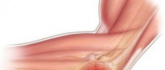
The neoplasm externally resembles a rounded dense nodule, which is located in a capsule formed from connective tissue. Thanks to this, it is relatively easy to get rid of the tumor using surgery.
Neuroma - causes of occurrence
Short description
Neuroma (schwannoma) is usually an encapsulated benign (WHO-1) tumor consisting of highly differentiated tumor Schwann cells.
Code according to the international classification of diseases ICD-10:
- D36.1 Peripheral nerves and autonomic nervous system
Epidemiology . Common tumor of peripheral nerves. Neuromas account for 8% of primary intracranial tumors and 20% of primary spinal tumors. Neuromas often develop in neurofibromatosis type 2 (see Neurofibromatosis). There is also a rare hereditary disease, schwannomatosis, which manifests itself as multiple subcutaneous schwannomas.
Anatomical localization . Most often, schwannomas develop from the sheaths of peripheral nerves in the head and neck, as well as the extensor surface of the extremities (sensory nerves are most often affected). The vestibular portion of the cochleo-vestibular nerve is the most common site of growth of intracranial schwannomas. Spinal neuromas are often localized intra-paraspinal in an “hourglass” pattern. Rare cases of schwannomas of intramedullary and intraventricular localization have been described.
Histopathology. The tumor is represented by spindle-shaped neoplastic Schwann cells, arranged alternately in the form of compact groups (“palisades”, Antoni A fibers) and areas of more loosely located cells (Antoni B). Clinical picture. Peripheral neuromas are characterized by a long asymptomatic course; usually the tumor is detected as a painless subcutaneous mass formation. Spinal schwannomas manifest themselves in the early stages of growth with symptoms of radiculopathy with pain, and later with myelopathy due to the mass effect. Neuromas of the VIII pair of cranial nerves are manifested by symptoms of damage to the structures of the cerebellopontine angle (ringing in the ear, hearing loss, paresthesia in the face).
Neuroma is caused by:
- genetic factors.
The likelihood of a tumor increases sharply if relatives suffered from this problem;
- difficult environmental situation
– exposure to harmful chemicals, radiation;
- cigarette addiction
.
Also, a neoplasm appears in people already suffering from another neoplasm.
Types of pathology
There are the following main types of disease:
Acoustic neuroma (aka vestibular schwannoma):
- occurs most often;
- the tumor affects the auditory nerve (usually the vestibular part, rarely the cochlear part). However, the neoplasm gradually grows and begins to penetrate the cerebelloptine angle;
- vestibular schwannoma can harm the facial nerve and even “reach” the trigeminal nerve. The optic and jaw nerves may also be affected;
- The tumor progresses very slowly (by a few millimeters per year). In rare cases, rapid, dangerous growth is observed (up to 30 mm in 12 months), due to which important parts of the brain (primarily the cerebellum) begin to be compressed;
- much more often it develops in only one ear (in two 5% of cases). Bilateral acoustic neuromas are usually caused by neurofibromatosis.
- appears at 40-50 years of age.
Vertebral neuroma:
- usually develops in the thoracic and cervical areas of the back;
- sometimes appears in the lumbar region;
- It is less common than acoustic, but also quite common.
Morton's schwannoma:
- this type of pathology appears on the foot (more precisely, its sole) of one leg, in the area of the 3rd and 4th toes;
- in rare cases, pathology develops on both feet;
- Morton's schwannoma is rare.
Types of neuromas by location and their corresponding symptoms
Let's consider the main types of pathology and what signs they are accompanied by.
Neuroma of the spine (cervical, thoracic)
Most often it affects the thoracic and cervical spine. In accordance with the generally accepted classification, it belongs to the extracerebral type. It develops on the spinal roots and compresses the spinal cord from the outside.
Symptoms that arise:
- Radicular syndrome. It is characterized by pain along the spinal cord, flaccid paralysis and impaired sensitivity in the affected area may be observed.
- Autonomic disorders. Symptoms depend on which nerve of the autonomic system was affected by the tumor. The functions of the pelvic organs (retention or incontinence of urine and stool), the gastrointestinal tract (abdominal pain, difficulty swallowing), and the heart (rhythm changes, bradycardia, angina) may be disrupted.
- Brown Sequard syndrome, when the spinal cord is compressed. Below the level of the tumor, spastic paresis occurs, flaccid paralysis at the level of the schwannoma, loss of sensitivity on the affected side, loss of sensation of temperature and pain on the opposite side.
- Discomfort between the shoulder blades, pain, loss of sensitivity.
Signs of pathology may appear and then go away. As the tumor grows, the symptoms become strong and constant. The pain is usually worse when lying down.
Brain neuroma
A cranial nerve schwannoma is a tumor that grows within the skull. Most often it develops on the trigeminal and auditory nerves. In 90% of cases it is unilateral. Symptoms usually include:
- intracranial hypertension;
- manifestations of compression of the surrounding brain matter;
- signs of nerve fiber damage;
- mental disorders;
- convulsions;
- ataxia;
- intellectual disabilities;
- hypotonia of the muscles of the legs and arms;
- dysfunction of the heart and breathing;
- change in visual fields.
Trigeminal neuroma
This is about 35% of all intracranial schwannomas, the second most frequently diagnosed schwannoma of the brain. Symptoms depend on the size of the tumor:
- first, sensitivity is impaired on the half of the head where there is a tumor;
- later, the masticatory muscles are affected and become weak;
- as the formation grows, nausea, vomiting, a bursting headache appear, and the cortex of the temporal lobe is compressed;
- at advanced stages, olfactory and gustatory hallucinations are added.
Acoustic neuroma (acoustic schwannoma)
Due to its location in the cerebelloptine angle, the tumor quickly compresses surrounding areas of the brain and nerves. Symptoms are divided into three types:
- damage to the cochlear nerve - ringing and noise in the ear on the affected side;
- Hearing deterioration - gradually, from high tones;
- when the tumor size is 2-3 cm, a toothache-like pain appears, the masticatory muscles atrophy;
- when the tumor reaches 4-5 cm, the abducens and facial nerves are affected, as a result of which taste in the tongue is lost, the salivary glands do not work properly, the sensitivity of the face on the affected side is impaired, strabismus with double vision occurs;
- vestibular disorders (size of formation 5-6 cm) – dizziness, fainting.
Schwannoma of the facial nerve
The consequences of such a neuroma are impaired taste in the first half of the tongue, numbness and asymmetry of the face, hypotonia of the facial muscles, and impaired salivation. Possible destruction of bone structures.
Morton's neuroma (foot)
It is a benign growth of fibrous tissue in the area of the plantar nerve. Mainly develops between the 3rd and 4th fingers. Symptoms include pain in the foot, sensation of a foreign body, increased discomfort when wearing tight shoes and exertion.
Cauda equina neuroma
Localized in the lowest part of the spinal canal. First it manifests itself as a unilateral radicular syndrome, then as a bilateral one. There is flaccid paresis of the legs, mosaic-type sensitivity disorders, difficulty with defecation and urination.
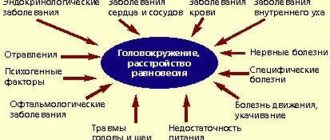
How does pathology manifest itself?
Often the disease is completely asymptomatic and is discovered by chance. This makes it difficult to treat neuroma. Signs of pathology depend on the location of the tumor:
For acoustic neuroma:
- tinnitus (tinnitus) develops;
- hearing deteriorates;
- coordination is impaired;
- severe dizziness appears.
If the facial nerve is damaged:
- tears flow;
- dry mouth;
- There is partial paralysis of the eyelids.
With neuroma of the dorsal vertebrae the following are observed:
- excruciating pain in the back;
- deterioration of sensitivity at the site of the lesion.
4. Morton's schwannoma causes various sensory problems:
- numbness is felt in the area of the feet;
- paresis occurs.
Etiology of neuroma
Scientific research has revealed a connection between the development of neuroma and excessive proliferation of lemmocytes. The reason for this phenomenon has not yet been established. The tumor itself is characterized by slow development and looks like a round or oval node, and sometimes even irregular in shape. Its surface is quite uneven, with bumps, covered with a capsule. The color of the tissue may vary, as it depends on the level of blood supply to the neuroma: it varies from pale gray to bluish. Inside, cysts of various sizes with fluid are detected; partial or complete degeneration into a cyst is possible.
Experts identify a number of factors that increase the risk of developing neuroma. These include the following:
- Exposure to chemical agents and poisons;
- Poor ecology, air pollution;
- Exposure to ionizing radiation;
- Frequent inflammatory processes of nerve tissue;
- Regular intake of carcinogens into the body through consumed food.
How is pathology diagnosed?
Treatment of neuroma is preceded by diagnosis. It is produced using:
- computer or magnetic resonance imaging examination. Magnetic resonance imaging is more effective. It is able to detect microscopic tumors, while computed tomography diagnostics detects only tumors whose diameter is at least 1 cm;
- electroneurography;
- audiograms (hearing test).
Diagnosis of acoustic neuroma
To establish a diagnosis in modern conditions, the following studies are used:
- pure tone and speech audiometry;
- assessment of auditory evoked potentials;
- vestibulometry;
- computer (CT) and magnetic resonance imaging (MRI), including with intravenous contrast.
Using tools in combination allows you to most accurately determine the location and size of the tumor, while each stage has its own combination of methods that is effective.
Treatment of the disease – surgical, radiation, conservative
Usually the neuroma must be removed; conservative treatment is not always effective. True, in the early stages, the tumor can actually be removed using radiation therapy (cyberknife).
Operation
Surgical treatment of neuroma involves a local opening of the skull (trepanation) so that the tumor can be reached.
It will not be possible to do without surgical intervention if:
- the tumor quickly increases in size (for example, after the patient has undergone radiotherapy);
- the disease began to lead to serious complications.
Surgical tumor removal is contraindicated if:
- the patient has problems with the heart or blood vessels;
- The patient is already elderly and has heart problems.
The prognosis is 99% favorable. Relapse of the pathology is a rare phenomenon. After the operation, the patient recovers within 6-10 months.
Possible complications are as follows:
- bleeding may begin;
- there is a small risk of infection;
- the patient's facial nerve may be affected by paresis (especially if the tumor exceeds several centimeters);
- hearing will worsen. If the tumor is larger than 2 cm, there is a high risk that its surgical removal will lead to partial hearing loss.
Radiation (non-operative) therapy
In addition to traditional surgery, there is a safer way to get rid of neuroma. We are talking about stereotactic radiosurgery (cyberknife) - irradiation of the tumor:
- The advantage of this operation is that the tumor is destroyed very precisely. The nerve tissue that surrounds it remains completely unharmed;
- disadvantage - this therapy is effective only if the neuroma is detected in the early stages and does not exceed 30 mm.
Radiation therapy is mandatory if:
- the tumor is located in an “inoperable place”;
- the patient is categorically against surgical intervention;
- the person has already reached old age and has heart problems.
Radiation therapy has side effects. After the procedure, the patient can:
· to feel sick;
· pain appears in the neck (where the stereotaxic frame was located).
Treatment of neuroma
Depending on the location, size and type of neuroma, the doctor prescribes the optimal type of treatment. Most often, the patient is referred for surgery. However, stopping tumor growth using non-invasive methods, especially when diagnosed in the early stages, has become possible with the development of new radiation treatment technologies, such as CyberKnife. At the same time, the functions of nearby nerves are preserved, which is extremely important for maintaining quality of life after treatment.
Radiation therapy is recommended if surgery cannot be performed or if the patient refuses it. With the introduction of the innovative CyberKnife system into practice, the possibility of non-invasive, fast, ultra-precise and comfortable treatment of neuromas of any location has become possible.
how to sign up for cyberknife treatment: - by calling the hotline - using the feedback form on the website.
call now!
Our consultants will answer all your questions, and experienced oncologists will determine the need for CyberKnife treatment.
See also: Treatment with CyberKnife
Diagnostics
Diagnostics. The method of choice is MRI. Treatment: surgical removal of the tumor. For certain indications (tumor size in maximum diameter less than 2.5 cm, age over 65 years, etc.), stereotactic radiosurgery (gamma knife, LINAC [linear accelerator]) is used as an alternative method for neuromas of the VIII pair of cranial nerves.
Forecast. Schwannoma is a benign encapsulated tumor that extremely rarely undergoes malignant transformation, so total removal of the tumor leads to a cure. The results of surgical treatment depend on the location of the tumor.
Synonyms • Lemmoblastoma • Lemmoma • Neurilemmoma • Perineural fibroblastoma • Schwannoglioma
ICD-10. D36.1 Benign neoplasm of other and unspecified locations - peripheral nerves and autonomic nervous system
Neuromas of other cranial nerves
Schwannomas (neurinomas) of the trigeminal (V), facial (VII), abducens (VI) nerves, as well as nerves of the caudal group - glossopharyngeal (IX), vagus (X), accessory (XI) or hypoglossal (XII) , occur much less frequently, than vestibular neuromas. And together they make up only a few percent of the total number of intracranial tumors. Also, only a few cases of the development of schwannomas from the roots of the oculomotor (III) and trochlear (IV) nerves . Given their rare occurrence and common approaches to diagnosis and treatment, these tumors are also combined under the general term non-vestibular schwannomas.
How do neuromas of other cranial nerves differ from vestibular schwannomas?
The clinical manifestations of any non-vestibular neuromas are first caused by dysfunction of the affected nerve (for example, in the form of numbness or impaired facial expression in the corresponding half of the face, with trigeminal or facial nerve neuromas, respectively). Subsequently, as they grow, they begin to put pressure on adjacent brain structures, also causing their dysfunction. Many neuromas (for example, trigeminal or caudal) can grow along the nerve root through the anatomical openings to the outer base of the skull, spreading into the soft tissues of the neck, pterygopalatine and infratemporal fossae. As the tumor develops to a large size, symptoms of increased intracranial pressure come to the fore, which are practically independent of the starting point of tumor growth.
How to diagnose non-vestibular schwannomas?
In addition to assessing neurological symptoms, magnetic resonance imaging (MRI) without contrast enhancement and with contrast . If the tumor is located in the projection of the anatomical openings of the skull base, MRI is necessarily performed in T1 mode with contrast and suppression of the signal from adipose tissue , because in this mode, the nature of the extracranial spread of the tumor can be most reliably assessed. Computed tomography is less informative in diagnosing tumors of the skull base when scanning in tissue mode. However, in the bone mode, one can see a pathological expansion of the anatomical openings of the skull base, which is an indirect sign of the presence of a pathological volume there. Spiral computed tomography in perfusion mode allows to differentiate schwannomas and other types of tumors of the skull base
What types of treatment are there for intracranial non-vestibular schwannomas?
Conservative “treatment” (wait & scan - wait and watch) . Considering the surgical inaccessibility of most non-vestibular schwannomas, as well as the potentially high risk of gross dysfunction of the affected nerves after surgery, the vast majority of neurosurgeons prefer not to operate, but to observe incidentally diagnosed and asymptomatic tumors. For example, in case of schwannomas of the facial nerve, the generally accepted neurosurgical tactic is to monitor tumor growth either until pronounced facial asymmetry appears (due to paresis of facial muscles) or until symptoms of intracranial hypertension appear against the background of a large tumor. This is due to the fact that surgical removal of such a schwannoma itself has an almost 100% risk of damage to the facial nerve.
Surgical removal of non-vestibular schwannomas traditionally remains one of the main methods of treating these tumors. However, it is associated with a very high risk of severe dysfunction (even loss) of the affected nerve, as a result of even partial removal of the tumor. Therefore, neurosurgeons prefer to operate on such tumors only in cases where dysfunction has developed naturally against the background of tumor growth, or when the size of the tumor and the resulting symptoms not only significantly disable the patient, but also threaten his life
Radiosurgery is currently a direct alternative to conservative (watchful waiting) tactics and surgical removal. According to the unanimous opinion of radiosurgeons, the successful use of Gamma Knife in the treatment of vestibular schwannomas, as well as the gradually accumulating experience in the treatment of non-vestibular tumors, allows us to clearly recommend radiosurgery in the early stages, without waiting for continued growth and the appearance (worsening) of neurological symptoms. Also, postoperative radiosurgery of tumor remnants is a good addition to surgical treatment, allowing traditional neurosurgeons to remove the tumor only partially, to eliminate brain compression and intracranial pressure, with good subsequent control of tumor growth. The effectiveness of Gamma Knife in the treatment of non-vestibular schwannomas is comparable to that in the practice of acoustic neuromas and is 97-99% with excellent functional results.
Clinical example 1 Patient, 43 years old. This example demonstrates not only a significant regression of a large (T4a according to Sami) acoustic neuroma (13 cc) - by 81% over 2 years, but also a safe and complete restoration of the function of the facial nerve, the impairment of which was noted at the onset of the disease. The positive effect of treatment lasts for 11 years
Clinical example 2 Patient, 52 years old. The onset of the disease is with the appearance of facial asymmetry on the left. The hearing on the left is not impaired. MRI shows a tumor of the left cerebellopontine angle
Rice. 1. Facial schwannoma (neurinoma of the facial nerve) of the left cerebellopontine angle at the time of stereotactic radiosurgery with Gamma Knife
Rice. 2. On the left - insufficiency of the function of the left facial nerve (asymmetry of the facial muscles) at the time of Gamma Knife radiosurgery. On the right - complete restoration of facial nerve function 3 months after radiosurgery