Science news. Myelination and its early diagnosis
What is myelin and why should it be measured?
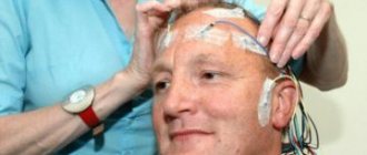
Myelination of nerve endings is the formation of a special layer of substance called “myelin” around the axial cylinder of the nerve fiber during its maturation. It's like insulating a wire. This amazing process begins at the 5th month of intrauterine development of the fetus and basically ends at the end of the second year of the child’s life. According to some data, the process of myelination continues until adolescence, and sometimes up to 40 years of age. Myelin, which covers nerve endings, increases the speed and accuracy of impulse transmission in the nervous system and protects nerve fibers from damage. Therefore, its deficiency causes various disorders in the functioning of the nervous system.
Russian neuroscientists are working on introducing a new approach to diagnosing the quantitative level of myelin. It consists of identifying biomarkers of maturation or damage to the nervous system of the body of children, including in the fetus from the 20th week of intrauterine development. These studies will provide invaluable assistance in the prevention of perinatal mortality and death of newborns. Detection of myelin levels will also be of great importance in preventing the development of severe diseases and defects of the central nervous system (CNS), cognitive disorders, including such a common disease today as early childhood autism.
From the history of the issue
It should be noted that the world's first non-invasive (bloodless, without penetration into the body and forced damage) method for assessing myelin was developed several years ago under the leadership of Tomsk State University and Washington University scientist Vasily Yarnykh. The new approach has already proven its effectiveness and is being implemented in several clinics in the Russian Federation. This method has become a great help for determining damage to nerve fiber sheaths in people who have suffered a stroke...
...And within the framework of the new project, a unique approach will be used to assess myelination in normal conditions and in various pathologies of the brain, both during the period of intrauterine development of a child and after his birth.
The essence of the new project
Myelination of nerve fibers is clearly visible in the picture obtained using magnetic resonance imaging (MRI). But MRI in the prenatal period (the period of intrauterine development) is an almost impossible task, since the fetus cannot be motionless for any period of time during the scan.
Therefore, to obtain an objective picture, special programs are used that allow combining MRI images (the so-called “Method of mapping the macromolecular proton fraction”) obtained against the background of fetal motor activity. The new method has already been tested in examining children in the first years of life and fetuses during intrauterine development. New data have been obtained on the features of the development of fetal brain structures at different stages of intrauterine development.
Thus, it has been established that the amount of myelin in the fetus in the second trimester (13-24 weeks) is still minimal. And first of all, it is formed in structures that are responsible for vital functions - breathing, blood circulation, cardiac activity.
But in the third trimester (25-40 weeks), the initial myelination of the structures responsible for movement and their coordination becomes noticeable.
When examining children 2-3 years old with pathology of the nervous system, a number of cases were identified where the new method helped to obtain additional diagnostic information that was very important for doctors.
Benefits of the new method
These studies are supported by the Russian Science Foundation and will be carried out for another three to four years. They will allow us to develop the technology for quantitative mapping of myelin in the prenatal period and the period after the birth of a child. Based on the results of the study, a normative quantitative digital atlas of fetal and child brain myelination, necessary for doctors and scientists, will be created.
The data obtained will provide doctors with additional information about the causes of developmental delays in children and will be useful for predicting their cognitive and intellectual abilities.
Prediction or diagnosis of incipient myelination disorders at an early age is also important from the point of view of identifying risk groups of patients with mental retardation, autism and other serious mental illnesses.
Such information can be used as a proactive preventive method: to prescribe early behavioral therapy and develop social adaptation skills.
What to do until this unique technique becomes widespread?
Only preventive measures!
Our center carries out a great deal of work on the prevention and early correction of developmental disorders in young children.
Expectant mothers receive information about the specifics of caring for a newborn at perinatal risk and its early development.
We work with young mothers to anticipate the pathological process in the baby from birth, when the changes are not yet visible to anyone. Parents sometimes do not believe in the possibility of developmental disorders in their baby, because “there are no manifestations yet.” And they miss precious time that you will never get back!
Our methodology of advanced and corrective development is aimed at stimulating the formation of age-related skills, the development of the motor , sensory , speech , communicative and emotional spheres of the child from birth. There's still time! After all, the myelin sheaths have not yet formed completely and have not blocked the enormous compensatory capabilities of the newborn’s body!
Therefore, thanks to our method, a healthy baby will receive training in all body functions, and a baby at risk of pathology will undergo stimulation of skills, which will restore damaged pathways and stimulate the appearance of new ones.
Magnetic resonance imaging (MRI) in St. Petersburg
In general, metabolic disorders constitute a group of numerous rare enzyme defects, often with an autosomal recessive pattern of inheritance. Some metabolic disorders lead to myelination disorders called dysmyelogenous diseases. Dysmyelogenous diseases are classified by enzyme defect as follows:
- Lysosomal storage disorders - metachromatic leukodystrophy, Krabbe disease (globoid cell leukodystrophy), Sal's disease, mucopolysaccharidosis
- Peroxisomal disorders - X-linked adrenoleukodystrophy; adrenomyeloneuropathy, Zellweger syndrome
- Metabolism disorders of amino acids and organic acids – Canavan disease
- Demyelination with an unclear metabolic defect - Pelizaeus-Merzbacher disease, Alexander disease, Sjögren-Larsson syndrome, leukoencephalopathy (mitochondrial, megalocephalic, etc.)
Impaired myelination is one of the main manifestations of these diseases. In some of them, for example Pelizaeus-Mirzbacher disease, the gray matter of the subcortical nuclei is also involved in the process. Clinically, dysmyelogenous diseases manifest themselves in childhood and begin with diffuse symptoms. Then they quickly progress from minor paresis and mild intellectual impairment to spasticity and dementia. Before the advent of MRI, diagnosis was very difficult in many cases, since brain biopsy was performed in exceptional cases. With MRI in St. Petersburg, metabolic disorders are found mainly in pediatric practice. In open MRI, dysmyelogenic processes may also be visible.
MRI of the brain is unusually sensitive for dysmyelogenous disorders, but is not very specific for differential diagnosis within the group. In all of these pathologies, T2-weighted MRI and MRI show hyperintense fields in the white matter.
With adrenoleukodystrophy on MRI, the process quickly spreads from the center of the occipital lobes forward through the internal and external capsules, semi-oval centers and into the subcortical white matter. Contrast-enhanced MRI of the brain sometimes shows enhancement at the edges of the lesion, consistent with an active process of myelin destruction . A low signal on MRI due to iron deposition is produced by the optic tuberosities, putamen and caudate nuclei. CT scans may show areas of calcification. The variant of the disease found in adults, adrenomyeloneuropathy, progresses more slowly and is accompanied by the destruction of myelin (demyelination) of the spinal cord, which is reflected on MRI , peripheral nerves, and adrenal dysfunction.
The morphological signs of Krabbe disease (globoid type of leukodystrophy) are distinguished by damage to the visual tuberosities and corona radiata. MRI has also described extension to the basal ganglia .
Alexander's disease (Rosenthal leukodystrophy) predominantly affects the central areas of the frontal lobes and is combined with encephalomegaly. Cysts are often found in the frontal lobes. The medulla oblongata is often affected, where the lesion on MRI must be distinguished from the tumor.
In Canavan disease (sponginous degeneration of white matter, Canavan–van Bogaert–Bertrand disease), the lesion begins in the occipital lobes and extends up to the corticomedullary junction. The last to be involved are the temporal lobes. Encephalomegaly and cerebellar atrophy are also observed.
MRI findings do not correlate well with the clinical severity of the disease. The role of MRI is not in a highly specific diagnosis (which requires a biopsy) or assessment of the severity of the process, but in differential diagnosis with diseases that have similar clinical symptoms.
Leave feedback.
MRI in St. Petersburg USA
Diagnosis and treatment of biotinidase deficiency in young children
Hereditary metabolic diseases (HMDs) are a large class of monogenic diseases caused by disorders of various proteins that perform various functions in cell metabolism. There are more than 600 nosological forms of NBO, which, in turn, are divided into 22 subclasses depending on the affected metabolic pathway. The total incidence of NBO in the population is quite high and amounts to 1 in 3–5 thousand live newborns [1, 3]. The majority of NBOs that occur with damage to the nervous system manifest in early childhood and are steadily progressive, leading to severe disability and maladjustment of patients. Clinical diagnosis of these diseases presents significant difficulties. They are associated with the low prevalence of individual nosological forms, the absence of specific symptoms in the early stages of the disease (poor weight gain, regurgitation, anxiety, sleep disturbances, muscle hypotonia/hypertension, weakness of eye contact, delayed psychomotor development), pronounced clinical polymorphism, and the presence of atypical forms [ 2, 3].
Before a correct diagnosis is made, children are usually observed in hospitals for various diseases: perinatal damage to the nervous system, consequences of intrauterine infection, epilepsy. The main criteria for suspecting the presence of a disease from the NBO group are: onset at a certain age, progressive delay in psychomotor development; seizures that are difficult to correct with basic antiepileptic therapy. In addition, in some diseases, other organs and systems are involved in the pathological process (hepato- and splenomegaly, damage to the skin and its appendages) [2, 3]. Biochemical and molecular genetic research methods play a priority role in their diagnosis.
Over the past 200 years, until the 30s. XX century, the identification of a hereditary disease was tantamount to a death sentence for the patient and his family, and empirical attempts to treat patients with these severe illnesses were unsuccessful. In the early 1930s. For the first time in the world, neurologist and geneticist S. N. Davidenkov, based on his own clinical experience and the achievements of experimental genetics, pointed out the fallacy of the opinion that NBO is incurable. However, the lack of information about the pathogenetic mechanisms of the development of these diseases at that time limited the possibilities of developing methods for their treatment, and all such attempts, despite the correct theoretical principles, remained empirical for a long time [1]. Currently, thanks to the successes of genetics in general and the significant progress of theoretical and practical medicine, many NBOs are treated with enzyme replacement or specific therapy [1, 18]. In particular, we are talking about diseases such as phenylketonuria, leucinosis (disease of “urine with the smell of maple syrup”), glycogenosis, lysosomal storage diseases (Gaucher disease, Fabry disease), homocystinuria, biotinidase deficiency (BN), etc. [11] . Therefore, issues of early diagnosis, treatment and prevention of these diseases are not only of great medical, but also socio-economic importance [5, 17].
Biotinidase deficiency
Enzyme deficiency leads to disruption of the release of biotin from dietary protein and the recycling of endogenous biotin. Biotinidase activity in the human brain is extremely low, so normal neuronal functioning requires a sufficient and constant supply of biotin across the blood-brain barrier. A decrease in biotin concentration in NB in the early stages of the disease leads to a decrease in the activity of pyruvate carboxylase, which causes the accumulation of lactate in the brain. This local lactic acidosis causes primarily neurological symptoms [12]. Ketoacidosis is a sign of prolonged biotin deficiency in the body and may not be detected in the initial stages of the disease. The cause of sensorineural hearing loss is the accumulation of organic acids, biocytin and larger biotinyl proteins [19]. A decrease in the level of protective fatty acids may be the cause of alopecia and skin rashes [11].
Clinical manifestations
NB manifests itself on average at 3–5.5 months of life. In rare cases, the disease debuts in adolescence or the first week of life. The severity of clinical manifestations of the disease depends on the level of biotinidase activity. Thus, high residual enzyme activity (from 25 to 30% of the norm) is characteristic of juvenile forms of NB [21]. With total NP (enzyme activity less than 5% of normal), the disease manifests itself in the first months of life.
At an early age, the most common initial symptoms are seizures: myoclonic, generalized or partial [9]. Sometimes the main clinical symptom is muscle hypotonia. In some cases, NP manifests itself as delayed psychomotor development, respiratory rhythm disturbances (Kussmaul-type breathing, laryngeal stridor, apnea), seborrhea, atopic dermatitis, alopecia areata and/or total alopecia, persistent conjunctivitis, sensorineural hearing loss [4, 8, 10, 13, 20].
Most patients have a combination of neurological and cutaneous disorders. When the disease debuts in adolescence, the initial symptoms are muscle weakness, spastic paraparesis, visual disturbances (decreased vision, the appearance of scotomas, atrophy of the optic nerves) [14, 15].
As a result of our research over 2 years, we were able to identify three patients with a precisely verified diagnosis: biotinidase deficiency. The diagnosis was confirmed by biochemical and molecular genetic methods: a total decrease in biotinidase activity in the blood serum, metabolic acidosis, an increase in the level of specific organic acids and lactate concentration in the blood and cerebrospinal fluid. When conducting DNA diagnostics, it was found that two patients are homozygous for the G98d7i3 mutation, and one is a compound heterozygous for the frequent G98d7i3/R538C mutations. It is known that the presence of these mutations in any combination is accompanied by early and severe manifestation of NB [7].
As an illustration, we present an extract from the medical history of one of the patients we observed.
Patient L.K., 5 months old, was in the department of psychoneurology and epilepsy of the Russian Children's Clinical Hospital. He was admitted with complaints of convulsions similar to serial infantile spasms, delayed psychomotor development, hearing loss, and increased sweating.
From the anamnesis it is known that the child was born from the 2nd pregnancy, which occurred against the background of the threat of miscarriage at 7–8 weeks, first birth, at the 39th week of pregnancy, independent, birth weight 2900 g, height 51 cm, estimated by Apgar scale - 9–10 points. He was discharged from the maternity hospital on the 5th day of life in satisfactory condition. Past diseases: otitis media, conjunctivitis. Tremors of the arms and chin were noted from birth. At 2 months, serial, flexor, symmetrical infantile spasms appeared up to 10 times a day. Anticonvulsant therapy was selected without a clear positive effect. During hormonal therapy with synacthen depot, seizures became less frequent, but persisted up to 5–7 times a day.
Upon admission, the condition was of moderate severity. The skin is clean and dry. Visible mucous membranes are pale pink. The hair on the head is thin and sparse. Subcutaneous fatty tissue is overdeveloped. The lower aperture of the chest is expanded. Nasal breathing is free. The borders of the heart are not enlarged. Heart sounds are rhythmic, there are no noises. The breathing in the lungs is puerile, there is no wheezing. The abdomen is soft, painless, accessible to deep palpation. Liver: + 1 cm from under the costal margin, spleen not enlarged. The external genitalia are developed according to the male type.
Neurological status: no cerebral or meningeal symptoms. Consciousness is clear. The shape of the head is round with prominent frontal tubercles. Head circumference – 42 cm.
Palpebral fissures - OD=OS, gaze fixes, briefly follows, pupils are round in shape, symmetrical, photoreaction is preserved, the face is symmetrical, decreased response to sound stimuli, installation nystagmus in the extreme abductions, pharyngeal and palatal reflexes are lively, dysphonia, salivation is not increased. Can't hold his head up and has difficulty turning on his side. Muscular hypotonia, with a moderate increase in muscle tone in the distal extremities. Tendon reflexes are symmetrical and lively. Pathological reflexes - on both sides. Reacts to painful stimuli. Delayed reduction of unconditioned reflexes: palmar-oral (+), Perez (+), Galanta (+).
Conscious, reacts adequately to examination, differentiates those around him, calms down in his mother’s arms.
A clinical analysis of blood and urine revealed no deviations from the norm.
Ultrasound examination of internal organs: without pathology.
Electroencephalographic study: typical data on the presence of local, diffuse and generalized epileptiform activity were not obtained during this study.
Computer audiometry: hearing loss is recorded according to the type of sensorineural hearing loss of I–II degrees.
Ophthalmoscopy: partial atrophy of the optic nerves.
| Figure 1. MRI of the brain of patient L.K., 5 months |
MRI of the brain: on a series of sagittal, frontal and axial MR tomograms in T1 and T2 modes, against the background of sharply expanded subarachnoid convexital spaces of the frontal, parietal, occipital and temporal regions and basal cisterns, a chronic subdural hematoma of both frontal and upper temporal regions is noted. There were no signs of hemorrhages in the brain substance. Conclusion: signs of delayed myelination of the medulla. Chronic subdural hematoma of the indicated area (Fig. 1).
An examination in the laboratory of hereditary metabolic diseases of the State Medical Genetic Research Center of the Russian Academy of Medical Sciences showed a decrease in biotinidase activity - 0.56 (4.40–12 nmol/min/ml).
Biotin was added to the antiepileptic therapy at a dose of 10 mg/day. 2 weeks after the administration of biotin, the condition improved: the child began to hold his head up and roll over from his back to his stomach, the seizures stopped, and there was a tendency toward normalization of biochemical parameters.
Upon re-admission after 3 months, against the background of specific therapy, positive dynamics were noted: no convulsions, the child turns well from back to stomach and vice versa, differentiates those around him, hearing, vision and hair growth on the head have improved, active babbling, the level of lactate and organic acids is normal (table).
The clinical manifestations in all the patients we examined are quite similar: the onset of the disease in the first half of life, a combination of neurological disorders with damage to the skin and its appendages (alopecia, dermatitis), seizures that cannot be corrected with basic antiepileptic drugs.
After the diagnosis was established, all patients were prescribed biotin at a daily dose of 10 mg/day, and anticonvulsant therapy was continued. After 1.5–2 weeks from the start of treatment, a paradoxical effect was observed in the form of regression of disorders identified before treatment. The seizures did not recur (with the exception of patient L.S., in whom the diagnosis was verified in the later stages of the disease). Sick children began to gradually acquire psychomotor skills and show an emotional reaction to examination: a smile, the ability to visually concentrate on faces, muscle tone improved, and hair growth was noted (Fig. 2). Such a high effectiveness of specific treatment can be explained by the normalization of metabolic processes in the body, which coincides with literature data [6].
| Figure 2. Patient L.S. before biotin therapy (a) and after (b) |
I would like to note that the outcome of the disease depends on timely diagnosis and prescription of specific therapy [16, 19]. Thus, with early diagnosis of the disease in patient P.N., 2 months after the start of pathogenetic therapy, psychomotor development corresponded to the age norm, while in patient L.K. a tempo delay in psychomotor development was observed. One of the patients, despite specific treatment, developed a severe neurological deficit (spastic tetraparesis, partial atrophy of the optic nerves, sensorineural hearing loss, delayed psychomotor development, rare seizures), which may be associated with the earliest malignant course of the disease (severe ketoacidosis and lactate -acidosis) in the study group, as well as its late detection (Table).
Conclusion
If a patient develops symptoms such as convulsions resistant to antiepileptic therapy, impaired psychomotor development, alopecia, or dermatitis in the first half of life, it can be assumed that the patient has a rare disease that belongs to the group of hereditary metabolic diseases - biotinidase deficiency.
The use of specific therapy with biotin in a dose of 10–30 mg/day for this pathology leads to relief of seizures, regression of neurological disorders, hair growth, restoration of acid-base balance and a decrease in the level of organic acids in the urine.
The outcome of the disease depends on the timely establishment of the correct diagnosis and the prescription of specific therapy.
Literature
- Bochkov N.P. Clinical genetics. - M., 2002. - P. 292.
- Temin P. A., Kazantseva L. Z. Hereditary disorders of the neuropsychic development of children. - M., 2001. - P. 193-217.
- Aicardi. Jean Diseases of the Nervous System in Childhood 2nd Edition, 1998.
- Baumgartner ER, Suormala TM, Wick H. et al. Biotinidase deficiency: a cause of subacute necrotizing encephalomyelopathy (Leigh syndrome). Report of a case with lethal outcome // Pediatr Res. 1989; 26: 260-266.
- Lawler MG, Frederick DL, Rodriguez-Anza S. et al. Newbom screening for biotinidase deficiency: pilot study and follow-up of identified cases // Screening. 1992; 1:17.
- Moslinger D., Muhl A., Suormala T. Molecular characterization and neuropsychological outcome of 21 patients with profound biotinidase deficiency detected by newborn screening and family studies. // Eur J Pediatr. 2003; 162: 46-49.
- Pomponio R., Hymes J., Reynolds T. et al. Mutations in the human biotinidase gene that cause profound biotinidase deficiency in symptomatic children: molecular, biochemical, and clinical analysis //Pediatr. Res.1997; 42: 840-884.
- Rahman S., Standing S., Dalton RN, Pike MG Late presentation of biotinidase deficiency with acute visual loss and gait disturbance. // Dev Med Child Neurol. 1997; 39: 830-831.
- Salbert BA, Pellock JM, Wolf B. Characterization of seizures associated with biotinidase deficiency. Neurology. 1993; 43: 1351-1355.
- Sander JE, Malamud N, Cowan MJ et al. Intermittent ataxia and immunodeficiency with multiple carboxylase deficiencies: a biotin-responsive disorder // Ann Neurol. 1980; 8: 544-547.
- Scriver CR, Beaudet AL, Sly WS // The Metabolic and Molecular Bases of Inherited Disease, New York: McGraw-Hill. 2001: 3935-3962.
- Suchy SF, Rizzo WB, Wolf B. et al. Fatty acids in biotin deficiency //Ann. NY Acad Sci. 1985; 447:429.
- Suchy SF, McVoy JS, Wolf B. Neurologic symptoms of biotinidase deficiency: possible explanation //Neurology. 1985; 35: 1510-1511.
- Wastell HJ, Bartlett K., Dale G., Shein A. 1998. Biotinidase deficiency: a survey of 10 cases // Arch. Dis. Child. 1998; 63: 1244-1249.
- Wiznitzer M., Bangert BA Biotinidase deficiency: clinical and MRI findings consistent with myelopathy. // Pediatr Neurol. 2003; 29: 56-58.
- Wolf B., Heard GS, Weissbecker KA et al. Biotinidase deficiency: initial clinical features and rapid diagnosis // Ann Neurol. 1985; 18: 614-617.
- Wolf B., Heard GS Screening for biotinidase deficiency in newborns: worldwide experience //Pediatrics. 1990; 85: 512-517.
- Wolf B., Heard GS Biotinidase deficiency //Adv Pediatr. 1991; 38:21.
- Wolf B. Biotinidase Deficiency: New Directions and Practical Concerns. // Curr Treat Options Neurol. 2003; 5: 321-328.
- Wolf B., Grier RE, Heard GS Hearing loss in biotinidase deficiency // Lancet. 1983; 10: 1365-1366.
- Wolf B, Grier RE, Parker WD et al. Deficient biotinidase activity in late-onset multiple carboxylase deficiency // New Eng. J Med 1983; 308:161.
S. V. Mikhailova E. Yu. Zakharova, Candidate of Medical Sciences E. S. Ilyina, Candidate of Medical Sciences A. S. Petrukhin, Doctor of Medical Sciences, Professor
RGMU, RDKB, MGSC, Moscow