How does the disease develop and what are the consequences?
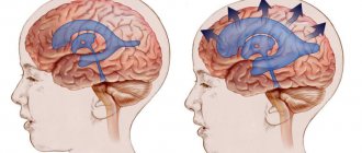
The activity of the endocrine glands, which produce cerebrospinal fluid, is disrupted. As a result, there is too much cerebrospinal fluid, and the small body is unable to ensure its normal outflow. On the left of the diagram is a normal brain, and on the right is one affected by hydrocephalus.
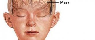
Hydrocephalic babies are easily recognized by their disproportionately large heads. Water cannot compress and increases the volume of the skull, and since in children under one year old rigid connections between the cranial bones have not yet formed, the increase in volume of the head occurs unhindered.
When hydrocephalus of the brain develops in an older child, excess fluid manifests itself exclusively as damage to the central nervous system.
The disease is quite dangerous: the cerebrospinal fluid does not leave the head and begins to put pressure on the nervous tissue, destroying neural connections. Without proper treatment, this usually leads to swelling of the brain and ultimately the death of the child.
COMPARATIVE ANATOMY
The brain structure of different vertebrate species is remarkably similar. When compared at the neuronal level, there are clear similarities in characteristics such as the neurotransmitters used, fluctuations in ion concentrations, cell types and physiological functions. Fundamental differences are revealed only when compared with invertebrates. Invertebrate neurons are much larger; often they are connected to each other not by chemical, but by electrical synapses, which are rarely found in the human brain. In the nervous system of invertebrates, some neurotransmitters are detected that are not characteristic of vertebrates.
Among vertebrates, differences in the structure of the brain concern mainly the relationship of its individual structures. By assessing the similarities and differences in the brains of fish, amphibians, reptiles, birds, and mammals (including humans), several general patterns can be derived. Firstly, in all these animals the structure and functions of neurons are the same. Secondly, the structure and functions of the spinal cord and brain stem are very similar. Thirdly, the evolution of mammals is accompanied by a pronounced increase in cortical structures, which reach their maximum development in primates. In amphibians, the cortex makes up only a small part of the brain, whereas in humans it is the dominant structure. It is believed, however, that the principles of functioning of the brain of all vertebrates are almost the same. The differences are determined by the number of interneuron connections and interactions, which is higher the more complex the brain is organized. See also COMPARATIVE ANATOMY.
Classification of the disease
In children, there are four main types of hydrocephalus of the brain:
- External - fluid accumulates in the subarachnoid space - the cavity between the pia mater and the arachnoid mater.
- Internal - cerebrospinal fluid remains in the cerebral ventricles. The substance of the brain begins to thin out.
- Open (communicating) - all ventricular systems of the brain expand, but there are no obstacles to the movement of fluid through the cerebrospinal fluid system.
- Closed (occlusive) - disruption of the liquor flow is caused by pathologies of the ventricles: their adhesions, neoplasms.
The open form of childhood hydrocephalus can be either external or internal. But occlusal in 99% of cases is internal.
In addition, dropsy is also divided according to the form of its flow:
- Acute – a rapid increase in intracranial pressure, the baby’s condition worsens in the period from one to three days.
- Subacute - normal circulation of cerebrospinal fluid is disrupted for three to six months, after which severe brain damage occurs.
- Chronic – the development of the disease also lasts up to six months, but there is no such serious damage to the brain. Characteristic of open hydrocephalus.
Causes of pathology
As a rule, this is a congenital disease. There are also acquired forms, but they are quite rare.
The causes of cerebral hydrocephalus in children are intrauterine infections, birth injuries, genetic disorders, and central nervous system defects.
Depending on the stage at which the disease developed, its causes vary:
- Fetal hydrocephalus in the vast majority of cases develops due to defects of the central nervous system. Infectious infections are to blame for the pathology of every fifth intrauterine hydrocephalus. Only a small proportion is due to genetic disorders.
- When the disease begins to develop immediately after birth, it is most likely due to infections in the womb. Such situations account for 75%. In 15% of cases, a newly born baby begins to suffer from dropsy due to a birth injury that led to meningitis or intracranial hemorrhage. The remaining 10% are malformations of either the spinal cord or brain, as well as disturbances in the functioning of blood vessels.
- In children aged one year and older, hydrocephalus is almost equally formed as a result of one of the following reasons: hemorrhage; brain tumor (spinal or brain); consequences of TBI; consequences of brain inflammation; genetic problems; malformations of cerebral vessels.
These generalized reasons also have their own classification.
So, among the infections that cause the disease, the following are common:
- rubella;
- cytomegalovirus;
- neurosyphilis;
- herpes virus type 1 or 2;
- toxoplasmosis;
- bacteria and viruses that cause meningitis;
- parotitis.
Defects responsible for the formation of dropsy:
- Narrowing of the canals connecting the cerebral ventricles.
- Arnold-Chiari syndrome (underdevelopment of the posterior fossa of the skull, due to which the brain structures located there do not fit into it).
- Anomalies of the cerebral venous system.
- Underdevelopment of the openings through which cerebrospinal fluid drains into the subarachnoid space.
- Dandy-Walker syndrome (anomaly of the development of the cerebellar spaces and cerebellum).
Also, among oncological diseases, hydrocephalus can be caused by:
- brain cancer;
- papilloma;
- tumor of the skull bones;
- brain ventricle tumor;
- vascular plexus meningioma;
- various forms of spinal cord oncology that impair fluid circulation.
Ventricles of the brain: structure, functions, diseases
The brain is the most complex organ in the human body, where the ventricles of the brain are considered one of the tools for interconnection with the body.
Their main function is the production and circulation of cerebrospinal fluid, due to which the transport of nutrients, hormones and the removal of metabolic products occurs.
Anatomically, the structure of the ventricular cavities looks like an expansion of the central canal.
What is a cerebral ventricle
Any ventricle of the brain is a special tank that connects with similar ones, and the final cavity joins the subarachnoid space and the central canal of the spinal cord.
Interacting with each other, they form a very complex system. These cavities are filled with moving cerebrospinal fluid, which protects the main parts of the nervous system from various mechanical damage and maintains intracranial pressure at a normal level. In addition, it is a component of the organ’s immunobiological protection.
The internal surfaces of these cavities are lined with ependymal cells. They also cover the spinal canal.
The apical portions of the ependymal surface have cilia that help move cerebrospinal fluid (cerebrospinal fluid, or cerebrospinal fluid). These same cells contribute to the production of myelin, a substance that is the main building material of the electrically insulating sheath that covers the axons of many neurons.
The volume of cerebrospinal fluid circulating in the system depends on the shape of the skull and the size of the brain. On average, the amount of fluid produced for an adult can reach 150 ml, and this substance is completely renewed every 6-8 hours.
The amount of cerebrospinal fluid produced per day reaches 400-600 ml. With age, the volume of cerebrospinal fluid may increase slightly: this depends on the amount of fluid absorption, its pressure and the state of the nervous system.
The fluid produced in the first and second ventricles, located in the left and right hemispheres, respectively, gradually moves through the interventricular foramina into the third cavity, from which it moves through the openings of the cerebral aqueduct into the fourth.
At the base of the last cistern there is a foramen of Magendie (communicating with the cerebellopontine cistern) and paired foramina of Luschka (connecting the terminal cavity with the subarachnoid space of the spinal cord and brain). It turns out that the main organ responsible for the functioning of the entire central nervous system is completely washed by cerebrospinal fluid.
Once in the subarachnoid space, the cerebrospinal fluid, with the help of specialized structures called arachnoid granulations, is slowly absorbed into the venous blood. Such a mechanism functions like valves that work in one direction: it allows fluid to pass into the circulatory system, but does not allow it to flow back into the subarachnoid space.
The number of ventricles in humans and their structure
The brain has several communicating cavities connected together. There are four in total, however, very often in medical circles they talk about the fifth ventricle in the brain. This term is used to refer to the cavity of the transparent septum.
However, despite the fact that the cavity is filled with cerebrospinal fluid, it is not connected to other ventricles. Therefore, the only correct answer to the question of how many ventricles are in the brain is: four (two lateral cavities, the third and fourth).
The first and second ventricles, located to the right and left relative to the central canal, are symmetrical lateral cavities located in different hemispheres just below the corpus callosum. The volume of any of them is approximately 25 ml, and they are considered the largest.
Each lateral cavity consists of a main body and canals branching from it - the anterior, inferior and posterior horns. One of these channels connects the lateral cavities with the third ventricle.
The third cavity (from the Latin “ventriculus tertius”) is shaped like a ring. It is located in the midline between the surfaces of the thalamus and the hypothalamus, and is connected inferiorly to the fourth ventricle by the aqueduct of Sylvius.
The fourth cavity is located slightly lower - between the elements of the hindbrain. Its base is called the rhomboid fossa and is formed by the posterior surface of the medulla oblongata and the pons.
The lateral surfaces of the fourth ventricle limit the superior cerebellar peduncles, and the entrance to the central canal of the spinal cord is located behind it. This is the smallest, but very important section of the system.
On the arches of the last two ventricles there are special vascular formations that produce most of the total volume of cerebrospinal fluid. Similar plexuses are present on the walls of two symmetrical ventricles.
Ependyma, consisting of ependymal formations, is a thin film that covers the surface of the central duct of the spinal cord and all ventricular cisterns. Almost the entire area of the ependyma is single-layered. Only in the third and fourth ventricles and the brain aqueduct connecting them can it have several layers.
Ependymocytes are elongated cells with a cilium at the free end. By the beating of these processes they move the cerebrospinal fluid. It is believed that ependymocytes can independently produce some protein compounds and absorb unnecessary components from the cerebrospinal fluid, which helps cleanse it of breakdown products formed during the metabolic process.
Functions of the ventricles of the brain
Each ventricle of the brain is responsible for the formation of cerebrospinal fluid and its accumulation. In addition, each of them is part of the fluid circulation system, which constantly moves along the cerebrospinal fluid pathways from the ventricles and enters the subarachnoid space of the brain and spinal cord.
The composition of cerebrospinal fluid is significantly different from any other fluid in the human body. However, this does not give reason to consider it a secretion of ependymocytes, since it contains only cellular elements of blood, electrolytes, proteins and water.
The liquor-forming system forms about 70% of the necessary fluid. The rest penetrates the walls of the capillary system and the ventricular ependyma. The circulation and outflow of cerebrospinal fluid is due to its constant production. The movement itself is passive and occurs due to the pulsation of large cerebral vessels, as well as due to respiratory and muscle movements.
Absorption of cerebrospinal fluid occurs along the perineural nerve sheaths, through the ependymal layer and capillaries of the arachnoid and pia mater.
Liquor is a substrate that stabilizes brain tissue and ensures full neuronal activity by maintaining optimal concentrations of essential substances and acid-base balance.
This substance is necessary for the functioning of the brain systems, since it not only protects them from contact with the skull and accidental impacts, but also delivers the hormones produced to the central nervous system.
To summarize, let us formulate the main functions of the ventricles of the human brain:
- production of cerebrospinal fluid;
- ensuring continuous movement of cerebrospinal fluid.
Ventricular diseases
The brain, like all other internal human organs, is prone to various diseases. Pathological processes affecting parts of the central nervous system and the ventricles, including, require immediate medical intervention.
In pathological conditions developing in the organ cavities, the patient’s condition rapidly deteriorates because the brain does not receive the required amount of oxygen and nutrients. In most cases, the cause of ventricular diseases is inflammatory processes resulting from infections, injuries or neoplasms.
Hydrocephalus
Hydrocephalus is a disease characterized by excessive accumulation of fluid in the ventricular system of the brain. The phenomenon in which difficulties arise in its movement from the site of secretion to the subarachnoid space is called occlusive hydrocephalus.
If the accumulation of fluid occurs due to a violation of the absorption of cerebrospinal fluid into the circulatory system, then this pathology is called aresorptive hydrocephalus.
Hydrocele of the brain can be congenital or acquired. The congenital form of the disease is usually detected in childhood. The causes of the acquired form of hydrocephalus are often infectious processes (for example, meningitis, encephalitis, ventriculitis), neoplasms, vascular pathologies, injuries and developmental defects.
Dropsy can occur at any age. This condition is dangerous to health and requires immediate treatment.
Hydroencephalopathy
Another common pathological condition due to which the ventricles in the brain may suffer is hydroencephalopathy. In this pathological condition, two diseases are combined at once - hydrocephalus and encephalopathy.
As a result of impaired circulation of cerebrospinal fluid, its volume in the ventricles increases, intracranial pressure increases, and because of this, brain function is disrupted. This process is quite serious and without proper control and treatment leads to disability.
Ventriculomegaly
When the right or left ventricles of the brain become enlarged, a disease called ventriculomegaly is diagnosed. It leads to disturbances in the functioning of the central nervous system, neurological abnormalities and can provoke the development of cerebral palsy. This pathology is most often detected during pregnancy at a period of 17 to 33 weeks (the optimal period for detecting pathology is 24-26 weeks).
A similar pathology often occurs in adults, but ventriculomegaly does not pose any danger to a mature organism.
Ventricular asymmetry
Changes in the size of the ventricles can occur under the influence of excessive production of cerebrospinal fluid. This pathology never occurs on its own. Most often, the appearance of asymmetry is accompanied by more serious diseases, for example, neuroinfection, traumatic brain injury, or a tumor in the brain.
Hypotensive syndrome
A rare phenomenon, usually a complication after therapeutic or diagnostic procedures. Most often it develops after a puncture and leakage of cerebrospinal fluid through the hole from the needle.
Other causes of this pathology may be the formation of cerebrospinal fluid fistulas, disruption of the water-salt balance in the body, and hypotension.
Clinical manifestations of low intracranial pressure: the appearance of migraine, apathy, tachycardia, general loss of strength. With a further decrease in the volume of cerebrospinal fluid, pallor of the skin, cyanosis of the nasolabial triangle, and breathing problems appear.
Finally
The ventricular system of the brain is complex in its structure. Despite the fact that the ventricles are only small cavities, their importance for the full functioning of human internal organs is invaluable.
The ventricles are the most important brain structures that ensure the normal functioning of the nervous system, without which the life of the body is impossible.
It should be noted that any pathological processes leading to disruption of brain structures require immediate treatment.
Risk factors
There are accompanying conditions that contribute to the development of hydrocephalus of the brain in a child:
- Early birth (gestation period did not exceed 34 weeks).
- A narrow maternal pelvis and, as a result, a difficult birth process.
- The newborn's light weight is less than one and a half kilograms.
- During childbirth, hypoxia or asphyxia was observed in the fetus.
- Active methods of obstetrics: manual techniques, vacuum, forceps.
- The mother suffered viral infections during pregnancy: herpes, influenza, ARVI, toxoplasmosis.
- Bad habits of the mother that manifested themselves during pregnancy.
- Untreated sexually transmitted diseases in the mother during pregnancy.
Most risk factors for hydrops are related to maternal health.
Even indirect reasons, such as a premature baby with low weight, indicate an abnormal pregnancy. Every problem with a woman’s health during pregnancy weakens the embryo’s body.
How does hydrocephalus manifest?
Depending on the form of development of the pathology and the age of the child, the external signs of the disease change.
For children under two years of age, in which in 95% of cases hydrocephalus is a congenital pathology, the following symptoms are characteristic:
- Severe course of the disease. A rapidly deteriorating condition caused by damage to brain structures.
- The main symptom is a rapid increase in head volume. Growth of 1.5 cm or more every month is typical, for at least three months in a row. Starting from the ninth month of life, growth drops to 8-9 mm.
- A child is born with a head whose girth is larger than the girth of the chest. If by six months the ratio does not change, and the head is still larger than the chest, there is reason to suspect hydrocephalus.
- The veins on the occipital, temporal and frontal parts of the head are clearly visible.
- At three months the child does not yet hold his head up and begins to sit up late, crawl, and walk.
- The fontanel on the top of the head is convex.
- The accumulation of cerebrospinal fluid primarily occurs in the frontal lobes of the brain. Head enlargement starts from the forehead.
- While fixating the baby's gaze, the baby's pupil twitches chaotically.
- The scalp becomes thin and has a painful shine.
- Overhang of the brow ridges over the facial bone - the eyes appear to be very deep-set.
A number of less characteristic signs can confuse the initial diagnosis of hydrocephalus in children and complicate treatment. For example, tearfulness, poor appetite, slow weight gain, frequent regurgitation, drowsiness, legs constantly bent at the knees, head tilting - all these symptoms are characteristic of hydrocephalus. But at the same time, they can be observed in dozens of other childhood diagnoses.
In the acute form of dropsy, when the disease progresses rapidly, other signs can be observed:
- convulsions;
- long cry on one note;
- loss of acquired physical skills (sitting, following other people's movements, vocal functions);
- vomit.
For children aged two years and older, other manifestations of hydrocephalus of the brain are typical:
- Headaches that begin in the morning (after sleep) and gradually subside in the evening. Nausea and vomiting often begin at the same time.
- Migraines that appear after stress, mental or physical work. Often accompanied by nosebleeds.
- A feeling of a rush to the head when bending over and bursting headaches.
- Pain in the fundus of the eye.
- Visual impairment: loss of sharpness, double vision.
- Urinary incontinence.
- Decreased muscle strength, accompanied by rapid fatigue.
- Cramps and fainting.
- Loss of coordination and uncontrolled movements of the limbs.
HOW THE BRAIN WORKS
Let's look at a simple example. What happens when we pick up a pencil lying on the table? The light reflected from the pencil is focused in the eye by the lens and directed to the retina, where the image of the pencil appears; it is perceived by the corresponding cells, from which the signal goes to the main sensitive transmitting nuclei of the brain, located in the thalamus (visual thalamus), mainly in that part of it called the lateral geniculate body. There, numerous neurons are activated that respond to the distribution of light and darkness. The axons of the neurons of the lateral geniculate body go to the primary visual cortex, located in the occipital lobe of the cerebral hemispheres. Impulses coming from the thalamus to this part of the cortex are converted into a complex sequence of discharges of cortical neurons, some of which react to the boundary between the pencil and the table, others to the corners in the pencil’s image, etc. From the primary visual cortex, information travels along axons to the associative visual cortex, where image recognition occurs, in this case a pencil. Recognition in this part of the cortex is based on previously accumulated knowledge about the external outlines of objects.
Planning a movement (i.e., picking up a pencil) probably occurs in the frontal cortex of the cerebral hemispheres. In the same area of the cortex there are motor neurons that give commands to the muscles of the hand and fingers. The approach of the hand to the pencil is controlled by the visual system and interoceptors that perceive the position of muscles and joints, information from which is sent to the central nervous system. When we take a pencil in our hand, the pressure receptors in our fingertips tell us whether our fingers have a good grip on the pencil and how much force must be exerted to hold it. If we want to write our name in pencil, other information stored in the brain will need to be activated to enable this more complex movement, and visual control will help improve its accuracy.
The example above shows that performing a fairly simple action involves large areas of the brain, extending from the cortex to the subcortical regions. In more complex behaviors involving speech or thinking, other neural circuits are activated, covering even larger areas of the brain.
Treatment of hydrocephalus in children
Due to the complexity of the disease and its danger, treatment for a child with hydrocephalus is prescribed not only by a neurosurgeon, but also by a neurologist.
Treatment can be conservative (that is, without surgery, only with the help of medications) or surgical.
During drug treatment, the first step is to reduce intracranial pressure using the drug Diacarb. It reduces the production of cerebrospinal fluid and promotes the removal of potassium from the body.
In the hospital, strong osmotic diuretics are prescribed - oral Glycerin and Mannitol. Salt diuretics, for example Furosemide, are also prescribed. It, like Diacarb, should be taken in conjunction with drugs that maintain normal potassium levels - Panangin or Asparkam.
Doctor's advice
Hydrocephalus - this conclusion can often be found after an ultrasound scan of infants. However, in most cases it is compensatory in nature, everything returns to normal by the first year of life. In addition to birth injuries, protracted labor, structural features, the cause of such dropsy is the immaturity of the nervous system. True hydrocephalus is characterized by the entire clinical picture of increased intracranial pressure - an increase in the size of the head, monotonous crying, and the presence of neurological symptoms. Conclusion Ultrasound is not a diagnosis, it is the result of an examination; the diagnosis is made based on a combination of complaints, anamnesis, examination, and research data.
Victoria Druzhikina Neurologist, Therapist
Any medicinal forms of treatment for acute hydrocephalus must be carried out under the strict supervision of doctors, and intermediate results must be checked using computed tomography and neurosonography.
Along with diuretic enzyme blockers, medications are prescribed that stimulate the functioning of brain neurons. For example, "Encephabol".
As for surgical intervention, the most effective method of surgical treatment of the brain in children is bypass surgery. Often this is the only way to save a child's life.
Shunting is the creation of a flow path (shunt) for cerebrospinal fluid to bypass an existing blockage. For closed hydrocephalus, it is used when tumors have already grown into the brain.
In the open form of the disease, three types of bypass surgery are provided:
- Ventriculoatrial.
- Ventriculo-peritoneal.
- Lumbo-peritoneal.
In addition to shunting, another type of surgical intervention is possible for closed forms of hydrops - dissection of arachnoid adhesions. This method involves the absorption of cerebrospinal fluid.
Watch the video about the causes and treatment of hydrocephalus:
The life span and quality of life of a child with hydrocephalus, his chances of recovery depend on many factors: the reasons that caused the development of the disease, the speed of diagnosis, the quality of treatment. But even a healthy baby can become a victim of the disease.
Don't forget about prevention. Protect your child from head injuries in every possible way; be sure to use protective equipment during active recreation. If the baby was born premature, be sure to undergo routine examinations with a neurologist, do an ultrasound and an MRI.
Women who are about to become mothers or planning to conceive a child should be aware of their health. Get rid of bad habits a few months before pregnancy. Get screened for infections. If you happen to get sick while pregnant, consult a geneticist and undergo an additional ultrasound.
Your unborn baby is sensitive to literally everything, so take care of him even before birth.
This article has been verified by a current qualified physician, Victoria Druzhikina, and can be considered a reliable source of information for site users.
Bibliography
1. https://docs.cntd.ru/document/499002598
Rate how useful this article was
3.5 4 people voted, average rating 3.5
Did you like the article? Save it to your wall so you don’t lose it!