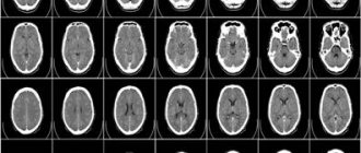
CT (computed tomography) is a method of diagnostic examination of the body. The instrumental test is carried out using special equipment - a computed tomograph, which is where its name comes from. In essence, this is a modern version of X-ray, in which a layer-by-layer image of an object of interest (for example, the head) is obtained. As a result, a three-dimensional picture is built, and not a flat projection, as is the case with classic x-rays.
CT is very close to MRI (magnetic resonance imaging). But CT is more often (though not always) used to study hard tissues (bones of the limbs, skull, etc.), and MRI - soft ones. In addition, there are technical differences related to the nature of the radiation, frequency and other parameters of the devices used to carry out the test.
Computed tomography is a modern research method that allows you to obtain an objective picture and make an accurate diagnosis. The advantages of this type of diagnosis include several points:
- very high speed of conduction - for example, by holding your breath for 20 seconds you can get a complete picture of the abdominal organs;
- large coverage area - you can examine several parts of the body and organs at once (for example, the stomach and chest at the same time);
- good spatial resolution allows you to obtain a detailed picture;
- radiation has a reduced load compared to x-rays;
- the procedure is absolutely painless and does not cause discomfort;
- CT provides objective data, so the risk of medical error is virtually eliminated.
However, this diagnostic method also has disadvantages:
- radiation is harmful to the body (with frequent exposure);
- CT scan is an expensive procedure;
- Tomography during pregnancy is possible only with the agreement of a doctor.
How is SCT research carried out?
The x-ray technician positions the patient in a special way, depending on the examination being performed. Next, the gantry (the device on which the tube and receiving sensors are installed) begins to rotate around the scanned area and makes slices of the area under study with certain parameters of power and tube passage step. When scanning the chest and abdominal organs, you will need to hold your breath several times, which the x-ray technician will warn you about. The scanning procedure lasts 2-4 minutes.
Difference between CT and SCT
With step-by-step CT, a single slice image is obtained during one rotation of the “tube-detector” system around the area of interest.
The table is then moved a predetermined distance and the rotation of the tube is repeated again to obtain the next image. The thickness of the resulting slice is 1 cm.
During diagnostics, a spiral tomograph collects data continuously due to the synchronous movement of the couch with the patient and the tube describing a helical trajectory.
This allows you to increase the speed of diagnosis, examine one anatomical area in 1 phase of the respiratory cycle and obtain sections 2-3 mm thick.
What can SKT do?
Computed tomography is simply irreplaceable in diagnosing the conditions of any bone structures, the condition of the lungs, and mediastinum in humans. SCT is also widely used in the study of abdominal organs (liver, spleen, gallbladder, pancreas), adrenal glands, kidneys, and pelvic organs. With the help of CT, it became possible to accurately detect any stones (for example, in the gall bladder, kidneys) and calcifications (for example, those changes that accompany tuberculosis, sclerosis).
Spiral computed tomography is leading in the diagnosis of complex bone injuries , multiple injuries (for example, road accidents) of bones and internal organs, in the diagnosis of complex or multiple injuries of the skull and acute injuries and hemorrhages of the brain.
SCT is also widely used by ENT doctors for a detailed assessment of the condition of the paranasal sinuses, auditory canals and temporal bones, since no other diagnostic method can compare with the accuracy and “depth” of computed tomography.
In studies of the spine using SCT, it is possible to determine the smallest damage in the bone structure, pathological calcifications in the ligamentous apparatus; to diagnose the condition of the intervertebral discs, it is best to use MRI.
The advisability of prescribing SCT in each individual case should be determined by the attending physician, who will subsequently interpret the results obtained from CT taking into account the entire picture and characteristics of the patient.
Advantages of SCT over MRI
A valuable property of SCT is the speed of scanning and the speed of obtaining diagnostic information necessary for making a diagnosis and starting treatment measures.
Unlike MRI, which takes up to 1 hour to visualize pathology, with SCT the same amount of information is obtained in 10-15 minutes.
The strength of MRI is considered to be the staging of cancer, the detection of diseases of soft tissues and the spinal cord, while bone structures are poorly visible.
SCT, on the contrary, clearly visualizes the osteoarticular apparatus, and is the method of choice for determining painful changes in this area.
In what cases is SCT prescribed?
The method is indicated for emergency diagnosis of acute conditions caused by head and torso injuries in unconscious patients with severe pain, when the patient cannot remain in a stationary position for a long time.
A routine scan is prescribed to identify:
- formations of a malignant and benign nature, determining the stage, metastatic damage to neighboring organs, lymph nodes;
- inflammatory, purulent-destructive processes;
- degenerative-dystrophic processes;
- congenital anatomical disorders;
- foreign bodies, stones;
- circulatory disorders;
- mechanical damage.
What does SKT “dislike”?
First of all, these are movements . Any movements during scanning of the study area distort the resulting images, of course, reducing their information content for the doctor. If in a series of images obtained, even just 1-2 scans are distorted by motion artifacts, then subsequent 3D reconstructions will already have defects and their information content for the doctor, of course, will be reduced.
Secondly, metal. Metal is to X-rays what a mountain is to the sun—it leaves a “shadow.” A large amount of metal in the examination area can significantly distort the resulting images. But if the metal is located in any place other than the area being examined, it cannot in any way affect the quality of the scans.
Computed tomography at the "Family Doctor"
“Family Doctor” uses new generation tomographs GE OPTIMA CT660 manufactured by GE Healthcare (a division of General Electric Corporation, USA). This device takes 64 image slices per revolution, which significantly speeds up the diagnostic process. A distinctive feature of the device is its high resolution, which allows you to create high-quality two-dimensional (2D) and three-dimensional (3D) reconstruction of images.
Other benefits of the GE OPTIMA CT660:
- improved scanner ergonomics, providing maximum patient comfort;
- setting scanning parameters is carried out in the presence of the patient, so he has time to get used to it;
- Diagnosis is carried out literally in seconds, while the radiation dose is optimized to ensure the lowest possible radiation exposure.
MSCT examination
Computed tomography services are provided in Polyclinics No. 5 (metro stations Barrikadnaya, Krasnopresnenskaya) and No. 15 (metro stations Baumanskaya).
What do you need to have with you for SCT?
Before the study, you must give your existing medical documentation to the x-ray technician or radiologist (a referral from the attending physician, which indicates the preliminary diagnosis, study area, clinical task, specific comments), data from previous studies (which directly relate to the reason for prescribing SCT to you) and other medical documentation, after analyzing which the radiologist will be able to formulate the most accurate diagnostic conclusion on SCT, taking into account the data of your medical. cards and recommendations of the attending physician. If you do not provide medical documentation in full, then the radiologist will evaluate only the directly obtained SCT scans, without taking into account previous injuries and diseases, any possible consequences that they could entail, and without taking into account possible concomitant pathologies.
Remember, there is no absolute and universal method of medical diagnosis: MRI will never replace SCT, SCT will never replace ultrasound, ultrasound will never replace radiography. All these methods complement each other and sometimes, data from previous studies have the decisive word in the correct interpretation of the resulting picture by a radiologist during computed tomography.
Make an appointment
Make an appointment by calling:
+38 (044) 393 0933
+38 (067) 230 2432
+38 (050) 332 3339
What will a CT scan of the lungs show?
It is known that computed tomography of the lungs is one of the most informative diagnostic research methods. How is a CT scan done? What does the doctor see? What is required from the patient to undergo the procedure? Nikita Valentinovich Monakov, a radiologist at Clinic Expert Irkutsk, answers these and other questions.
— Nikita Valentinovich, what is computed tomography (CT)?
— This is a special type of X-ray examination, the essence of which is layer-by-layer scanning of tissues by shining them with X-rays. Based on a series of such images from multiple sections, the doctor receives targeted three-dimensional (3D) images of the area under study and the pathological focus.
CT is one of the most informative and highly accurate methods. It allows you to determine the location and size of the pathological focus, and evaluate the results of treatment. The sections can be made very thin, less than a millimeter, so the doctor has very detailed information about the pathology.
CT is used both as a method of primary diagnosis and as a clarifying technique in addition to other diagnostic methods - say, X-ray or ultrasound.
— In what cases can a doctor recommend a computed tomography scan of the lungs?
— CT scans of the lungs are prescribed to assess the extent of the upcoming surgical intervention, when assessing the effectiveness of radiation or chemotherapy, as well as in cases where other methods do not allow identifying the pathological focus.
— What does a CT scan of the lungs show?
— Using computed tomography, you can see the lung tissue, bronchial tree, lymph nodes, blood vessels (including the aorta, pulmonary veins). CT allows you to identify focal and diffuse processes, determine their nature, prevalence, localization, and degree of severity of changes. In addition, during the examination of the lungs, the condition of the esophagus, heart, and bone frame of the chest is assessed. The method allows you to measure the density of both the lung tissue itself and the specific pathological focus located in it. The sensitivity of CT in detecting cysts, space-occupying formations, and metastases in the lungs is 98 percent.
“X-ray and computed tomography of the chest, bronchoscopy will be useful in detecting pulmonary echinococcosis.” Quote from the material “Attention, echinococcosis!”
— How is a CT scan of the lungs done?
— Like any other types of tomography, this procedure is non-invasive and completely painless. The patient simply lies down on a special couch, which slowly moves through the detector frame of the tomography machine. The only condition: you need to lie still. The entire procedure lasts from 5 to 20 minutes.
— How to prepare for a CT scan of the lungs?
— The patient fills out a questionnaire about possible contraindications, after which he must undress to the waist and remove all metal objects and jewelry. Before the study, it is necessary to show the direction, results of previous studies (ultrasound, CT, MRI, radiography; films, disks, descriptions - if all this is available), medical documentation (specialist reports, extracts from the medical history, outpatient records, etc.).
— Computed tomography is based on x-rays. In this regard, the question may arise: is a CT scan of the lungs dangerous?
— The radiation dose a patient receives during a CT scan depends on the person’s build: the larger the patient, the higher it is. On new devices - say, like those in our clinic - the radiation dosage is calculated and controlled automatically. According to children's protocols, this load is lower. CT scans do not pose any danger to human health.
— Taking into account the fact that computed tomography is a radiation research method, the question arises: how often can a CT scan of the lungs be done without harm to health?
— This type of research is prescribed by a doctor, and only he decides whether it is necessary or not. If a doctor needs to assess the dynamics of some pathological process, such as pneumonia, then the standard provides for a second CT scan two weeks after the first.
— Are there any contraindications to CT scan of the lungs?
— There are no absolute contraindications. CT scans should not be performed on pregnant and lactating women and small children. But in each specific situation, the decision on the need for a tomography is made by the doctor, weighing all the risks.
— If CT is a radiation research method, then why not do an MRI of the lungs? After all, magnetic resonance imaging is also a modern type of diagnostics, and there is no radiation exposure.
— CT is the “gold standard”. MRI is an alternative, third method for detecting lung diseases. MRI can be recommended for certain diseases, such as cystic fibrosis, pulmonary embolism, and can also be considered an alternative method for tumors and pneumonia in children, as an addition to other methods (radiography, CT). MRI may also be recommended if CT results are ambiguous and tumor invasion into the mediastinum or pleura is suspected.
Modern MRI makes it possible to visualize bronchiectasis, thickening of the bronchial walls, the presence of fluid levels, consolidation and destruction.
— Nikita Valentinovich, what is a CT scan of the lungs with contrast?
— There are practically no differences from conventional CT. The patient is injected intravenously with a contrast agent, which during examination allows the doctor to obtain an even more complete picture. Before contrast, the patient must undergo a serum creatinine test, and, depending on the result, a decision is made whether he can have a CT scan with contrast or not.
There are relative contraindications for the administration of a contrast agent. These are a reaction to a previously administered contrast agent, a multivalent allergy with severe reactions, asthma requiring treatment, and diabetes mellitus.
Contraindications to contrast-enhanced CT include hyperthyroidism, thyrotoxicosis, and treatment with radioactive iodine.
Contrast-enhanced CT can be performed if the patient has seasonal allergies, mild to moderate reactions to various allergens (including iodine, seafood), and non-exacerbated asthma.
Interviewed by Igor Chichinov
For reference
Monakov Nikita Valentinovich
In 2012 he graduated from Irkutsk State Medical University, Faculty of Pediatrics.
In 2013-2014 – internship in radiology at IGMU.
2016 – professional retraining in ultrasound diagnostics at the State Budgetary Educational Institution of Further Professional Education of the Irkutsk State Medical Academy of Postgraduate Education.
2018 – advanced training in radiology with a course in computed tomography of the Federal State Budgetary Institution National Medical Research Center named after. V. A. Almazova, St. Petersburg.
2018 – advanced training in MRI, ChUDPO IPKMK, Voronezh.
Who deciphers the results and when they can be obtained.
The resulting SCT images must be examined by a radiologist (a doctor who specializes in CT, MRI, X-ray studies and has the appropriate certificate). It is necessary to “read” all the scans, study the provided medical information. documentation, compare it with the obtained CT image, describe the study, draw a diagnostic conclusion, shoot film with scans. On average it takes about 1-3 hours. At ACMD, we undertake to prepare the results for delivery by the end of the Clinic’s working day.
After the examination, you receive films with scans and a diagnostic report, which will be signed by a radiologist. Also, the diagnostic report may contain recommendations regarding your further actions (which doctor to contact with these results, what types of further examination are recommended, and so on).
How to prepare for SCT
SCT diagnostics does not require any special preparation. In general, patients are recommended to:
- 24 hours before the procedure, do not drink alcohol;
- at least do not smoke for 4-5 hours;
- do not drink or eat anything for the previous 3 hours;
- Do not take all metal objects with you - this restriction also applies to dentures, as they can distort the results of the examination.
If you have previously had an ultrasound or MRI, you must have the results with you.
The doctor should be warned in advance about possible allergic reactions to the medications used or that you suffer from claustrophobia.
Consultations and questions regarding your SCT study.
A radiologist is not a clinician (a doctor who treats you) and, as such, cannot provide any advice or recommendations regarding your treatment or diagnostic procedures.
To make objective comments on an SCT study, it is necessary to fully know the patient’s objective data, the picture of his previous studies, the characteristics of his current condition, anamnestic data, and much more.
You can ask all your questions to your attending physician or physician, whose consultation will be recommended to you by a radiologist.
If you still have any specific questions about computed tomography, you can ask them in the specialized MRI and SCT diagnostics section on our Forum.
Check out comprehensive body check packages with up to 30% discount
Contraindications and complications
In some cases, computed tomography is contraindicated:
- pregnancy;
- lactation period;
- severe heart pathologies;
- plasma cell dyscrasia;
- renal failure;
- diabetes mellitus of decompensated type;
- myeloma.
As for complications, in most cases they are not observed. But if X-ray contrast agents are used, side effects may occur. In this case, it is better to clarify with the doctor in advance the specifics of the diagnosis, especially if the patient has chronic diseases or is recovering from surgery.
Contrast techniques for SCT.
When examining the abdominal organs, the patient will be asked to drink 500–1000 ml. drinking water with a contrast agent dissolved in it in order to “separate” the stomach and intestines from nearby organs and tissues, as well as to ensure that intestinal loops do not create additional “interference” for the radiologist during the study. This is the so-called oral contrast.
The same technique of intravenous administration of a contrast agent is very often used in order to “highlight” areas with pathological changes, to study the blood flow of tumor formations, or specifically to study blood vessels.
How is the examination performed?
SCT in medicine is a diagnosis that is not particularly complex. The patient is placed on a special table, which will gradually and smoothly be immersed inside the round chamber where the sensors and emitters are located.
The doctor usually observes the processes behind the glass
You need to be prepared for the fact that the doctor may ask you to hold your breath for a while. This is necessary in order to avoid blurring the results obtained. The scanner responsible for processing information is located in a separate room behind glass. Specialists are also located in the same room.
During the examination, young children are allowed to have their parents present only if they wear a protective lead apron.
Sometimes the examination requires the use of contrast agents. If a person experiences atypical reactions in the form of itching, urticaria, shortness of breath, or swelling after the administration of such drugs, then it is necessary to immediately inform the doctor.
The duration of spiral CT is not the same. For some, the procedure will only take a few minutes, while for others it will take half an hour. It depends on what area is being studied.
Based on the images obtained, the doctor must make a diagnosis
The radiologist issues a report indicating the results of the examination. The attending physician will interpret the data obtained, clarify the diagnosis and determine the necessary therapy.
Is the SCT procedure painful and is it harmful? What are the contraindications? Where to get a CT scan in Kyiv?
The SCT examination is absolutely painless; the scanner itself does not directly touch your body.
The method is based on X-ray radiation. Naturally, during an SCT study, the patient is exposed to x-rays, therefore, when prescribing this study, the doctor must have fairly compelling reasons and take into account the negative impact of x-rays on the body.
An absolute contraindication for SCT is pregnancy and breastfeeding. Also, SCT is strongly not recommended for children without significant reasons. In other cases, you should consult your doctor.
a computed tomography (CT) scan in Kiev at the ACMD clinic, a 64-slice tomograph (the radiation level is reduced significantly!)
When SCT is contraindicated
Not everyone can undergo SCT diagnostics. This type of examination is not recommended:
- women during pregnancy;
- people with pacemakers;
This examination is not performed during pregnancy.
- suffering from psychopathologies (schizophrenia, depression, etc.);
- children under 4 years of age;
- overweight patients;
- if you are allergic to iodine or other elements included in the contrast agent;
- when it is impossible to ensure immobility for the patient;
- with intestinal hyperpneumatization.
Spiral CT is an informative and effective research method that allows you to detect pathology in the early stages. Thanks to this, it is possible to begin timely treatment and significantly increase the chances of a favorable outcome of the disease.