The central nervous system regulates the functioning of the body as a whole, so violations of its function are always dangerous for humans. Early diagnosis of central nervous system diseases significantly increases the chances of recovery and reduces the risk of complications. In order to identify pathological changes in nerve cells, modern hardware examination methods are used: computer and magnetic resonance imaging. To determine the optimal diagnostic method, you need to know how CT differs from MRI of the brain.
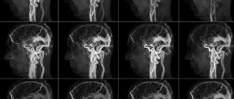
CT angiography of cerebral vessels, three-dimensional image
Which is better: CT or MRI of the brain?
Tomography involves scanning the human body using X-rays or electromagnetic pulses. As a result of scanning the brain, a special computer program converts the response of the examined tissues into a series of layer-by-layer photographs reflecting changes in the structure, shape, and size of the organ being studied.
Each method has its own advantages; the decision to prescribe CT or MRI is made by the attending physician. The brain has a complex structure, pathological changes in any part lead to disruption of the vital functions of the whole organism, so the doctor chooses a diagnostic method based on the clinical manifestations of the process.
CT is used to determine the condition of solid elements, scan the skull bones, identify traumatic lesions, hematomas, and large areas of atherosclerosis. Computed tomography is informative regarding dense structures, and the examination process itself takes less time than MRI. The cost of CT is much lower than similar scanning using a magnetic field. The method is well suited for identifying neoplasms that develop with the involvement of bone elements. The use of contrast in brain examination increases the information content of tomography, allows for a detailed study of the state of the vascular system of the brain and differentiates between hemorrhages and bleeding in cases of skull injuries.
Comminuted fracture of the skull bones and a focus of contusion of the left hemisphere of the brain on CT
MRI is more informative when studying the condition of soft tissues and nerve fibers. The method gives a detailed picture of the state of the cerebral cortex, reflects the nature of the blood supply to this area, and reveals minor changes in the structure of the substance. Such scanning is indispensable in diagnosing malignant tumors in the early stages. Vascular MRI angiography visualizes disturbances in the blood supply to various areas and the consequences of ischemia of cerebral structures.
Brain cavernomas on MRI
The use of a magnetic field is harmless to the body, so MRI scanning can be performed repeatedly. This advantage is used when necessary to observe the process over time and monitor the effectiveness of treatment measures. The method is suitable for diagnosing brain pathologies in pregnant women, children and people who have contraindications to the use of X-ray radiation.
Contraindications to CT and MRI
CT
implies the presence of intense radiation, therefore it is strictly excluded for pregnant and breastfeeding women.
MRI is contraindicated in the following cases:
- the subject suffers from claustrophobia (in this case, open MRI may be used);
- nervous pathologies that prevent the patient from remaining immobile for a long time;
- the body weight of the subject is more than 200 kg.
- Metal fragments.
- Artificial pacemaker.
- Hearing aid or implants in the cochlea.
- Metal implants.
- Fixed metal dental bridges and/or crowns.
- Surgical clips, for example, in the area of the aneurysm.
- Surgical staples.
- Lateral column stimulators.
- Kava filters.
MRI examination cannot be carried out
in patients with severe impairment of vital functions requiring constant hardware and other correction, as well as in people with fear of confined spaces and in patients with inappropriate behavior. There are no such contraindications for CT scanning.
How does an MRI differ from a CT scan of the brain?
The main difference between MRI and CT scan of the brain is the use of relevant physical phenomena to visualize the state of body tissues.
In a CT scan, the scan is done using x-rays. Hard and soft structures absorb ionizing radiation with different intensities, resulting in dark and light areas appearing on the film. They correspond to areas of reduced or increased hardness. By studying changes in the density of the structure of bone and cartilage tissue, it is possible to recreate a picture of the state of the studied area.
Unlike CT, MRI uses a magnetic field of varying intensity for scanning (which determines the power of the tomograph). It affects the location of water dipoles in the cells of the medulla. The response from fluid-saturated structures will be higher, so the information content of the method increases when scanning soft tissues. MRI of the brain shows minute changes in the brain matter, reflects demyelination processes, and reveals the growth and development of atypical cells.
Thanks to the use of a magnetic field, MRI does not have a negative effect on the human body, which distinguishes this method from computed tomography.
conclusions
Which is better, MRI or CT?
There is no clear answer to this question. Each of these methods has both its advantages and disadvantages. In some cases it will be more effective to use CT, in some MRI, and in some cases both MRI and CT will be needed.
Thus, we come to the conclusion that MRI and CT are fundamentally different diagnostic methods. The choice of one or another method of diagnosing the body depends on the specific case.
Head of the X-ray department Elena Pavlovna Smolkina
In what cases is MRI of the brain prescribed?
Indications for head tomography are various pathological processes affecting the central nervous system and skull bones. The doctor analyzes the patient’s symptoms and complaints to choose the optimal diagnostic method: CT or MRI of the brain.
Magnetic resonance examination is carried out with the following clinical picture:
- headaches, dizziness, short-term loss of consciousness;
- the presence of paresis, decreased sensitivity of the tissues of the face, torso, and limbs;
- suspicion of the presence of neoplasms;
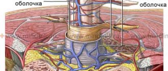
- signs of inflammation of the meninges;
- hearing impairment;
- decreased visual acuity;
- the appearance of sudden seizures;
- tingling, pain in the facial area;
- ischemic lesions of areas of the brain.
MRI is prescribed for conservative and surgical treatment of central nervous system diseases to monitor effectiveness. For malignant tumors, the method allows one to monitor the process of tumor development and determine the results of therapeutic and surgical effects on malignant areas.
Thanks to MRI, the following pathological processes affecting various parts of the brain are determined:
- benign and malignant formations of soft tissues, brain matter and its membranes, cranial nerves, vascular tumors;
- inflammatory processes – encephalitis, meningitis, abscess;
- organic lesions – encephalopathy, etc.;
- congenital developmental anomalies;
- cerebral ischemia in the acute stage and consequences of hemorrhagic stroke;
- degenerative processes - Alzheimer's, Pick's, Parkinson's diseases;
- demyelinating pathologies - multiple sclerosis, etc.
Diseases accompanied by changes in the structure of the brain matter are detected in the early stages thanks to magnetic resonance imaging. Scanning parts of the brain in the axial, sagittal and frontal planes provide a clear picture of the condition of soft tissues and fluid structures. Based on a series of photographs of the study area, a 3D model of the study area can be constructed.
Germinoma of the brain, MRI image
In what cases is a CT scan of the brain prescribed?
Tumors of the cerebral cortex can deform and thin the bones of the skull and grow into nearby sinuses. In this case, computed tomography reveals changes in solid structures, allows you to localize the process and determine its nature.
Brain injuries are often associated with damage to bone tissue, which performs a protective function. The formation of hematomas and hemorrhages can also be a consequence of mechanical damage.
Such conditions require prompt diagnosis and effective treatment. To identify pathological changes in the bone and cartilaginous structures of the skull and assess the nature of the blood supply to the brain, the doctor may prescribe computed tomography, which is a fast and accurate diagnostic method.
CT is indicated in the following cases:
- open and closed skull injuries;
- headaches, dizziness of unknown etiology;
- history of inflammatory processes in the area of the meninges;
- previously diagnosed strokes, cysts, brain abscesses;
- suspicion of the presence of neoplasms;
- changes in the shape of the skull;
- unexplained neurological symptoms (convulsions, fainting);
- suspicion of the presence of a foreign body in the cranial cavity;
- possible hematoma, cerebral edema;
- hemorrhagic stroke in acute and subacute stages;
- suspicion of the existence of cerebral vascular anomalies (aneurysms, arteriovenous malformations).
The presence of contraindications to the use of a magnetic field in the diagnosis of diseases of the central nervous system is also the reason for prescribing CT.
Using computed tomography, the following pathologies are determined:
- fractures, cracks of the skull bones;
- hemorrhages and hematomas due to any reason;
- benign and malignant formations involving bone and cartilaginous structures;
- cerebral aneurysms;
- strokes.
A CT scan of the brain is performed with contrast, which allows one to see the characteristics of the blood supply to this area and identify pathologies of the vascular system.
Consequences of hemorrhagic cerebral stroke on CT
What is the difference between EEG and MRI?
The difference between electroencephalography and magnetic resonance scanning lies in the basic principle of the technique. The subject of EEG study is brain activity.
The electroencephalograph records neuronal signals arising as a result of metabolic processes in nervous tissue and the movement of positively and negatively charged particles (ions). The consequence of these physical and chemical processes is the formation and change of a bioelectric impulse. The potential difference between deep and superficial points of the brain allows us to draw conclusions about the state of intracranial vessels and cerebral structures.
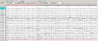
Encephalogram of a healthy child (6 years old)
Pathological changes in the central nervous system may be caused by dysfunction of the musculoskeletal system, liver, kidneys, or cardiovascular system. Venous stagnation, increased pressure in the aorta, and destabilization of the acid-base balance provoke hypoxia, cerebral edema, ischemic and hemorrhagic phenomena. In this case, a change in the bioelectric signal of neurons is observed.
EEG and MRI help determine the development of pathological processes. As a result of electroencephalography, the doctor receives waves of various amplitudes recorded on paper. The signal is received through sensors installed on the patient's head. An electroencephalogram allows you to assess the coherence of the brain parts and determine the nature of functional disorders.
The procedure can be performed in a sitting position; a special cap with electrodes is placed on the patient’s head. For better signal transmission, a transparent hypoallergenic gel is used. The session lasts 10-15 minutes, during which the patient should limit movements of the eyes, mouth, and facial muscles. The main requirement is a stable psycho-emotional state of the subject.
Unlike EEG, magnetic resonance imaging involves scanning the brain with visualization of cerebral structures. The force field induced by the apparatus causes vibrations of hydrogen atoms in water molecules. The resonance of charged particles is detected using a radio pulse receiver with a built-in analog-to-digital converter. The data is transmitted to a computer monitor in the form of monochrome layer-by-layer photographs taken in transverse, frontal and lateral projections. If necessary, 3D reconstruction of the studied area is possible.
What is the difference between EEG and MRI? In the first case, the doctor receives a graph of brain activity (encephalogram). In the second, tomography allows you to see the state of intracranial anatomical formations and pathological changes in cerebral structures.
Brain oligodendroglioma on MRI
The procedure takes from 10 to 30 minutes. The patient lies down on a mobile table, which is rolled into a wide tunnel. To immobilize the head and body of the subject, special fastenings and bolsters are used. Staying still is necessary to prevent defects from appearing in the photographs.
Tomograms visualize inflammatory, neoplastic, degenerative processes. Contrast enhancement is used in the diagnosis of neoplasms and vascular pathologies.
The object of study during magnetic resonance scanning is the signs of organic brain damage. The encephalogram shows the features of physiological processes.
What is more informative, CT or MRI of the brain?
The information content of each method depends on the area of application and the task set by the attending physician.
Computed tomography visualizes the condition of bone and cartilage structures, with its help the nature of damage in head injuries is determined, the condition of blood vessels is assessed and some brain tumors are identified. For soft tissues, the method is less informative, since they have weak radiopacity.
The reliability of CT scanning can be increased by using a contrast agent administered intravenously. The solution enters the bloodstream and highlights the vascular system of the scanned area in the image. Such a study is relevant in the presence of neoplasms that have their own special network of arterioles, aneurysms, especially if a rupture is suspected, arteriovenous malformations, arterial sinus anastomosis.
MRI displays in detail the condition of soft tissues, membranes of the brain and its substance, and nerve fibers. The quality of the image when scanning with an electromagnetic field depends on the saturation of the tissue with liquid. The use of a contrast solution increases the information content of the method in relation to focal brain pathology.
Magnetic resonance imaging detects malignant processes at an early stage.
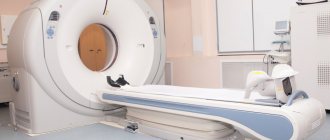
CT device
A computed tomograph works using x-rays, although the emitter itself is significantly different from a conventional x-ray machine. In a tomograph, this is a special tube that rotates around the patient’s body or is stationary at a certain angle
It creates a conical beam of rays towards it. Opposite the emitter there is a receiving detector that receives the rays passing through the patient's body. The information enters a computer, where it is processed using almost the same programs as on an MRI.
The difference between CT and MRI of the brain in terms of information content is not too great. Both systems provide information in the form of black-and-white images; on many, it is possible to simulate a three-dimensional image, highlight individual areas at any depth of the organ, record all the information on a digital medium, and then compare the patient’s condition over time.
In the case of CT, the doctor not only sees tissues, but can study their X-ray density, which changes with diseases; In the case of MRI, the doctor evaluates the images only visually. Quite often, an MRI or CT examination is prescribed by the attending physician, but, as a rule, it would be better for him to do this in consultation with a radiation diagnostician: in a number of cases, instead of an expensive MRI, a cheaper, but no less informative computed tomography can be used.