Ultrasound diagnostics is one of the safest and most effective ways to detect various diseases of internal organs. This technique is used in various fields of medicine, including assessment of the condition and possible pathologies of blood vessels. In this case, Doppler ultrasound or ultrasound is performed, which is used to examine the neck and head. Early diagnosis allows you to choose gentle and effective treatment methods and prevent the development of complications. First you need to understand what an ultrasound scan of the vessels of the head and neck shows, why this method is effective in diagnosing many diseases.
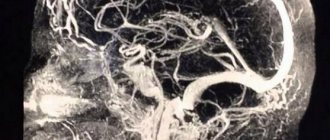
The Doppler ultrasound effect is based on the ability of ultrasound waves to be reflected from blood cells. As a result, electronic impulses appear that appear on the device screen in the form of images of veins and arteries with blood flow.
This ultrasound examination allows you to assess the condition of the large arteries that supply the brain, carotid and vertebral arteries, determine the intensity of blood flow, and also identify traces of obstruction.
How is Dopplerography performed?
This examination is carried out in the presence of certain symptoms. Preliminary preparation is necessary so that the results of the ultrasound examination give an accurate picture of the patient’s condition.
There are two types of procedure:
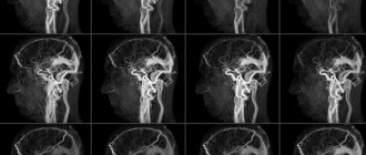
- Transcranial. The sensor is installed on the skull bones in those places where they are thinnest. The procedure gives an accurate picture of the vessels located directly in the brain.
- Extracranial. The technique is used to examine large vessels passing through the neck (carotid arteries, jugular veins, etc.). The sensor is located at each site separately.
Vascular Doppler
- fast fatiguability,
- reduced performance;
- sleep disturbance;
- pain, especially in the neck; numbness or tingling of the skin;
- noise in ears.
TYPES OF VASCULAR DOPPLER
Depending on the location of the problem in the body, the patient is prescribed an ultrasound scan:
- arteries and veins of the upper and lower extremities;
- hearts;
- pregnancy test;
- Doppler of head and neck vessels.
Several studies can be performed at once during one visit to the Doctor Ost MC.
Ultrasound of the vessels of the head and neck
The blood supply to the brain through the great vessels is assessed.
During Dopplerography of the vessels of the neck, otherwise the procedure is called ultrasound of the BCA (brachiocephalic arteries), extracranial veins and arteries are examined, that is, those located outside the cranium.
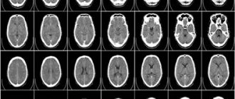
Doppler of head vessels - transcranial search for pathologies in the brain.
Indications for ultrasound scanning of head and neck vessels:
- frequent headaches, migraines;
- poor coordination, dizziness, fainting;
- noise in ears;
- increased intracranial pressure;
- sleep problems;
- fragility of blood vessels, high cholesterol, thrombosis, aneurysms;
- osteochondrosis in the cervical region;
- decreased visual acuity.
DOPPLER OF LOWER LIMB VESSELS
It is used to evaluate the veins and arteries in the legs: their shape, structure, conductivity, and functionality are examined.
Doppler ultrasound of the vessels of the lower limb is performed when:
- varicose veins;
- regular swelling and pain;
- tendency to seizures;
- decreased sensitivity;
- the appearance of ulcers or spots on the skin;
- increased sensitivity to cold.
As part of a complete examination of the circulatory system, vascular Doppler of the extremities includes evaluation of the arteries and veins in the arms.
Appointed if:
- suffers from high blood pressure;
- changes are possible due to genetic abnormalities;
- there is a predisposition to life-threatening diagnoses: strokes, heart attacks;
- the patient has diabetes and high cholesterol;
- there are bad habits.
ADVANTAGES OF USG OF VESSELS OF THE LIMB AND HEAD
- large-scale assessment of the functionality of the circulatory system;
- painlessness: the patient does not feel anything during the procedure;
- no contraindications: Doppler of the vessels of the head is prescribed even for newborns, and ultrasound of the vessels of the lower extremities can be performed even if there are lesions on the skin;
- high information content
- Ultrasound scanning of the vessels of the limbs and head does not require special preparation from the patient; it can be performed even on patients hospitalized at the Doctor Ost Medical Center.
- efficiency of research. The data is displayed on the monitor, and the specialist evaluates the picture based on several parameters. The conclusion is issued within a few minutes after the procedure.
WHAT VASCULAR DOPPLER SHOWS
As an additional study, ultrasound examination of the vessels of the head, neck or extremities is prescribed:
- to study the circulatory system of a newborn in a period from several days to one month. It is easier for children to carry out a full-scale study with Doppler ultrasound of the brain vessels, since the bones of the skull are still very thin. Genetic and neurological abnormalities are identified, which are easier to correct in the early stages;
- Doppler ultrasound of the head vessels is also prescribed in case of injuries or falls;
- It is possible to examine older children for the diagnosis of autism and speech delays. What does vascular Doppler show in this case:
- oxygen starvation of the brain,
- changes in the structures of the main arteries and veins,
Indications for the procedure
Doppler sonography is indicated in the following cases:
- Problems with coordination and other symptoms indicating a malnutrition of the brain (the patient is dizzy, has tinnitus, etc.).
- Unstable heart rhythm.
- Hypertonic disease.
- Transient ischemic attacks.
- IOP.
- Previous stroke or heart attack.
- There are suspicions of atherosclerosis.
- Diabetes.
- Fatigue quickly for no apparent reason.
- Cervical osteochondrosis.
This procedure is also indicated if heart surgery is to be performed, if there are pulsating formations in the neck, or if thrombosis is suspected. In children, it is performed in case of serious postural problems and delayed development of the child.
How is it carried out?
Doppler ultrasound is prescribed for suspected vascular pathology. The study is carried out in an office equipped with a modern ultrasound scanner with special sensors. The procedure is completely painless and does not cause discomfort. The doctor applies a special gel to the area of study and uses a sensor to process control points proportional to the projections of the vessel being examined.
The sensor processes the signals and sends them to a computer, which converts them into graphic images and displays them on the monitor. The procedure can last 30-60 minutes, depending on the volume and area of research. The following indicators are assessed:
- wall thickness and diameter of the vessel;
- consistency of vascular valves;
- nature of blood flow;
- resistive and ripple index;
- maximum and minimum speed of blood flow.
15-20 minutes after the ultrasound examination, the patient receives a medical report on a special form. It is also possible to record the result to a flash drive or disk.
Are there any contraindications?
Ultrasound waves are absolutely safe for humans, so there are no special contraindications to this type of ultrasound. The only exception is a violation of the integrity of the skin. The procedure involves contact of sensors with the head or neck, so the examination is postponed until the wound heals.
However, there are a number of special cases that make duplex examination of blood vessels difficult:
- cardiac arrhythmia, which changes the speed of blood circulation through healthy arteries and veins;
- the area under study is hidden under bone tissue;
- slow blood flow in the patient.
These are not contraindications, but in this case changes in the location of the sensors are required. It will be necessary to navigate non-standard conditions, which requires professionalism on the part of the doctor.
What is Doppler ultrasound (Doppler ultrasound)
Doppler ultrasound (Doppler ultrasound) of the veins of the lower extremities is one of the most accessible research methods, based on the Doppler effect (named after the Austrian physicist and mathematician Doppler), which consists in recording a measurement of the speed of reflected sound waves from moving blood cells. Allows you to study only one function - the patency of the vessel based on the graph of the blood flow.
Ultrasound Doppler for examining the veins of the lower extremities
It is performed blindly, the sensor is placed on the approximate projection points of the vessel. If a violation of the patency of a vessel is detected, it is impossible to clarify the cause of this violation, because there is no visualization of the vessels. Currently, it is practically not used during a consultation with a phlebologist, as it is not very informative.
Features of ultrasound examination
As a rule, Doppler sonography is planned in advance, but it can be performed urgently. The procedure itself is painless, so patients tolerate it calmly.
The examination is carried out according to the following scheme:
- The patient lies on his back. The head should be relaxed (a flat pillow is placed under it).
- You need to remain calm, breathe evenly and, if possible, not move.
- A special gel is applied to the area under study to prevent the penetration of air between the skin and the sensor.
- The movement of the sensors is controlled by a specialist. As a result, an image of the vessels appears on the monitor screen.
The study lasts no more than 1 hour. During the study, the doctor may ask the patient to do some action: turn his head, squeeze a vein in a certain place, breathe less often, etc.
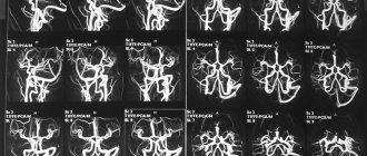
Dopplerography of extracranial vessels begins with examination of the right lower segment of the carotid artery. The sensor is then moved up the neck. Next, the color mode is activated, which makes it possible to identify a number of pathologies: deformations of the walls of blood vessels, impaired blood flow in certain areas, etc.
How is an ultrasound examination of the veins of the lower extremities performed?
The leading role in the diagnosis of varicose veins of the lower extremities today is occupied by ultrasound (ultrasound examination of the veins of the lower extremities). And there are many reasons for this: firstly , this procedure does not require any special preparation of the patient, secondly, it is absolutely painless, therefore does not require anesthesia, thirdly , the speed of the result obtained, and finally , fourthly , efficiency and accuracy this result!
Ultrasound diagnosis of varicose veins is carried out by a phlebologist, ultrasound doctor, Ph.D. Raskin V.V.
What can she show?
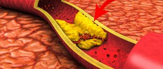
Duplex scanning allows you to determine areas of narrowing of the lumen of blood vessels, the direction of blood flow, its speed, as well as the condition of the vascular walls. This study can show the location of cholesterol plaques and the presence of blood clots, which is very important when diagnosing atherosclerosis.
A decrease in the elasticity of the walls of the arteries, as well as their thickening, indicate that the patient has hypertension (high blood pressure). If a change in blood flow is diagnosed, this indicates the presence of obstacles. For example, an aneurysm is a sac-like protrusion of a vessel.
What pathologies can be diagnosed?
Doppler ultrasound has certain limitations in diagnosing diseases. For example, she will not be able to study small veins - for this she needs to perform angiography. But there are a number of pathologies that can be identified:
- Atherosclerosis. By examining the head and neck using ultrasound, it is most often possible to diagnose atherosclerotic disorders. This condition is indicated by echogenicity, the presence of plaques, and thickening of the vascular walls. Moreover, if the lumen of the artery narrows by more than 20%, then the patient is diagnosed with “stenosis.”
- Thrombosis. Thrombosis is characterized by blocking of the arterial lumen by a blood clot. On Doppler ultrasound, inflammation of the vessel wall with a homogeneous formation inside is clearly visible.
- Compression of blood vessels. The examination allows you to see areas where blood flow is impaired due to the impact of swollen tissue on the channels. This allows you to diagnose cervical osteochondrosis and identify problems with blood circulation after injuries.
In addition, these ultrasound examinations can detect a number of other diseases, including vasculitis, post-traumatic dissections of the arterial walls and other pathologies.
Doppler ultrasound is a safe diagnostic method that can be performed in any case if there is a medical need for it. Timely implementation of the procedure allows you to identify the disease at an early stage of its development.
Price list for ultrasound examination of vessels
| № | Name of service | price, rub. |
| 1 | Color duplex scanning of the veins of the lower extremities/upper extremities. | 2 100 |
| 2 | Color duplex scanning of the arteries of the lower extremities / upper extremities. | 2 100 |
| 3 | Ultrasound color duplex scanning of the vessels of the lower extremities (veins and arteries). (UZDG, UZDS, UZAS, DS of vessels...) | 3 200 |
| 4 | Triplex scanning of the vessels of the upper extremities (veins and arteries). | 3 200 |
| 5 | Duplex scanning of the brachiocephalic vessels of the neck for children over 1 year old and adults. (BCA of veins and arteries, USDG of extracranial, extracranial parts of the brain). | 2 100 |
| 6 | Transcranial color duplex scanning of brain vessels. TCD for children over 1 year old and adults. Intracranial examination of the vessels of the head. | 2 500 |
| 7 | Duplex scanning of the vessels of the brain and neck with functional tests for children from 1 year and adults. | 3 200 |
| 8 | Renal vessels and abdominal aorta (duplex scanning of veins and arteries). | 1 800 |
| 9 | Triplex scanning of the abdominal aorta and inferior vena cava | 1 500 |
| 10 | Ultrasound of the pelvic vessels (Doppler study of blood flow in the uterus and appendages). Transvaginally. | 1 700 |
| 11 | Neurosonography with Doppler analysis. NSG or ultrasound of the brain for infants. | 1 500 |
| 12 | Ultrasound video recording on DVD. | 500 |
| 13 | You can order duplex scanning of blood vessels and other studies at home, see the page “Ultrasound at home” |