Benefits of MRI
Magnetic tomography allows you to obtain a three-dimensional image of the areas under study in three projections. During the procedure, the device takes many slices, the thickness of which can be set individually and is usually 2-4 mm.
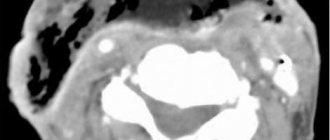
Images taken using a tomograph
Obtaining a large number of sections allows you to examine the entire organ and detect even the slightest abnormalities and pathologies.
Who is decoding the images?
The magnetic resonance imaging procedure makes it possible to obtain results both in the form of conventional images and in the form of three-dimensional images. MRI results can also be recorded on various removable media (disk, flash drive).
Typically, the patient receives MRI results in the form of images. The doctor who does the MRI is called a radiologist. Next, the patient turns to a radiologist for a complete interpretation of the images. A radiologist interprets images obtained using MRI, CT and X-rays. A typical decryption takes no more than 3 days, this duration is explained by the fact that:
- the doctor needs to study all the patient’s documents provided;
- analyze the obtained MRI results with the patient’s documents;
- digitize X-ray film into images;
- describe the pictures;
- give a conclusion.
The attending physician gives a full explanation of the images to the patient. In private clinics, interpretation of MRI results occurs a little faster; usually the interpretation results are ready by the end of the day.
The procedure helps to accurately determine a person's diagnosis. However, finding a good radiologist to interpret is quite difficult.
How MRI images are interpreted:
- Before decoding begins, the images are placed in front of a light source.
- The doctor determines abnormalities in the image by comparing the resulting image with images in the anatomical reference book.
- Records deviations from the norm (size, color, shape).
- The dark areas in the image are lesions with fluid inside, and the light areas are often tumors.
What types of tomographs are there?
Modern magnetic tomographs are available in various variations with a wide variety of characteristics.
All tomographic devices are divided into:
- open;
- closed.
Despite the fact that conducting a study in an open tomograph is usually considered more comfortable for the patient, closed devices have greater power and detail. If the patient does not have a strong fear of closed spaces and does not have weight restrictions, it is recommended to conduct the study in a closed-type apparatus.
Tomographs are also divided according to the strength of the magnetic field radiation, the unit of measurement of which is called Tesla. Magnetic tomographs can be:
- low-floor – power up to 1.0 T;
- high-field – radiation intensity above 1.0 T.
Low-field tomographs do not provide a clear and detailed picture. A study using a high-field tomograph will allow you to examine the diagnosed area with the highest accuracy.
Modern high-field tomograph
The DiMagnit clinic has a closed-type tomograph from Philips, whose power is 1.5 Tesla. Using the device, it is possible to obtain images of the highest quality and detail.
Should you be afraid of the procedure?
Some patients are worried before the test. But their fears are in vain - magnetic resonance imaging is absolutely painless, and the effect of magnetic radiation on the body is safe.
Unlike other types of radiation diagnostics, MRI does not use ionizing radiation. The magnetic field does not have a carcinogenic or mutagenic effect on the cells of the body. Magnetic resonance scanning can be performed as often as needed.
Whole body MRI: when is it necessary?
“Look from head to toe.” How would you imagine such a survey?
As it turned out, this is not science fiction, but a completely feasible task for a method such as magnetic resonance imaging. Now you can do such a check-up - scan your body “from head to toe” in many diagnostic scanners.
What can a full body MRI show? We asked the chief physician of MRT Expert Elets LLC, Vladimir Vladimirovich Tulinov, to answer this and other questions.
- Vladimir Vladimirovich, let’s first remind our readers what the MRI method is and what principle is it based on?
Magnetic resonance imaging is a high-tech diagnostic method based on the magnetic resonance method. What is it essentially? Every cell in the body contains hydrogen atoms. When a body is placed in a magnetic field, the nuclei of these atoms vibrate, generating signals with certain characteristics. These signals are converted by the MRI device and software into images that the doctor sees. Since the number of hydrogen atoms in tissues is different, then, accordingly, the images of tissues and organs will be visually different.
- Is it true that much more often patients come to have an MRI examination of one area of the body? If so, in what cases might whole-body MRI be appropriate?
Statistically, yes, people are more likely to come in to have one area examined. This can be explained by the fact that often a person comes to us at the direction of the attending physician, who may already have some diagnostic assumptions and they may be associated with specific organs and systems.
If a person’s complaints are quite “blurry”, he cannot say exactly what and where exactly is bothering him or hurting him, then in such cases, an MRI of the whole body can help bring clarity.
In addition, such an extended study may be appropriate for systemic diseases (for example, ankylosing spondylitis), when several anatomical areas are affected, or when searching for tumor metastases, when it is not known exactly in which organs they are present.
In addition to the actual medical indications, full MRI can be used as a preventive examination to detect diseases in early, often asymptomatic stages. The advantage of the method is that it allows you to look at all internal organs and systems in one diagnostic study.
-What specific areas are examined in a whole body MRI?
This is the brain with its arteries, all parts of the spine (cervical, thoracic, lumbar and sacrococcygeal), soft tissues of the neck, organs of the abdominal cavity, retroperitoneal space and pelvis.
-Magnetic resonance imaging does not cause radiation exposure and is safe for the body. Whole body MRI is no exception? Or are there any differences in this regard from research in one area?
No, there are no differences in terms of security. There is no radiation exposure to the patient’s body undergoing such a check-up.
- Vladimir Vladimirovich, please tell us how to properly prepare for this study?
There are no fundamental features. However, given that with a whole-body MRI, the doctor examines, among other things, the abdominal cavity and pelvis, recommendations for preparing for the study, in particular, of these areas are followed.
They are as follows:
- last meal - 4 hours before the study;
— 2 hours before the test, no-spa or drotaverine is prescribed (the dosage is selected by the doctor);
- 30 minutes before the test or immediately before it, you need to drink a glass of clean still water. This can, in addition, be done by agreement with the doctor during a “break” if the procedure is done with a break.
- How and for how long is a full body MRI performed?
It is no different from MRI of one area, i.e. The patient is placed on the machine table and the examination is performed.
The procedure itself lasts 1 hour 45 minutes. At the same time, you need to understand that it is not at all necessary to lie down all this time without a break, since physically it is quite difficult. In this regard, the procedure can be carried out with a break; technically this is quite feasible.
-Are there any special contraindications to whole body MRI? Or are these just well-known contraindications to MRI in general?
There are few contraindications, I will name some of them:
Among the absolute ones:
— artificial pacemaker, pacemaker, neurostimulator;
— Ilizarov apparatus;
— prosthetic heart valves, with the exception of prostheses permitted for MRI diagnostics;
- insulin pumps.
Relative:
- fear of enclosed spaces (true claustrophobia, much more common is far-fetched - a person believes that he has such difficulties);
- first trimester of pregnancy;
- psychomotor agitation;
— the need for continuous monitoring of vital physiological parameters (for example, blood pressure, respiratory rate, electrocardiogram) and/or continuous resuscitation measures.
I would especially like to say about the relevance of the fear of closed spaces (claustrophobia), since the examination is quite lengthy. If a person has such a problem, it is recommended to consult with his attending physician to decide on the possibility of examination. But in many cases of fear before an MRI examination, we are always ready to help.
For reference:
Tulinov Vladimir Vladimirovich
Graduate of the Faculty of Medicine of the Ryazan State Medical University. Academician I.P. Pavlova 2010.
In 2011, he completed an internship at Kursk State Medical University with a degree in Radiology.
Practicing MRI diagnostics doctor, member of the Association of MRI Diagnostics Doctors.
Currently, he is the director and chief physician of MRI Expert Yelets.
The difference between MRI and CT and ultrasound
Magnetic resonance diagnostics has a number of advantages compared to ultrasound and computed tomography.
Ultrasound examination allows you to obtain a two-dimensional image of the area under study, but does not allow you to see a three-dimensional image of soft structures.
Computed tomography can be compared to MRI in terms of image clarity, but has a number of serious contraindications. CT is more often used to visualize hollow organs and bone structures, while MRI is much more effective at visualizing soft tissue.
If you decide to go to a private clinic
If the patient decides to go to a private clinic for magnetic resonance imaging, he will need to get a doctor’s referral. The following must be indicated in the direction:
- survey authority;
- presence of contraindications;
- performing MRI with or without contrast agent;
- results of a preliminary examination of the patient.
It is impossible to undergo the procedure even in private clinics without a referral. If the referral is received and there are no contraindications, then you will have to pay for an MRI in a private clinic. There are advantages to private clinics:
- no long queues;
- the possibility of performing MRI at night (there is a 50% discount for such a procedure at night).
In a private clinic, the procedure is performed by a doctor with a higher medical education (specialists of the highest categories, doctors of medical sciences). Work experience is at least 5 years, a relevant certificate is required. The medical center has documents and permits. It is advisable to first find out patient reviews about the clinic.
What does an MRI show?
Magnetic resonance imaging is successfully used to diagnose diseases:
- thyroid gland;
- liver;
- gallbladder and ducts;
- pancreas;
- kidney;
- spleen;
- joints;
- spinal cord;
- vessels of the head, neck, abdominal region;
- pelvic organs;
- soft tissues;
- etc.
All of the above anatomical structures are perfectly visualized on MRI images. The diagnostic results make it possible to accurately identify abnormalities in the functioning of the organs being examined.
What is a head MRI?
Magnetic resonance imaging (MRI) is a non-invasive test that helps doctors diagnose and treat diseases.
MRI involves the use of powerful magnetic fields, high-frequency pulses and a computer system that allows you to obtain a detailed image of organs: soft tissues, bones and almost all structures inside the human body. The resulting images can be studied on a computer monitor, transmitted electronically, printed or copied to storage media. Ionizing (X-ray) radiation is not used in MRI.
Detailed images allow doctors to accurately assess the condition of various organs and systems and identify certain diseases that may be indistinguishable when using other examination methods, for example, X-ray, ultrasound or computed tomography. In modern clinical practice, MRI is the most sensitive method for imaging organs and tissues of the head, especially the brain.
Up
In what cases is MRI prescribed?
The wide capabilities of magnetic resonance diagnostics make its use indispensable in the following cases:
- the need for a primary diagnosis;
- conducting a comprehensive survey;
- preparation for surgery;
- monitoring the effectiveness of the therapy and treatment methods used.
In each individual case, the choice of diagnostic technique is made by the attending physician. Magnetic resonance imaging is more often used than other methods to detect diseases and injuries of soft tissues.
The MRI technique is indispensable for diagnosis:
- Neoplasms.
Magnetic resonance scanning can clearly identify the boundaries and size of the tumor and the extent of its invasion into soft tissue. No other radiation diagnostic technique is capable of providing such a clear and detailed picture of diseases.
MRI also makes it possible to determine the nature of the tumor with a high degree of probability. Malignant neoplasms have unclear boundaries and grow into surrounding tissues. Benign neoplasms, as a rule, are clearly differentiated from healthy tissues.
- Brain diseases.
The greater accuracy of magnetic resonance diagnostics makes it possible to visualize such small anatomical structures as the pituitary gland and the sella turcica. Also, MRI with contrast of the brain shows high efficiency for diagnosing demyelinating diseases (multiple sclerosis, Parkinson's disease, etc.), as it allows one to clearly see the structure of altered nerve tissues.
Brain images obtained with contrast-enhanced MRI of the brain are particularly clear because magnetic waves are difficult to image solid anatomical structures and there are no artifacts from the skull in the brain images.
- Diseases of the intervertebral discs.
MRI images of the spine
Magnetic resonance imaging is the only diagnostic method that allows you to see intervertebral discs. Even modern diagnostic methods, such as computed tomography, allow you to see only the space between the vertebrae, while MRI gives a complete picture of the condition of the discs, the possible presence of hernias and protrusions.
The use of MRI is not limited only to the above diseases, but is used when it is necessary to identify and monitor a wide range of pathologies, congenital developmental anomalies, consequences of injuries and previous surgical interventions.
If an MRI of the brain showed...
There is probably no person in the world who has never had a headache in his life. “Can you do an MRI of the brain?” is one of the most common questions received by our diagnostic centers. But even more questions arise for those who have undergone the examination. Serious diagnoses are not always hidden behind the terms used by MRI diagnostic doctors when describing images. That’s why we always insist: don’t take your fears with you. We are always there to help you figure it out and refer, if necessary, to a doctor of the required specialization; explain what is hidden behind this or that, sometimes frightening name.
In this article, we have collected questions regarding magnetic resonance imaging of the brain and its vessels that patients ask us most often.
Answered by Mikhail Kosygin – radiologist at MRT Expert Lipetsk LLC
— What does a brain MRI diagnose?
MRI of the brain allows us to identify space-occupying formations of various origins, congenital developmental anomalies, ischemic and hemorrhagic strokes, hemorrhages in the area of the meninges, inflammatory and demyelinating processes, and much more.
We talked about the topic of MRI of the brain on the pages of our website earlier. They raised the question “why undergo an MRI of the brain when nothing hurts” and talked about “how MRI will help a patient with a traumatic brain injury and its consequences.”
— Will MRI help identify the causes of high blood pressure?
If the increase in pressure is a consequence of arterial hypertension of idiopathic (i.e., unclear) origin, then this cannot be determined during an MRI study. But there is symptomatic arterial hypertension, which is not an independent disease, but accompanies, as a symptom, another disease. Based on their origin, such diseases are divided into four groups: renal, endocrine, neurogenic and hemodynamic. Most of them can be identified on MRI and it can be assumed that they are the cause of the patient’s increased blood pressure.
— What is the quadrigeminal plate in the brain?
The quadrigeminal midbrain is a plate-shaped formation located in the roof of the midbrain. The quadrigeminal tubercles are independent structures. In this case, the superior colliculi function as subcortical formations and work as centers of the visual analyzer. The lower tubercles serve as subcortical formations, working as centers of auditory analyzers.
— What to do if an MRI showed the presence of an angioma?
Brain angioma is a vascular formation of benign origin and looks like a tangled tangle of blood vessels. Such formations come in various sizes and degrees of fullness.
If MRI reveals an angioma, we recommend monitoring the formation once a year in order to prevent the occurrence of extremely rare, but still occurring complications in the form of hemorrhages.
Answered by Alexander Samaev, radiologist at MRI Expert Lipetsk LLC
— What is the pathology of the intracranial arteries of the brain?
The arteries supplying blood to the brain have cervical (extracranial) and intracranial (intracranial) sections. Consequently, this is a pathology of the intracranial sections of the arteries supplying blood to the brain.
— Should there be focal changes on MRI in ischemic stroke?
Ischemic stroke can manifest itself in both focal (up to 1.0 cm) and focal (more than 1.0 cm) changes and even larger areas. Therefore, the presence of focal changes on MRI depends on the caliber of the affected vessels (the main large arteries of the brain or small capillaries are affected).
— What is caudal displacement of the cerebellar tonsils?
Normally, the lower part of the cerebellum, which is the tonsils, is located at the level of or above the foramen magnum (i.e., the hole through which the spinal cord exits).
Caudal displacement of the cerebellar tonsils is a displacement below the level of this foramen, into the lumen of the spinal canal. The condition is diagnosed by space-occupying formations in the brain and indicates that there is not enough space in the skull.
— What are the dangers of a pineal gland cyst?
We find this formation quite often during MRI of the brain and classify it as an accidental finding. That is, the patient came for the study because he was bothered by some symptoms associated with this disease. A pineal gland cyst most often does not manifest itself with any symptoms.
However, if it begins to grow rapidly, this can lead to disruption of liquor dynamics and be the cause of internal hydrocephalus.
Therefore, if the diagnosis reveals a pineal gland cyst, then the recommendation to monitor it is justified. There is another reason for constant dynamics in this pathology - in the initial stages, tumors of the pineal gland can look the same as a cyst.
Answered by Vladimir Mikolaichuk – radiologist at MRI Expert Lipetsk LLC
— In what cases is it necessary to study the veins of the brain?
Examination of the veins (or MR venography) is necessary if thrombosis of the sinuses (“large veins”) of the brain is suspected; for the diagnosis of vascular malformations (so-called structural and developmental anomalies); when assessing the degree of germination of a brain tumor into a venous vessel.
In order to assess the nature of the changes, there is a need to compare MR venography data with the picture revealed during an MRI study of the structure of the brain.
For example, the narrowness of the sinus of congenital origin and its thrombosis may look almost identical when performing MR venography. But with an MRI examination of the brain, we will see a pathological signal from a thrombosed vessel.
— Is it possible to detect meningitis using MRI?
Yes, you can. If a patient has meningitis, an MRI of the brain with a contrast agent will identify structural changes in the membranes of the brain, accompanied by intense accumulation of contrast.
But we see a similar picture in other diseases. For example, with sarcoidosis, cancer, intracranial hypotension and a number of other pathological conditions, therefore, an important factor in making a diagnosis will be a comparison of MRI diagnostic data with the general clinical picture, anamnesis and, of course, laboratory test data.
***
Especially for our patients, the #GKExpert Encyclopedia was created on the company’s official public pages on social networks Vkontakte, Facebook, Odnoklassniki, within which a team of doctors from the medical group answers questions from our patients. You don't know what's wrong with you? us.
When to Apply Contrast
Magnetic resonance diagnostics can provide a very high degree of clarity of the resulting images. In most cases, the use of contrast is not required.
But when it comes to diagnosing tumors and small anatomical structures, a contrast agent can still be used.
The staining agents are made from the rare earth metal gadolinium and are injected intravenously into the patient during an MRI.
MRI contrast agents are much better tolerated than CT contrast agents. This makes the use of the dye safe even for patients with kidney pathology and does not require a preliminary test for creatinine, which is necessary for CT diagnostics with contrast.
MRI with contrast is used in the following cases:
- suspicion of a neoplasm;
- the need for differential diagnosis of a malignant tumor;
- study of the pituitary gland;
- the need to diagnose demyelinating diseases.
The use of contrast allows one to obtain a comprehensive picture of the disease, its course and the effectiveness of the therapy used.
In what areas is head MRI used?
MRI of the head is used to detect the following conditions and diseases:
- Brain tumors
- Brain development disorders
- Vascular abnormalities of the brain, such as aneurysms
- Eye and inner ear diseases
- Stroke
- For injuries (as determined by the doctor)
- Pituitary gland diseases
- Some chronic nervous system diseases, such as multiple sclerosis
- Determining the cause of headaches
In addition, MRI is used to detect pathological changes in brain tissue in patients with dementia.
Up
Contraindications for MRI
Despite the fact that magnetic resonance diagnostics is a safe technique, the study has a number of absolute contraindications, in the presence of which diagnostics are prohibited:
- presence of a pacemaker, neurostimulator, insulin pump;
- vascular clips on the arteries of the brain;
- the patient’s inability to maintain a stationary position for various reasons;
- early childhood up to 5 years;
- the patient’s weight is more than 130 kg and body girth is more than 150 cm;
- first trimester of pregnancy;
There are also a number of conditions in which MRI examination is carried out with caution:
- severe pain syndrome, in which it is difficult for patients to remain in a motionless position for a long time;
- fear of confined spaces;
- psychical deviations;
- second and third trimester of pregnancy.
The presence of various prostheses and implants in the patient's body may be a contraindication to MRI if they are made of metals sensitive to magnetic radiation. Modern medical devices are most often made of titanium and other materials that are inert to the effects of magnetic fields. Their presence in the body does not interfere with MRI.
How is the reception carried out?
Before the procedure, the patient is in the waiting room. He removes all metal things, fills out documentation, communicates with the doctor. The doctor establishes contraindications for the procedure, measures the patient’s body weight and height in order to accurately adjust the tomograph.
Information to be established before the procedure:
- Do you have a pacemaker?
- Is a metal hemostatic clamp installed on the cerebral vessel?
- The presence of hearing devices, various prostheses (dental, joint, heart valves), braces, metal crowns, metal structures for stabilizing the spinal column, springs that dilate coronary vessels, wire sutures on the sternum, devices for subcutaneous administration of insulin through a catheter are checked.
- Can a person wear a wig, will he be able to lie on his back, will the procedure cause discomfort?
- Is the study patient pregnant?
- Was contrast ever introduced?
- Was there any surgery on the ears, eyes, or brain, and what operations were performed in the last six months?
- Are there blood pathologies?
- Are there any bullets in the body?
- Are there any piercings or tattoos on your body?
- Are there metal foreign bodies in the eyes?
- What substances lead to allergic reactions?
By the way, tattoos heat up during examination, since they have metal connections.
Before diagnosis, an X-ray must be taken to identify metal objects inside the body; if they are found, an MRI is not performed.
Before conducting the study, the specialist is provided with the test results:
- General, biochemical blood test.
- Blood test for clotting.
- Rheumatic tests.
- Blood test for hormones, infections, tumor markers.
- ECG (electrocardiography).
- EEG (electroencephalography).
- Ultrasound (ultrasound) and other examinations as indicated.
to contents ^
How is an MRI performed?
Before the procedure, the radiologist questions the patient about the presence of contraindications to the study. The patient is asked to remove any metal accessories, including clothing with metal fittings, and lie down on a couch, which is then placed in the CT scanner tube.
During the diagnosis, it is strictly forbidden to move, as this may affect the clarity of the resulting images.
Scanning procedure
High-field tomographs produce a fairly high level of noise, which can cause some discomfort to patients. The medical department will provide you with headphones that will play pleasant music, drowning out the sounds of the operating device.
The tomograph scans the patient’s body in different projections and instantly transmits the images to the computer screen. The interpretation of the results by the research physician begins even before the procedure is completed.
Getting Results
Scan results are available immediately after the end of the study. Many of the images obtained are carefully examined by a radiologist, and a detailed report is drawn up describing both the normal anatomy of the area under study and possible abnormalities and pathologies.
15-30 minutes after the procedure, the patient is given a written report and a computer disk with the resulting images.
Magnetic resonance imaging is a modern, safe type of radiation diagnostics that allows you to obtain accurate and quick results and thoroughly examine the area under study. MRI helps to identify many diseases and abnormalities even at the initial stages of their development.
| MRI of the pancreas in Rostov-on-Don |
| MRI of the abdomen with contrast |
| MRI of the spine, what does it show? |
| MRI of ureters |
| MRI of the abdominal cavity, which organs are checked? |
| What does an MRI of the knee show? |
Which doctor issues a referral for examination and how to get it for free
To make a diagnosis, magnetic resonance imaging is performed. MRI gives accurate readings. In addition to the advantages of the examination, a significant disadvantage is the high price of MRI.
A doctor may prescribe a procedure if he suspects the presence of neoplasms, disruptions in the nervous system, or the presence of pathologies. You can get this examination for free using a medical policy, since MRI is included in the list of free health insurance services in Russia. The patient receives a referral from his/her attending physician. You can get an MRI for free in government institutions. Private medical clinics do not provide this service.
Not every insurance policy includes a free MRI. The high price is the reason that not all insurance companies add MRI to their list of free procedures. If insurance still requires payment for diagnostics, then there is no need to rush to rejoice. Usually in any state clinic there are several free MRI procedures, and the lucky ones are carefully selected. Everyone else will still have to pay for the procedure or use an alternative research option (X-ray, ultrasound).
Patient steps to take a free MRI:
- First, ask the insurance company for a list of free services provided. This can be done by calling the insurance company's hotline.
- Get a referral for an MRI from your doctor.
- Have the following documents:
- doctor's referral;
- passport;
- SNILS;
- insurance policy.
If it is not possible to get a free MRI, then they undergo a free examination using CT, ultrasound or X-ray. When a patient chooses to be examined using an MRI, he or she will be on a waiting list for more than one month. By law, the wait is no more than 30 days. In addition to the regular queue, there is an “urgent” queue. The wait there is no more than 2-3 days. The urgent queue includes patients for whom every day of waiting is important for the disease. Such cases include:
- tumors and the presence of metastases (relapses);
- stroke;
- heart attack;
- pathologies of cerebral vessels;
- heart diseases;
- pathologies of the heart, liver, kidneys, etc.
- prescription of treatment after surgery.
A referral for an MRI can only be issued by the attending physician or local doctor. More precisely, MRI can only be PET. Positron emission tomography is a study of the human body using radionuclides.