Author: TainT
October 27, 2021 12:14
Community: Science
Tags: macro photography science neuroscience photo it's interesting
6379
22
The brain is a whole universe. What is there, what is being done and how it all works today we can see with the help of a microscope and macro photography. Let's lift the curtain and take a look into this wonderful world.
0
See all photos in the gallery
"Snail" of a newborn rat
0
This stunning image, which took eighth place in the 2021 Nikon Small World competition, shows us the main organ through which we hear: the snail. Hair cells are shown in green, the deviation of which triggers a nerve impulse that ultimately arrives in the auditory cortex. Red - nerve cells of the cochlea, “collecting” signals from hair cells.
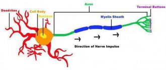
Real and idealized neurons
To begin with, we will get acquainted with a generalized neuron , which, however, does not always satisfactorily reflect the real state of affairs. In fact, there is a huge variety of neurons, neuroactive substances, and potential information processing mechanisms.
Our first simplification will be to consider only the integrative neuron . This classical neuron receives signals from other nerve cells using depolarizing potentials arising on the dendrites. If the total dendrite potential exceeds -50 mV, then a rapidly propagating action potential wave passes along the neuron's axon.
A classical neuron transmits a signal by conducting an action potential wave along the axon from the cell body to the axon terminals. At the terminals, the neurotransmitter is released into the synaptic cleft. The neurotransmitter causes depolarization of the postsynaptic membrane .
Neuroscience focuses on the connections and interactions of neurons. It is convenient to start considering such connections with a generalized neuron.
Despite the amazing complexity of the brain, the ideas that help us study it are relatively simple. But simple components are capable of forming large systems. There are a huge number of ways to combine neurons into cascades, circuits and networks.
Signal transmission along the axon, (a) Signal transmission along the axon is ensured by the passage of ions through adjacent sections of the membrane. In the figure, area 2 is depolarizing, while area 3 has already generated an action potential and is currently hyperpolarized. The action potential will go further due to the depolarization of area 1.
(b) Myelinated axons are wrapped in specialized Schwann cells. The axon membrane is open only at the nodes of Ranvier.
(c) The action potential of the myelinated axon is regenerated at the nodes. Myelinated axons have an increased conduction velocity compared to unmyelinated axons.
Dividing neuronal stem cells
0
The skin, liver, heart, kidneys, lungs and blood can form new cells to replace damaged ones. Until recently, experts believed that this ability to regenerate did not extend to the central nervous system, consisting of the brain and spinal cord. However, over the past five years, neuroscientists have discovered that the brain does change throughout life: new cells are formed to cope with emerging difficulties. This plasticity helps the brain recover from injury or disease, increasing its potential.
#gallery | Are your neurons just as beautiful?
When Greg Dunn received his PhD in neuroscience from Penn in 2011, he bought himself a sensory deprivation chamber as a gift. This is a tank into which light, sounds and smells do not enter. Placed in a solution of Epsom salts in a tank, a person is in a state of weightlessness. So, the newly minted doctor moved from the world of science to the world of creativity and meditation.
He now works as an artist and lives in Philadelphia. Dunn says that when he was a student, he was greatly inspired by the beauty of neurons working as spots of color. The Golgi spot, for example, consists of one or two black neurons on a gold background.
“They embody the same zen that is close to me,” Dunn says.
But don’t rush to classify a neuroscientist as a crazed mystic.
What he once saw under a microscope reminded him of the laconic elegance of papyrus and other forms of Asian painting. So he started drawing neurons in a similar style. His own developed paint application techniques involved blowing drops of ink onto a surface. The ink spilled out the way neurons grow, Dunn notes, drawn by random forces and around microscopic obstacles.
“I like the concept of drawing on similar forces to create,” says the artist.
Dunn has sold his work to research laboratories and hospitals, and his paintings are popular among neurologists, neuropathologists and others interested in studying the brain, as well as among people with neurodegenerative diseases.
“I think by looking at the images, people begin to understand the essence of things that concern them.”
By the way, you might be interested in reading about the ten most common myths about the human brain.
The images in the gallery are created by the artist's imagination, but strengthened by his knowledge of brain anatomy.
“One of my frustrations in graduate school was the need for absolute adherence to truth, principles and facts,” Dunn laments. “I am inspired by anatomy, but I am not a slave to it.”
So in the image above:
Bark in metallic pastel
This painting shows a cross-section of the motor cortex, an area that is involved in planning movements. You should be familiar with this famous layer of V pyramidal neurons.
Gold, palladium, mica, enamel and paint on an aluminum panel.
Cerebellum
This is the cerebellum, the area of the brain responsible for movement, balance and motor memory.
Gold, paint and enamel on an aluminum panel.
Maki-e neurons (蒔絵)
Dunn notes that there is no real biological description for this picture.
It is simply subjective representative physical anatomy. Gold and paint on an aluminum panel.
Developing cerebral cortex
This image shows the growing cortex at 15 weeks gestation.
Gold, enamel, metal powder and paint on an aluminum panel.
Retina
This sumi-e style painting depicts the layered structure of the retina.
Visual processing begins with the photoreceptors (top) and progresses in the ganglia (bottom) before the information is sent to the thalamus and visual cortex for further processing. Ink on silk brocade.
Bark
This picture shows the layered structure of the cerebral cortex, where sensory and motor information is processed.
Enamel on composite gold leaf.
Hippocampus
And here is a cross-section of the hippocampus, an area of the brain that is involved in learning and memory.
Enamel on composite gold and aluminum sheet.
NG2+ cells
NG2+ (yellow gold) cells are special glia, a type of cell that helps neuronal synapses regulate synaptic transmission.
Here they are shown among the pyramidal cells in the hippocampus (white gold). Gold and paint on a metal panel.
Hypothalamus
This is a compressed cross-section of the hypothalamus, a region of the brain responsible for appetite, temperature, circadian rhythm and sleep.
Gold and paint on stainless steel.
pyramidal cells
These are neurons that integrate information received from their dendrites, which branch at the bottom of the cell.
Pyramidal cells transmit information to other neurons through the axon, a large branch that extends upward. Enamel on composite gold leaf.
Source: wired.com
Immune cells heal brain after bleeding
0
In a new paper in Nature Communications, Houston neuroscientists show how immune cells called neutrophils can repair the brain after a hemorrhagic stroke. Neutrophils are known as the "infantry" in the body's war against infection. It turns out that they have another function: a new study shows that these immune cells may play a crucial role in protecting the brain from stroke, and can also be used in the treatment of intracerebral hemorrhages.
Once again about everything
At the end of the twentieth century, an approach to studying the brain emerged that simultaneously considers two important characteristics: how much a neuron (or neural network, or synapse) encodes and transmits useful information and how much energy it spends [6], [18], [19] . Their ratio is a kind of criterion for the energy efficiency of neurons, neural networks and synapses.
The use of this criterion in computational neuroscience has provided a significant increase in knowledge regarding the role of certain phenomena and processes [6], [18–20], [26], [30], [43], [55]. In particular, the low probability of neurotransmitter release at the synapse [18], [19], a certain balance between inhibition and excitation of a neuron [55], and the selection of only a certain type of incoming information due to a certain combination of receptors [50] - all this helps to save valuable energy resources.
Moreover, the very determination of the energy consumption of signaling processes (for example, generation, conduction of action potentials, synaptic transmission) makes it possible to find out which of them will suffer first in case of pathological disruption of nutrient delivery [10], [25], [56]. Since synapses require the most energy to operate, they are the first to fail in pathologies such as ischemia, Alzheimer’s and Huntington’s diseases [19], [25]. In a similar way, determining the energy consumption of different types of neurons helps to determine which of them will die before others in the event of pathology. For example, with the same ischemia, the interneurons of the cortex will fail first [9], [16]. Due to their intense metabolism, these same neurons are the most vulnerable cells during aging, Alzheimer’s disease, and schizophrenia [16].
In general, the approach to determining energetically efficient mechanisms of brain function is a powerful direction for the development of both fundamental neuroscience and its medical aspects [5], [14], [16], [20], [26], [55], [64]. ].
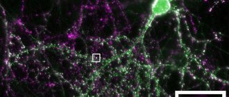
Cerebellar granule cells
0
Here is a snapshot of the cells of the granular layer of the cerebellum. Granule cells are small neurons, about 10 micrometers in diameter.
0
In the vast majority of cases, the cause of vision loss is the death of a large number of photoreceptor cells in the retina, which convert light into electrical nerve signals. Retinal cells that are insensitive to photons remain intact.
Pathways
The nervous system has its spheres of influence throughout the body. With the help of conductive fibers, nervous regulation of systems, organs and tissues is carried out. The brain, thanks to a wide system of pathways, completely controls the anatomical and functional state of every structure of the body. Kidneys, liver, stomach, muscles and others - all this is inspected by the brain, carefully and painstakingly coordinating and regulating every millimeter of tissue. And in case of failure, it corrects and selects an appropriate model of behavior. Thus, thanks to the pathways, the human body is characterized by autonomy, self-regulation and adaptability to the external environment.
Brain pathways
A pathway is a collection of nerve cells whose function is to exchange information between different parts of the body.
- Association nerve fibers. These cells connect various nerve centers located in the same hemisphere.
- Commissural fibers. This group is responsible for the exchange of information between similar centers of the brain.
- Projection nerve fibers. This category of fibers articulates the brain with the spinal cord.
- Exteroceptive pathways. They carry electrical impulses from the skin and other sensory organs to the spinal cord.
- Proprioceptive. This group of pathways carries signals from tendons, muscles, ligaments and joints.
- Interoceptive pathways. The fibers of this tract originate from internal organs, blood vessels and intestinal mesenteries.