Magnetic resonance imaging combines high information content, safety and non-invasiveness. MRI of cerebral vessels is the most optimal radiation method for studying the vascular system. The contrast agent used during the study practically does not cause allergic reactions and is well tolerated.
In the absence of contraindications, the procedure is not associated with risks to the patient’s health. Electromagnetic radiation, which provides reading of information about the vascular system, has indicators that do not exceed standard values for humans in any research mode.
MRI of cerebral vessels provides detailed information for making a diagnosis in complex cases and with a confusing clinical picture - many diseases have similar symptoms. The blood supply to the head and brain comes from branches of the aorta passing through the neck. At this level, diseases or damage to the vascular system are often noted that affect cerebral circulation. To accurately determine the cause of painful conditions, an MRI of the vessels and arteries of the neck is performed in conjunction with a study of the brain vessels.
The examination allows you to study the condition of the main groups of cerebral vessels:
- internal carotid artery and its branches (ophthalmic, anterior and middle cerebral arteries);
- vertebral arteries located in the cervical spine, basilar, posterior cerebral arteries;
- Circle of Willis - a system of connected arteries at the base of the brain that increases the reliability of the blood supply to the brain;
- veins of the brain and venous sinuses of the dura mater.
Carrying out MRI of the vessels of the head and neck with a contrast agent allows you to visualize the smallest vessels and determine disorders of microcirculation and metabolic processes.
When is the examination carried out?
Before undergoing an MRI of the head and neck vessels, it is advisable to consult a neurologist. The doctor will assess the patient’s symptoms and condition and refer him for the most appropriate study.
The examination is usually carried out if the patient observes the following phenomena for a long time:
- prolonged headaches of unknown cause that cannot be treated with known drugs;
- periodic dizziness, loss of consciousness, fainting, darkening of the eyes, difficulty in spatial orientation;
- unstable blood pressure;
- decreased vision;
- numbness of the arms, shoulder girdle;
- actively developing osteochondrosis of the cervical spine with corresponding symptoms;
- herniated disc in the cervical region;
- subluxation, instability of the cervical vertebrae;
- traumatic brain injury, spinal injury;
- suspicion of cancer or mass formations in the brain.
The value of MRI of the head and neck vessels lies in the possibility of making an early diagnosis. The examination helps to start therapy in a timely manner and prevent the progression of the disease. Having received the necessary treatment, the patient can undergo the procedure again an unlimited number of times without risk to health. The doctor monitors the effectiveness of therapy and, if necessary, makes adjustments or changes tactics.
MRI of cerebral vessels is actively used by neurosurgeons to plan operations. Information about the location and structural features of the vascular system allows you to avoid unforeseen situations and make precise interventions. MRI is important in the postoperative period to monitor recovery processes.
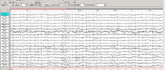
Rheoencephalography of cerebral vessels
REG (rheoencephalography) is a non-invasive method for diagnosing cerebral vessels, recording changes in the electrical resistance of vascular walls when a high-frequency pulse passes through them.
Provides an opportunity to check the vessels of the head, their reactivity, tone, elasticity, level of resistance of vascular tissue, possible blockage and characteristics of blood supply. REG is indicated for suspected diseases:
- vascular pathologies;
- circulatory disorders;
- atherosclerosis;
- stroke;
- vascular stenosis;
- clarification of the effect of drugs;
- traumatic brain injuries.
To perform rheoencephalography, rheographs with a number of channels from 2 to 6 are used, recording the corresponding number of vessels. The patient is sitting or lying down. Electrodes are installed on his head using rubber bands. A special paste can be applied underneath them to improve contact with the skin and reduce resistance. Electrodes are attached depending on the vessel being examined. So, if you need to check the internal carotid artery, the electrode is placed on the bridge of the nose and mastoid process.
Some electrodes send electrical signals, others receive them and transmit them to the rheograph. The latter processes and transmits to a monitor or print in the form of a graph - a rheoencephalogram. The procedure lasts several minutes.
During the examination, the patient may be given a small dose of nitroglycerin under the tongue, Papaverine, or asked to change body position or turn his head. These measures will allow you to see a change in tone and blood supply.
The advantage of rheoencephalography is its simplicity, safety for the patient, and the ability to monitor blood flow for a long time. Disadvantages include the inability to measure cerebral blood flow and determine the exact cause of circulatory disorders.
Before the study, you should not smoke, drink alcohol, coffee, or energy drinks. If the patient is taking medications, the doctor must be informed about this. Some may need to be stopped for a while.
Rheoencephalography is not prescribed for children under 7 years of age, as well as for wounds and injuries at the places where the electrodes are attached.
What will the examination show?
MRI of the vessels of the head and neck is performed in angiography mode - in this case, only areas of the vascular system are visible in the images, without surrounding tissues. The examination makes it possible not only to assess the condition of the arteries and veins, but also to determine the speed of blood flow in the vessels in real time. Based on the scanning results, a three-dimensional 3-D model of the area under study is constructed.
Signs of which diseases will be shown by MRI of cerebral vessels:
- stroke - hemorrhagic (leakage of blood from the vascular system into the brain tissue or meninges) or ischemic (cessation of blood supply to a part of the brain, more common);
- arterial aneurysm - pathological expansion and thinning of the vascular wall with the risk of rupture;
- arterial dissection as a result of congenital weakness of the arterial wall or damage by a pathological process;
- thrombosis, thromboembolism;
- inflammatory diseases of the vascular system – phlebitis, arthritis, systemic vasculitis;
- vascular tumors – hemangioma, hemangioblastoma, angioreticuloma;
- atherosclerosis;
- narrowing of the lumen of the vessel due to structural features or compression from the outside by a space-occupying formation;
- congenital developmental anomalies and structural features - absence, decrease or increase in diameter, bifurcation of some parts of the vascular system, arteriovenous malformation.
MRI of the vessels and arteries of the neck will show structural defects and pathological processes leading to impaired cerebral circulation:
- dissection of the carotid and vertebral arteries - damage to the artery wall as a result of injury, bending, with further formation of a hematoma between the walls of the vessel, narrowing of the lumen of the artery and disruption of the blood supply to the brain;
- pathological tortuosity of the carotid artery;
- developmental anomalies and acquired lesions of the vertebral artery;
- neoplasms;
- aneurysm of a section of the vascular system;
- pathologies in the area of the carotid sinus.
Methods for studying cerebral vessels
A specialist can make a reliable diagnosis using ultra-sensitive medical equipment that provides reliable data. A doctor’s mistake can cause drug therapy to be ineffective, and precious time will be lost.
- How to check the blood vessels of the brain, what methods are used
Before the manipulations, the blood is examined. This is necessary to identify pathologies that affect its coagulation. A medical specialist uses special equipment to examine the brain. You can check the vessels of the head without interfering with their functioning.
In cases of serious illnesses and cancers, doctors carry out the most accurate and detailed examination by introducing chemicals into the blood. The equipment monitors the movement of fluid through the vessels; on the monitor you can see the most complete picture of the existing pathology.
Research using high frequency sound waves
Brain examinations are typically performed using high-frequency signals. Methods for studying the brain are varied and are represented by the following types.
Echoencephalography
Improper functioning of the brain and blood vessels is analyzed using high-frequency sounds directed at human tissue. The measurement is carried out with an oscilloscope. High-frequency sound transmits reflected signals, as a result of which a special diagram-drawing is displayed on the monitor screen.
The study allows us to assess how affected individual areas of the brain are and how the detected disorders affect the activity of the body as a whole. To conduct an examination using this method, no preparation is needed.
The effect of the device on a person is not accompanied by painful sensations.
Ultrasound Dopplerography (USDG)
This is the latest technological development of scientists. The essence of the method is that high-frequency sound penetrates tissue to a depth of 9 cm. Medical equipment analyzes extremely accurately and finds pathology in the smallest vessels. There is no need to make any special preparations for this study.
The hardware installation determines the speed of blood flow in the neck and head. Using Doppler, you can determine the filling of blood vessels with blood. If a pathology is detected, then you can understand how serious it is. This technique does not involve interfering with the functioning of the body. With its help, you can record how effective the vascular treatment is.
Ultrasonography (USG)
- Treatment of cerebral vessels: folk remedies, traditional medicine
During the examination, the arteries of the head and neck are examined simultaneously. This technique is used to analyze the degree of cholesterol contamination of blood vessels.
The device detects the presence of blood clots. This is very important, since at any moment a detached blood clot can lead to death.
Duplex scanning of head and neck vessels
The latest developments in medicine have made it possible to obtain color images of the body from the inside. Studying individual parts of the body allows us to most accurately determine the cause of improper blood movement through the vessels. This study helps doctors make the correct diagnosis and prescribe optimal treatment.
Neurosonography (NSG)
This method allows you to examine the vascular system of infants up to one year old. The head is examined through the fontanelle, which is not yet closed. Used for birth injuries. The state of the liquor-conducting system and blood flow parameters are studied. The specialist looks to see if brain development is normal. The study of cerebral vessels is of great importance, since defects detected in time allow timely therapy. The equipment does not cause discomfort to the child.
Rheoencephalography (REG)
The essence of the special equipment is to pass an electric current through the brain tissue. The examination is painless, the patient does not feel anything. High-frequency electric current makes it possible to conduct research on the brain.
The method helps to find out where the vessels are damaged and how deeply, to detect neoplasms in the head, and to assess the consequences of injury to brain tissue. High-frequency technology provides data on the presence of blockages in blood vessels and bleeding in the brain.
Contraindications for magnetic resonance imaging of the vascular system of the head and neck
The ban on MRI of head and neck vessels is due to the peculiarities of the interaction of electromagnetic radiation with certain metals and magnets. The examination is prescribed with caution in certain patient health conditions.
MRI of cerebral vessels is contraindicated if the patient has the following devices in the body:
- pacemaker and other stimulating implants and pacemakers, cochlear implant - implants may fail under the influence of a magnet;
- metal intravascular stents, artificial heart valves;
- endoprostheses, foreign metal bodies in the body;
- metal clips installed in the vessels of the brain after surgery - under the influence of a magnetic field they can move and cause bleeding.
Not every material prohibits MRI of vessels and arteries of the neck and head. Metals that react to a magnetic field by displacement and heating: stainless steel, iron, nickel, cobalt. In other cases, the procedure is not prohibited.
In some patient conditions, examination is allowed by the doctor’s decision on an individual basis:
- pregnancy up to 12 weeks - the fetus is especially sensitive to external factors;
- serious condition of the patient, severe pain;
- epilepsy, seizures, mental illness;
- body weight should not be more than 130 kg - the devices have restrictions on the patient’s weight and waist circumference.
The procedure can be performed in patients who find it difficult to maintain a stationary body position under general anesthesia or in an open-type apparatus. During the entire period of using MRI, there have been no cases of fetal pathology developing after the procedure in the first trimester of pregnancy, however, doctors are careful. If the patient suffers from a mild form of claustrophobia, he may take sedatives before the examination.
MRI of the vessels of the head and neck with the use of contrast agents is contraindicated in pregnant women at any stage, as well as in patients with renal failure.
Preparation
It is not necessary to follow a specific diet, fast or cleanse the intestines before conducting an EEG, but the study is carried out after following several rules for preparing for it:
- It is up to the doctor to decide whether or not to cancel the planned medication intake. You need to consult him about this in advance.
- 12 hours before the examination, you need to stop taking products containing caffeine or energy drinks: coffee, chocolate, tea, cola, energy drinks.
- Wash your hair, do not apply any products (spray, conditioners, masks, oils) to your hair after washing, as this will ensure insufficient contact of the electrodes with the scalp.
- You need to eat a couple of hours before the procedure.
- The EEG is carried out in a calm state, that is, you cannot be nervous or worried during the study.
- If the doctor needs to detect seizure activity in the brain, he may ask the patient to sleep for a short amount of time before the test. In this case, you cannot get to the medical facility while driving.
- Do not undergo testing if you have ARVI.
- Do not perform the examination with your hair on your head.
The study is not contraindicated for children and pregnant women, but during these periods it is performed without functional tests.
If an EEG needs to be performed on a child, then first:
- parents need to explain to him the essence of the procedure, that it will not hurt;
- practice putting on a cap (for a pool, a sports one), presenting it in the form of a game of pilots, tank crews, divers;
- practice breathing deeply;
- wash your hair, do not braid your hair, remove your earrings;
- before leaving the child, feed and calm him down;
- take with you delicious food and drink, toys and books (to calm you down, distract you from the procedure).
Progress of the procedure
This type of diagnosis is usually carried out during the day, but sometimes a sleep EEG is more informative.
The patient goes into a special room, isolated from light and sound; a special cap with electrodes is put on his head, he sits in a comfortable chair or lies down on a couch. Only he remains in the room; communication with doctors is maintained using a microphone and camera.
Several times the patient is asked to close and open his eyes to evaluate the artifacts that appear on the encephalogram during blinking. During the diagnostic procedure, the eyes remain closed.
If at any point during the procedure a person needs to change position or go to the toilet, he informs the researcher. Diagnostics is paused.
Various tests can be used to diagnose hidden epilepsy:
- With a flash of bright light;
- With monotonous light switching on and off;
- With hyperventilation, for which the patient is asked to breathe deeply several times (against this background, he may feel dizzy, but this will stop as soon as he breathes as usual);
- With a loud sound;
- With falling asleep - independently or with the help of a sedative.
In all these cases, a seizure or its equivalent may develop.
The procedure lasts from 45 minutes to 2 hours during the daytime. After its completion, the person can return to their usual activities.
Preparing for the study
First of all, you should make sure that there are no contraindications, and if there are any, collect all the technical documentation describing the devices and endoprostheses implanted into the body. The examination becomes possible after analyzing the composition of metal implants.
It is advisable to have with you a doctor’s referral, extracts from medical documents, expert advice, as well as the results of previous studies, if any. A patient who is scheduled to receive a contrast agent should undergo a kidney function test in advance. Women should be sure that they are not pregnant.
There is no need for special preparation for MRI of vessels and arteries of the neck and head. If an examination with contrast is prescribed, you must arrive on an empty stomach.
Commonly Used Methods
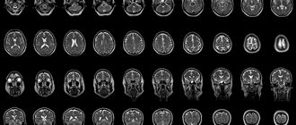
CT scan
Diagnosis of brain diseases using CT is based on calculating the intensity of penetration of X-rays through brain tissue. Thus, it is possible to obtain their detailed cross-sectional image. Modern devices for computer diagnostics have a low level of radiation, which does not affect the accuracy of the results.
Such a brain examination may be prescribed if the patient has a history of:
- dizziness;
- headache;
- loss of consciousness;
- convulsions;
- strokes;
- speech and memory disorders;
- auditory and visual impairments.
This method of diagnosing brain pathologies is contraindicated for children and pregnant women. If intravenous administration of a contrast agent is necessary, the following are added to the list of contraindications: renal and liver failure, diabetes mellitus, heart disease, asthma, thyroid pathologies, allergic reaction to iodine.
If there are no contraindications, computed tomography is performed as many times as necessary to make a diagnosis and monitor the effectiveness of therapy.
CT scan does not require special preparation of the patient. Unless before the procedure with contrast, you should not consume food or liquid for 4 hours. The examination lasts from 15 minutes to half an hour. At this time, the person is on a movable table, which moves into the tomograph. The patient is not allowed to move, and sometimes will need to hold his breath at the command of the medical staff.
Magnetic resonance imaging
It’s not for nothing that in foreign films about the work of medical institutions, doctors constantly refer patients to MRI – today it is one of the leaders among brain research methods. Visualization of the state of the organ occurs thanks to the magnetic field constantly maintained in the tomograph. Electromagnetic waves are passed through it, the energy flow from which is repelled by hydrogen atoms, which are present in all cells of the human body. Computer equipment converts the data into images of brain tissue.
MRI is effective for diagnosing a wide range of diseases: from vascular pathologies to tumors.
Modern tomographs emit low-frequency electromagnetic fields and are equipped with a number of specialized computer programs, which allows one to obtain a detailed picture of the functioning of the brain.
Examination with a tomograph is contraindicated in the following cases:
- the patient is mentally unstable, has acute pain syndrome or is in a coma;
- The patient’s body contains metal and ferromagnetic implants, pins, clips on blood vessels, and permanent crowns on teeth.
- The patient's skin has tattoos made with paint containing metal particles.
Pregnant women are required to inform the doctor about the presence of pregnancy and its duration before an MRI.
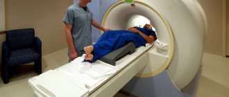
As with a CT scan, for the examination a person must lie down on a movable table, where his body will be secured with special belts, and sensors that send and read a signal are attached to his head. Afterwards the table moves into the tomograph. Depending on the number of programs used for scanning, the duration of the procedure is 15-40 minutes. All this time the person must lie motionless.
The principle of using a low-frequency electromagnetic field makes MRI completely safe for children and adults.
Magnetic resonance angiography
This technique for studying the brain is based on the same principles as MRI, but its main task is to identify pathologies of the vascular bed. Modern equipment is designed to obtain a three-dimensional image of the entire network of brain vessels, as well as to isolate thin sections of individual vessels and nerve trunks.
Positron emission tomography
Using this method, you can examine the brain in order to record in a three-dimensional projection all the functional processes occurring in it. Analysis of the metabolism of brain structures occurs at the cellular level, therefore PET is the best way to distinguish benign from malignant neoplasms in the early stages. This type of tomography is also used to obtain information about the consequences of injuries, abnormalities in brain function, and to assess the condition of patients after strokes.
PET cannot be performed during pregnancy and breastfeeding, as well as diabetes.
You should not eat 4-6 hours before the procedure. It is recommended to have a protein-free dinner the night before. Part of the examination is the intravenous administration of a radiopharmaceutical. The scanning itself lasts 30-75 minutes.
How is the examination carried out?
First of all, you need to get rid of metal items of clothing, the neck and ears should be free of jewelry. Bags, money, plastic cards, telephones, and electronic devices must be left outside the office. The patient receives disposable comfortable clothing for a comfortable procedure.
The examination without contrast agent lasts about 20 minutes; the introduction of contrast agents doubles the examination time. The patient is positioned on a table, which smoothly slides into the body of the tomograph, where the scanning will take place. During the examination, the neck and head should not make any movements. Fixation is ensured by a special coil.
When performing MRI of vessels and arteries of the neck and head, the tomograph operation is accompanied by unusual sounds, so the patient receives earplugs or headphones before the examination. The body of the tomograph is equipped with a fresh air supply system, as well as an intercom through which the doctor can give the command to hold your breath or slightly change the position of your head. If the patient becomes ill, it is possible to urgently contact the staff and stop the scan. After completing the examination, the patient changes clothes and can await the result.
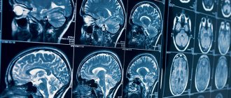
What the results look like
After performing an MRI of the vessels and arteries of the neck and head, the doctor receives layer-by-layer images of the vascular pattern with the ability to construct a three-dimensional model. The patient can pick up the images on electronic media or in printed form. The photographs are accompanied by a report from a radiologist.
Normally, the internal carotid arteries are located symmetrically, have clear contours, are not displaced, and are not compressed. The cerebral arteries arise in a typical place, the diameter is not changed, and there is no pathological tortuosity of the vessels. The cerebral veins and sinuses should be of normal diameter, without areas of blood flow disturbance, filling defects or deformations.
On MRI images of the vessels and arteries of the neck you can see signs inherent in certain diseases:
- an aneurysm is a round formation with a high signal intensity; when a thrombus attaches, a layered structure appears;
- vascular malformations appear as linear or tortuous structures with dilated vessels;
- dissection of the arterial wall is characterized by the appearance of a crescent-shaped hematoma located along the vessel, while the lumen of the artery is narrowed evenly or in the form of a rosary;
- atherosclerosis of the vascular system looks like an area of increased intensity on the vessel wall, the lumen is narrowed;
- hemangioma is a high-intensity formation with clear edges and a lobular structure.
Despite a detailed description of all identified pathological formations, the conclusion of a radiologist is not a diagnosis. The MRI result of the area of the cerebral vascular system should be shown to the doctor who ordered the study. Clinics offer the opportunity to consult with the radiologist who performed the procedure. If necessary, he will recommend contacting specialized specialists - a neurologist, oncologist or surgeon.
Where to get a magnetic resonance imaging scan
The search for a clinical diagnostic center can be significantly speeded up if you use the help of our website. The service contains information about a large number of clinics, ranging from contact details to discount offers. If necessary, our specialist will advise you free of charge over the phone about operating hours, the availability of tomographs of the required type and the time available for making an appointment.
A patient who wants to undergo magnetic resonance imaging of the vascular system of the head and neck can independently filter the list of medical institutions by rating, cost and features of the procedure (24-hour clinics, pediatric MRI).
Methods for studying cerebral vessels
In medical practice, there are several research methods to check the vascular system of a patient’s brain:
- Using an ultrasound machine, the following procedures can be performed:
- Ultrasound Dopplerography. Thanks to this study, it is possible to determine: The presence of plaques that do not allow blood to circulate properly inside the vessels of the brain;
- The speed of blood movement through the vessels of the head and cervical spine;
- Size of carotid, cerebral and vertebral arteries.
- Duplex scanning. Thanks to this study, the doctor can:
- Construct a color diagram of blood movement in the vessels of the brain;
- Identify diseases of the vascular system of the head that are still at an initial stage, for example, atherosclerosis, stenosis, aneurysms, occlusions.
Echoencephalography allows you to check:- The condition of both the inside of the brain and the periosteal space of the skull;
- How strong is the pulsation in the brain? Due to this verification, the patient's intracranial pressure is determined.
- Neurosonography pris given to children who have not yet turned one year old. Because the brain of small patients is checked through the fontanelle, which has not yet become overgrown in them. In the process of such a study, a specialist can determine the following:
- Blood circulation in the brain;
- No tumors or cysts in the brain;
- Does the little patient have any diseases related to the functioning of the brain, for example, high cranial pressure, a tendency to epilepsy or encephalopathy. Unfortunately, such unpleasant diseases can appear due to possible birth injuries or due to the fact that the baby did not have enough oxygen while passing through the birth canal.
- Using X-ray tests called angiography. There are several types of such checks:
- Magnetic resonance angiography allows you to check the patient’s cerebral vessels for the following diseases: Strokes;
Heart defects;- Increased pressure inside the skull;
- Atherosclerosis of blood vessels in the neck or brain;
- Vegetovascular dystonia;
- Vasculitis;
- Stenosis;
- Aneurysm.
- Magnetic resonance angiography has several subtypes:
- Diffusion-weighted magnetic resonance imaging will be prescribed to the patient if he is suspected of ischemic brain disease;
- Diagnostics of the main cerebral arteries;
- Sinusography allows you to study the condition of the cerebral veins and the collectors of this venous system. This procedure will be performed on the patient in order to prevent the formation of blood clots in the vessels of the brain.
- It should be borne in mind that such diagnostics also have their contraindications:
- The patient is overweight (more than 150 kg);
- Metal implants;
- The presence of artificial joints;
- Electrical pacemakers.
- Computed angiography. Such a study is carried out according to the following algorithm:
- The patient is given local anesthesia;
- A catheter is placed into the brain vessel through which a special substance (x-ray contrast) will flow;
- After all the preparatory work, the diagnosis itself is carried out using an angiograph;
- This X-ray machine takes pictures of the brain vessels;
- After all the necessary actions have been carried out for the patient, the place where the catheter was located should be tightly bandaged.
- The patient needs to know that such an examination should be carried out only on an empty stomach, and after diagnosis it is necessary to drink a lot in order to remove from the body the substance that was introduced through the catheter during the examination.
- Rheoencephalography is a diagnostic method that can be used to:
- Assess blood circulation in the brain;
- Determine at what speed blood moves through the vessels;
- See what condition the walls of the blood vessels are in.
Anyone who undergoes any examination of the cerebral vessels should know that a diagnosis based on these diagnostic studies can only be made by a doctor who, if necessary, will prescribe specific treatment or send the patient for additional examinations of other organs of the body.
- Vascular diseases of the head and neck: treatment with drugs and folk remedies