Methods of functional diagnostics in neurology. Part 1
What methods of functional diagnostics exist, what do they allow you to find out, what are the advantages of these methods, what is an EEG, when is it worth doing an electroencephalogram, says Dmitry Aleksandrovich Ovchinnikov, neurologist, functional diagnostics doctor.
Ovchinnikov Dmitry Alexandrovich Doctor of functional diagnostics.
Hello. My name is Dmitry Aleksandrovich Ovchinnikov. I am a neurologist, functional diagnostics doctor at the Scandinavia clinic. Today we will talk about functional diagnostic methods in neurology. Neurology is a branch of medicine that deals with the diagnosis and treatment of disorders of the nervous system. Functional diagnostics is a very diverse group of methods that are united by one principle. Assessment of not only and not so much the static state of the system, but the functioning and functioning of organs. In this way, functional diagnostic methods compare favorably with other groups of techniques, such as radiation diagnostic methods, where the structure is assessed primarily at each specific point in time, or laboratory diagnostic methods, which evaluate the composition of tissues and fluids, but only at one specific point in time. It becomes clear why functional diagnostics is so important in neurology, because the nervous system is an extremely dynamically changing structure, the main task of which is to respond to external and internal stimuli and change its functioning over time.
Let's briefly look at the functional diagnostic tools available to neurologists. A large group of functional diagnostic methods are united by their electrophysiological principle. Nerve cells in the course of their work are capable of being excited, changing the polarity of their membranes and generating an action potential. Fixation and registration of this action potential allows a number of functional diagnostic methods to study nerve tissue and draw certain conclusions about the functioning of the nervous system. These methods include electroencephalography, electromyography, evoked potential methods, and a number of other electrophysiological techniques.
One of the electrophysiological methods of functional diagnostics in neurology is electroencephalography, or EEG for short. Electroencephalography is a technique for assessing the bioelectrical activity of the brain. During the study, electrodes are placed on the patient's head to record brain activity in various areas of the brain. Also during the research, functional tests are carried out, such as photostimulation or tests with changes in the gas composition of the blood, followed by stimulation of the brain with hyperoxygenation. The results of measuring this activity are currently processed by a computer and displayed on the monitor in the form of graphs. The doctor is able to perform various types of analysis of this recording so that the conclusions are as comprehensive and profound as possible. The studies are carried out in a shaded room, but do not involve any discomfort. Lasts from 15 to 30 minutes.
There are also methods for long-term recording of electroencephalograms, but they are used less routinely. With some exceptions, an electroencephalogram does not require any preparation, but on the day of the study it is recommended to wash your hair to reduce skin resistance to electric current.
A serious advantage of the electroencephalogram over other research methods is the ability to assess the functioning of the brain over a certain period of time. What no other technique can provide, such as MRI, which allows a very precise and very comprehensive assessment of the structure and anatomy of the brain, but cannot assess its functioning during a specific drive. Or such as computed tomography, which evaluates primarily the bone structure of the skull and the structure of the brain without detailed differentiation. Thus, various types of electroencephalogram are the gold standard in the diagnosis of epilepsy and various epi-syndromes. No other technique currently used in clinical practice is capable of identifying these deviations.
The experience accumulated by the Soviet, Russian and international scientific schools allows the use of EEG not only in the diagnosis of epilepsy. Encelography makes it possible to indirectly judge violations of recoordination, the work of the hypothalamic-pituitary system, the functioning of the brain and cerebral cortex as a whole, and the relationship between the work of the cortex and subcortical structures. Certain changes in the encephalogram can give the doctor the opportunity to judge the functioning of the cognitive component and the exhaustion of the nervous system. Thus, today an electroencephalogram is one of the few methods that allows us to evaluate not only structural changes in the brain, but also changes in its functioning.
Indications for an encephalogram at the moment are loss of consciousness of any origin, functional disorders of the nervous system, such as neurotic, psychological, emotional, psychosomatic complaints of patients. Assessment of the consequences of traumatic brain injuries, assessment of the consequences of neuroinfections. Theoretically, we can evaluate the functioning of the hypothalamic-pituitary system and its impact on brain function. Previously, electroencelography was used to assess changes in the structural properties of nervous tissue to search for focal changes, such as tumors and hematomas. However, at the moment, MRI copes much better with these tasks.
In addition to the indications described above, it is advisable to conduct electroencelography when there are changes in brain function caused by vascular disorders, disorders caused by chronic changes in cerebral blood flow.
Publication date: 04/10/17
Diagnosis and prevention of diseases of the nervous system.
Currently, neurologists have a large number of instrumental research methods in their arsenal that allow them to assess the functional state of both the central and peripheral nervous systems. To choose the right diagnostic direction, correct treatment, assess the prospects of therapy, and prognose the course of the disease, the clinician must be familiar with the methods of functional diagnostics and have an idea of the results that can be obtained using one or another method.
- Electroencephalography (EEG) is a method of studying the brain based on recording the electrical potentials of the cerebral cortex. EEG makes it possible to identify in a patient, first of all, the presence or predisposition to epileptic manifestations, such as fainting and prolonged loss of consciousness with and without convulsions, which is the main danger at work; in addition, this study reveals indirect signs of the presence of neoplasms (benign, malignant tumors , cysts, post-traumatic changes). Using an EEG, you can assess the nature and severity of brain disorders.
Electroencephalogram monitoring is a method for assessing the functional activity of the brain in various diseases and conditions over a long period of time (several hours).
- Evoked potentials (EP) are a method of functional diagnostics based on recording electrical potentials of the brain in response to a stimulus:
- auditory EPs - used to diagnose damage to the auditory nerve and brain stem;
- visual EPs - used to study visual functions;
- somatosensory EPs - used to assess the conductive functions of the nervous system from the periphery to the cerebral cortex;
- cognitive - used to study the cognitive function of the brain, the volume of RAM;
- sympathetic skin potential - used to study the autonomic nervous system (sympathetic, parasympathetic).
This method helps in diagnosing multiple sclerosis, clarifying the level of damage to the nervous system, diseases of peripheral nerves, and diagnosing tumors, injuries and other pathological processes in the brain and spinal cord.
- Electroneuromyography (ENMG) is a method that allows you to assess the functional integrity of the neuromuscular system. It is used to determine the level of damage to the neuromuscular system, to diagnose muscle diseases and lesions of the spinal cord, peripheral nerves, and brain stem.
- Magnetic stimulation of the nerves of the brain is a method for determining the level of disturbance in the conduction of motor impulses. The advantage of the method over electrical stimulation is that the magnetic field is able to pass through any anatomical structures without changes (i.e. the signal does not weaken when passing through various media) and excite nerve tissue, in addition, the magnetic effect is painless.
- Rheoencephalography and rheovasography (REG, RVG) is a method of studying blood vessels based on recording the electrical resistance of body tissues. It is used in the diagnosis of diseases with disorders of arterial and venous circulation, cerebrovascular accidents, vegetative-vascular dystonia, intracranial hypertension, and the consequences of traumatic brain injury.
- Color duplex scanning of extracranial (extracranial) vessels and transcranial duplex scanning of intracranial vessels is a method of studying vessels and vascular blood flow using ultrasound. It is used in the diagnosis of vascular stenosis, congenital and acquired anomalies of the head and neck vessels, consequences of traumatic brain injury, and cerebrovascular accidents.
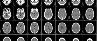
- Magnetic and computed tomography are one of the leading methods for diagnosing diseases, allowing one to detect pathological changes at a very early stage. CT is based on the use of X-rays, and MRI is based on a magnetic field. This method allows you to diagnose inflammatory and tumor processes in tissues, degeneration, traumatic injuries, developmental defects, and vascular disorders.
- Polysomnography (PSG) is a method of long-term recording of various body functions throughout sleep. The method includes monitoring of brain biopotentials (EEG), electrooculogram, electromyogram, electrocardiogram, heart rate, air flow at the level of the nose and mouth, respiratory efforts of the chest and abdominal walls, oxygen fluctuations in the blood, and motor activity during sleep. The method allows you to study all pathological processes that occur during sleep: apnea syndrome, heart rhythm disturbances, changes in blood pressure, epilepsy.
- The echoencephaloscopy method is a method of ultrasound diagnosis of disorders in the brain and allows one to judge the presence and degree of displacement of the midline structures, which indicates the presence of additional volume (intracerebral hematoma, hemispheric edema). Currently, the significance of the method is not as great as before; it is primarily used for screening assessment of indications for emergency neuroimaging (computed tomography (CT) or magnetic resonance imaging (MRI).
One of the most important prerequisites for human health and longevity is a strong and healthy nervous system. Therefore, to keep it that way, it is important to follow some rules:
- Control your mood
Mood is a driving force that can lead to the achievement of a designated goal. When you get angry, start counting. Don't say anything because you might regret it later. If possible, it is better to immediately engage in physical activity or take a walk in the fresh air.
- Do not quarrel
Anger and hatred are manifested as a result of excited disputes. Arguing while eating is most unfavorable and can even lead to stomach ulcers. As soon as a person feels that the argument has become heated, he should immediately stop it.
- Don't annoy others and don't let them annoy you
Remember that not everyone you interact with shares your point of view. Contact people you find unpleasant as little as possible, but don’t hate. Stay away from them, and if you have to be in their company, minimize communication with them. If even once you allow yourself to be a scapegoat for someone, he will constantly continue to torment you. But if you ignore him or walk away silently, he will leave you alone. This is human psychology.
- Don't isolate yourself
Express your concerns to others. If you feel that you have a legitimate resentment against someone, you should approach that person and calmly explain everything. Bring this principle into all your relationships. By accumulating everything in yourself, you build a stone wall, which depletes the nervous system and leads to a loss of strength. In this case, you need to share it with someone. Perhaps such a conversation will help you see the problem from the other side and find a compromise solution.
- Don't take on an unbearable burden
People who strive to do things that are impossible are constantly under tension, which has a bad effect on the health of the nervous system.
- Smile and laugh more often
An old saying goes: “If you laugh, the whole world laughs with you. And if you cry, you’re the only one crying.” Laughter improves blood circulation in the abdominal cavity and heals the nervous system thanks to the joyful state of mind of the laugher.
- Don't take words to heart
If you were rude, forget it. Don't make a big deal out of this. Be wise and prudent enough not to allow this poison to poison your soul. Don’t look for perfection in people, but try to see only the good in them.
- Learn to live with yourself
It’s great when you have a lot of true friends, but besides this, it’s important to learn to live alone with yourself. You should always be your own good company and never bore yourself. In solitude, you begin to know yourself better, and this will help you know others better.
- Stay busy
The one who is busy is the one who is happy. Idleness gives rise to gossip, quarrels and other negative phenomena. Find something you enjoy and then life will seem like a great adventure. Live every day, making every minute count.
- Don't torture yourself
Get rid of grief and other emotional shocks by forgetting them. Direct your thoughts in a different direction, get away from the painful habit of finding solace in torturing yourself with grief. With the help of willpower, sweep away sad, bad thoughts and replace them with happy memories. We must learn to control our emotions and not let them control us.
- Don't worry unnecessarily
Worry can destroy us. Anxiety is often inevitable. But here too we must be guided by a clear mind and common sense. You won't achieve anything by worrying; on the contrary, the more we worry, the more we create tension on our nervous system and deprive ourselves of the strength we need to overcome difficulties.
Doctor of the Department of Functional Diagnostics Byako I.V.
Features of functional diagnostics
Doctors know for sure that all systems and organs are interconnected. For example, a change in the functional state of the nervous system entails a variety of symptoms. Often, without proper examination, it is impossible to make an accurate diagnosis.
Functional diagnostics becomes especially important in the case of ineffective treatment for a chronic disease or in the absence of visible somatic pathologies along with persistent patient complaints. Modern methods make it possible to find out how systems and organs cope with the load. Assessing the functional state of the cardiovascular, nervous and respiratory systems are the main objectives of the examinations.
It is extremely important to undergo examination under the supervision of a competent specialist - often the research results do not have an unambiguous interpretation. After all, each body’s systems and organs work differently. To make a diagnosis, you will have to take into account the anamnesis, clinical picture, and the results of functional tests. With the right approach, functional diagnostics allows you to clarify the diagnosis and develop an effective treatment strategy.
additional research methods
Category: Nursing in neurology/Basic principles of examination and treatment of neurological patientsThe following additional research methods are most often used in neurology: radiography of the skull and spine, spinal puncture, ophthalmological examination, electroencephalography, rheoencephalography, Dopplerography, echoencephaloscopy, myelography, myography, pneumoencephalography, computed tomography and magnetic nuclear tomography.
.
Spinal (lumbar) puncture
- used for the diagnosis of inflammatory diseases of the central nervous system, volumetric processes of the brain and spinal cord and hemorrhages in the medulla and under the membranes of the brain. The nurse must prepare instruments, sterile material and the patient himself for the puncture and assist the doctor during the puncture.
Ophthalmological examination
carried out by an ophthalmologist. The vessels of the fundus, the optic nerve head, visual fields, visual acuity and color perception are examined.
X-ray of the skull
widely used for injuries and a number of non-traumatic brain diseases. The study is carried out in two projections - frontal and lateral. On craniograms, attention is paid to the size of the skull, the integrity of the bones of the vault and base of the skull, the severity of sutures, fontanelles, which is especially important in childhood. Signs indicating increased intracranial pressure are important: the presence of finger-like indentations and destruction of the dorsum sella, as well as excessive expression of the vascular pattern.
X-ray of the spinal column
- one of the main diagnostic methods. On a spondylogram, attention is paid to the shape and size of the vertebral bodies, the presence of bone growths, the structure of bone tissue and the severity of intervertebral spaces, the condition of the vertebral processes.
Electroencephalography (EEG)
consists in recording the biopotentials of the brain with their subsequent visual or computer processing. Normally, EEGs are a combination of waves of unequal duration and amplitude. There are alpha rhythm (oscillation frequency - 16 per second), beta rhythm 14-35, delta rhythm 1-3 and theta rhythm 4-7 oscillations per second. Sharp waves “peaks” and discharges of slow high-voltage waves characterize increased convulsive readiness in epilepsy; with brain tumors in the area of their localization, the alpha rhythm may be suppressed, delta waves or convulsive discharges may appear. EEG is important in assessing the depth of anesthesia and determining “brain death”.
Rheoencephalography (REG)
- a method for studying cerebral circulation, based on recording changes in the resistance of head tissue to electric current, caused by fluctuations in blood supply, the tone of cerebral vessels and the state of venous outflow. By positioning the electrodes, the carotid and vertebrobasilar basins are examined. REG is especially informative for cerebrosclerosis, hypertension, and cerebrovascular accidents.
Echoencephalography (EchoEG)
is based on the ability of ultrasonic waves to be reflected from various formations located inside the skull. The study determines the position of the structures along the midline, which normally deviate from no more than 2 mm. If the midline structures are deviated by more than 2 mm, this indicates the presence of a pathological process in the cranial cavity.
Dopplerography (USDG)
is an ultrasound examination of intravascular blood flow, patency of the vascular bed, blood flow velocity. The method allows you to analyze blood flow not only in the great vessels, but also in the intracranial vessels. The method is used to diagnose vascular pathology of “brain death”.
Electromyography (EMG)
registers muscle biopotentials, which makes it possible to study their functional state, the nature of the conduction of nerve impulses along the nerves and conductors of the spinal cord. The changes are most indicative of damage to the central, peripheral neuron in the muscles themselves.
Pneumoencephalography
consists of introducing into the subarachnoid space of the spinal cord through a lumbar puncture 15 to 70 ml of air or oxygen, which rise up into the cranial cavity, fill the ventricles and subarachnoid space. After which they take an X-ray of the skull and judge the location and nature of the pathology. The nurse's tasks for this examination are the same as for a lumbar puncture.
Myelography
carried out to diagnose pathological processes in the spinal canal (spinal cord tumors, prolapsed intervertebral discs, adhesions). The method involves introducing a contrast agent through a lumbar or suboccipital puncture into the subarachnoid space of the spinal cord. Before performing myelography, the patient is given cleansing enemas in the evening and in the morning. During the procedure, the nurse assists the doctor.
Carotid and vertebral angiography
is performed by injecting 10-15 ml of contrast agent into the carotid or vertebral artery, which makes it possible to obtain an image of the main vessels of the brain on craniograms, the condition of which is used to judge the presence of a pathological process. Angiography is especially valuable in diagnosing vascular pathology (aneurysm, thrombosis) and brain tumors. The duty of the nurse is to test the patient's sensitivity to the contrast agent. On the eve of the study, the nurse administers 2 ml of contrast agent intravenously to the patient, monitors his reaction and informs the doctor. Angiography is performed on an empty stomach. After the examination, the patient is taken on a gurney to the ward, where his condition is monitored. Angiography, like pneumoencephalography and myelography, is an invasive and difficult research method, in which it is necessary to keep in mind the possibility of severe complications.
Computed tomography, magnetic nuclear tomography
— these are the most modern methods for diagnosing neurological and neurosurgical pathologies. They are based on the irradiation of organs or parts of the body, respectively, with X-rays or magnetic rays, followed by their registration and data processing using computers. These methods make it possible to obtain an image of the brain and spinal cord with all its structures and tissues in various planes and sections. These data are indispensable in the diagnosis of hematomas, strokes, tumors, multiple sclerosis, encephalitis, abnormalities of brain development and hydrocephalus.
Positron emission tomography
— this method makes it possible to observe metabolic processes in brain tissue, which is especially valuable for studying the dynamics of pathological processes during drug treatment.
See examination of neurological patients
Saenko I. A.
Sources:
- Bortnikova S. M., Zubakhina T. V. Nervous and mental illnesses. Series 'Medicine for you'. Rostov n/d: Phoenix, 2000.
- Nurse's Handbook of Care/Ed. N. R. Paleeva. - M.: Medicine, 1980.
- Martynov Yu. S. Neurology: Textbook. ed. 4th, rev. and additional - M.: RUDN, 2009.
Diffuse diseases of the peripheral nervous system causing general weakness
Localization of the problem
Finding the location of lesions in patients with diffuse damage to the peripheral nervous system is often not difficult, since spinal reflexes are usually reduced in all 4 limbs. The peripheral nervous system consists of nerve roots, nerves, the neuromuscular junction, and muscles. Further differentiation by neurological examination alone between these structures and identification of the area of disease in the nerve roots, nerves, neuromuscular end plates or muscles is often difficult or impossible. Additional research is needed, such as electromyography, biopsy of muscle and nerve tissue and laboratory tests.
Diseases leading to polyneuropathy in small animals
| Vascular | Ischemic neuromyopathy (thromboembolism caused by cardiomyopathy or trauma). Cats, dogs |
| Inflammatory, infectious | Acute polyradiculoneuritis (Coonhound paralysis) (cats, dogs), chronic recurrent polyradiculoneuritis (cats, dogs), neosporosis (dogs), toxoplasmosis (dogs) |
| Traumatic | — |
| Abnormal | — |
| Metabolic | Hypothyroid polyneuropathy (dogs), hypoglycemic polyneuropathy (dogs), diabetic neuropathy (cats, dogs), hyperchylomicronemia (cats), hyperoxaluria (cats). Vincristine-induced polyneuropathy (cats, dogs), thallium-induced polyneuropathy (cats, dogs), chronic intoxication with organophosphorus compounds (cats, dogs) |
| Idiopathic | Autonomic dysfunction (Dysautonomia) (cats, dogs) |
| Neoplastic | Paraneoplastic polyneuropathy caused by bronchogenic carcinoma, mammary adenocarcinomas, malignant melanomas, osteosarcoma, thyroid adenocarcinoma, mastocytomas (cats, dogs) |
| Degenerative | Spinal muscular atrophy ( Lapland dogs, English spaniel, English pointer, German shepherds, Rottweiler, Cairn terrier); giant axonal neuropathy (German shepherds); sensory neuropathy (boxers, long-haired dachshunds, Jack Russell terriers, English pointers, long-haired collies (Collie Rough), huskies); hypertrophic neuropathy (Tibetan mastiffs), globoid leukodystrophy (Cairn terriers, West Highland white terriers, cats). Sphingomyelinosis (Siamese), distal sensorimotor polyneuropathy (Rottweilers), distal polyneuropathy (Burmese cats), glycogen storage disease Type IV (Norwegian Forest cats), Congenital hypomyelinating neuropathy (Golden Retrievers) |
Diseases that cause damage to synapses in small animals
| Vascular | — |
| Inflammatory, infectious | Acquired myasthenia gravis (cats, dogs), tick paralysis (cats, dogs) |
| Traumatic | — |
| Abnormal | Inherited myasthenia gravis (cats, dogs) |
| Metabolic | Botulism (cats, dogs) |
| Idiopathic | — |
| Neoplastic | Paraneoplastic myasthenic syndrome (thymomas) (cats, dogs) |
| Degenerative | — |
Diseases causing polymyopathy in small animals
| Vascular | Ischemic neuromyopathy (thromboembolism) (cats, dogs) |
| Inflammatory, infectious | Idiopathic polymyositis (cats, dogs). Neosporosis (dogs), toxoplasmosis (dogs), systemic lupus erythematosu-polymyositis (dogs), dermatomyositis in collies, Leptospira icterohaemorrhagiae (dogs), Clostridium spp. (dogs), Hepatozoon (Hepatozoon canis is a single-celled parasite that causes disease in dogs) |
| Traumatic | — |
| Abnormal | — |
| Metabolic | Hyperadrenoscorticism (steroid myopathy) (dogs), hypothyroidism (dog), hyperthyroid myopathy (cat), hypokalemia (dogs and cats), hyperkalemia (dogs and cats), malignant hyperthermia (dogs), exercise-induced myopathy (dogs and cats) , hypernatremia-myopathy (dogs), mitochondrial myopathy (dogs and cats), myopathy caused by vitamin E and selenium deficiency (dogs and cats) |
| Idiopathic | — |
| Neoplastic | Paraneoplastic myositis (bronchogenic carcinoma, myeloid leukemia, tonsil cancer) (dogs and cats) |
| Degenerative | Muscular dystrophy (Devon Rex, Golden Retriever, Irish Terrier, Samoyed, Rottweiler, Miniature Schnauzer, Welsh Corgi, cats); hereditary myopathies (Labrador retriever); myotonic myopathy (chow chow, Staffordshire terrier, Great Dane, Rhodesian Ridgeback, cats); glycogen storage diseases (dwarf breeds, German Shepherd, Akita, Swedish Lapphund, Norwegian forest cat, English Springer Spaniel); nemaline myopathy in cats; Central core disease (CCD) in Great Danes, degenerative myopathy of Bouviers des Flandres |

