The structural unit of the nervous system is the nerve cell, or neuron. Even if one neuron is isolated from the rest of the nervous system, it retains the ability to perceive information, analyze it, and respond adequately. The nerve cell that is involved in the perception of information is called an afferent neuron, or sensory neuron. It is one of the main links of the reflex arc. More details about these structures are provided later in the article.
Neuron structure
Fundamentally, the structure of the afferent neuron is no different from other cells of the nervous system. It has two main components: the body and processes. The shoots, in turn, are of two types:
- dendrites - have a relatively short length;
- an axon is a long extension of a neuron, always one per cell.
The function of dendrites is to transport nerve impulses to the nerve cell body. Due to this, information from the environment enters the nervous system. Dendrites also make it possible for neurons to communicate with each other.
A long process - an axon - carries information from the cell body to other parts of the body. In this way, information spreads beyond the nervous system.
The body of the afferent neuron consists of several parts:
- a shell consisting of phospholipids and proteins;
- nucleus with genetic material;
- the Golgi complex, which performs the function of packaging carbohydrates;
- endoplasmic reticulum, where protein synthesis occurs;
- mitochondria, in which ATP energy is generated;
- lysosomes, which perform the functions of “swallowing” foreign particles.
All these elements work together and are in constant interaction.
How do they work together and how are they different?
Afferent neurons typically have two axons that carry electrochemical signals to the spinal column or brain. Once there, the signal travels through a network of interneurons and through the efferent neuron. Afferent-efferent pairs of neurons that pass through the spine control reflexes (such as the knee-jerk reaction).
Finished works on a similar topic
Course work Afferent and efferent nerve conductors and their role in psychology 430 ₽ Abstract Afferent and efferent nerve conductors and their role in psychology 230 ₽ Test work Afferent and efferent nerve conductors and their role in psychology 210 ₽
Receive completed work or specialist advice on your educational project Find out the cost
Afferent neurons are designed to respond to various stimuli. For example, an afferent neuron designed to respond to heat detects excess heat and sends an impulse through the central nervous system. The efferent neuron then causes the muscles to contract to move the body away from the heat. The skin has sensory receptors for heat, cold, pleasure, pain and pressure.
Afferent neurons have round and smooth cell bodies, while efferent neurons have satellite bodies. Afferent neurons are found in the peripheral nervous system, and efferent neurons are located in the central nervous system. Axons in afferent neurons move from the ganglia (clusters of nerve cells that contain afferent and efferent neurons) to the spinal cord. The long axon is actually connected to the efferent neuron.
Afferent neurons have a single long myelinated dendrite, whereas efferent neurons have shorter dendrites. The dendrite in an afferent neuron is what is responsible for transmitting nerve impulses from the receptors to the cell body, while in an efferent neuron the impulses travel through the dendrite and exit through the neuromuscular junction that is formed between the effectors and the axon.
Concept and structure of the reflex arc
A reflex is a universal response of the body to irritation. The unconditioned reflex is transmitted hereditarily and is the same for any person. Conditional is one that is acquired during life. Each person can have his own conditioned reflex.
Afferent and efferent neurons are the main components of the reflex arc.
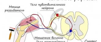
At its core, a reflex arc is a path that overcomes a nerve impulse to a working organ or muscle. There are three main components of the arc:
- analyzer, or afferent, part;
- central, or contact, part;
- executive, or efferent, part.
According to modern concepts, there are not three, but five parts of the reflex arc. It begins with a receptor - the end of a sensitive neuron. In addition to the receptor, the arc includes the following elements:
- sensitive, or afferent, neuron;
- interneuron;
- motor, or efferent, neuron;
- the organ that responds to stimulation is the effector.
That is, for a reflex arc to form, there must be at least three nerve cells. An exception is the tendon reflexes, which contain only two cells - sensory and motor.
Analyzer part
An afferent neuron is a neuron that perceives information from the internal and external environment of the body using a receptor. Thus, the analyzer part consists of a receptor and a sensitive pathway.
There are several classifications of receptors. Depending on their location they are divided:
- on exteroceptors - those that are located in the skin and mucous membranes;
- proprioceptors - those located in muscle fibers, tendons and ligaments;
- interoreceptors - those that are located in the internal organs.
Depending on the type of stimulus that the receptor perceives, they are divided:
- to thermoreceptors - they react to changes in temperature, they are found on the skin and tongue;
- baroreceptors - react to pressure changes, located in the aortic arch, carotid sinus;
- chemoreceptors - react to changes in chemical composition, located in the stomach and intestines;
- photoreceptors - react to light, located in the retina;
- pain receptors - provide the perception of pain, found in the skin, peritoneum, capsule of internal parenchymal organs, periosteum;
- phonoreceptors - respond to sounds, located in the inner ear.
The sensitive pathway is the same afferent neuron with a body and processes. The axon of a nerve cell enters the central nervous system through the dorsal horns of the spinal cord, which are also called sensory. Directly in the spinal cord it transmits information to the interneuron.
Neurons
A neuron is a complex, highly specialized cell with processes capable of generating, perceiving, transforming and transmitting electrical signals, and also capable of forming functional contacts and exchanging information with other cells.
Each neuron has only 1 axon, the length of which can reach several tens of centimeters. Sometimes lateral processes extend from the axon - collaterals . Axon endings typically branch and are called terminals . The place where the axon extends from the cell soma is called axonal hillock .
In relation to the processes, the soma of the neuron performs a trophic function, regulating metabolism. A neuron has characteristics common to all cells: it has a membrane, a nucleus and a cytoplasm in which organelles are located (endoplasmic reticulum, Golgi apparatus, mitochondria, lysosomes, ribosomes, etc.).
In addition, the neuroplasm contains special-purpose organelles: microtubules and microfilaments , which differ in size and structure. Microfilaments represent the internal skeleton of the neuroplasm and are located in the soma. Microtubules stretch along the axon along the internal cavities from the soma to the end of the axon. Biologically active substances spread through them.
In addition, a distinctive feature of neurons is the presence of mitochondria in the axon as an additional source of energy. Adult neurons are not capable of division.
Types of neurons
There are several classifications of neurons based on different characteristics: the shape of the soma, the number of processes, the functions and effects that the neuron has on other cells.
Depending on the shape of the soma, they are distinguished: 1. Granular (ganglionic) neurons , in which the soma has a round shape; 2. Pyramidal neurons of different sizes - large and small pyramids; 3. Stellate neurons ; 4. Fusiform neurons .
Based on the number of processes (by structure) there are: 1. Unipolar neurons (single-processed) , having one process extending from the cell soma, are practically never found in the human nervous system; 2. Pseudounipolar neurons (false unipolar), such neurons have a T-shaped branching process, these are cells of general sensitivity (pain, temperature changes and touch); 3. Bipolar neurons (bipolar) having one dendrite and one axon (i.e. 2 processes), these are cells of special sensitivity (vision, smell, taste, hearing and vestibular stimulation); 4. Multipolar neurons (multi-process) , which have many dendrites and one axon (i.e. many processes); small multipolar neurons are associative; medium and large multipolar, pyramidal neurons - motor, effector.
Unipolar cells (without dendrites) are not typical for adults and are observed only during embryogenesis. Instead, in the human body there are pseudounipolar cells, in which a single axon divides into 2 branches immediately after leaving the cell body. Bipolar neurons are present in the retina and transmit excitation from photoreceptors to ganglion cells that form the optic nerve. Multipolar neurons make up the majority of cells in the nervous system.
According to the functions they perform, neurons are: 1. Afferent (receptor, sensory) neurons - sensory (pseudo-unipolar), their somas are located outside the central nervous system in ganglia (spinal or cranial). Along sensory neurons, nerve impulses move from the periphery to the center.
The shape of the soma is granular. Afferent neurons have one dendrite that connects to receptors (skin, muscle, tendon, etc.). Through dendrites, information about the properties of stimuli is transmitted to the soma of the neuron and along the axon to the central nervous system.
Example of sensory neurons: a neuron that responds to skin stimulation.
2. Efferent (effector, secretory, motor) neurons regulate the work of effectors (muscles, glands, etc.). Those. they can send orders to the muscles and glands. These are multipolar neurons, their somas have a stellate or pyramidal shape. They lie in the spinal cord or brain or in the ganglia of the autonomic nervous system.
Short, abundantly branching dendrites receive impulses from other neurons, and long axons extend beyond the central nervous system and, as part of the nerve, go to effectors (working organs), for example, to skeletal muscle.
Example of motor neurons: spinal cord motor neuron.
The cell bodies of sensory neurons lie outside the spinal cord, while motor neurons lie in the anterior horn of the spinal cord.
3. The betting neurons (contact, interneurons, associative, closing) make up the bulk of the brain. They communicate between afferent and efferent neurons and process information coming from receptors to the central nervous system.
These are mainly multipolar stellate-shaped neurons. Among interneurons, neurons with long and short axons are distinguished.
Example of interneurons: neuron of the olfactory bulb, pyramidal cell of the cerebral cortex.
The chain of neurons consisting of sensory, intercalary and efferent neurons is called the reflex arc. All activities of the nervous system, as defined by I.M. Sechenov, is of a reflex nature (“reflex” means reflection).
According to the effect that neurons have on other cells: 1. Excitatory neurons have an activating effect, increasing the excitability of the cells with which they are connected. 2. Inhibitory neurons reduce cell excitability, causing an inhibitory effect.
Nerve fibers and nerves
Nerve fibers are processes of nerve cells covered with a glial sheath that carry out nerve impulses. Through them, nerve impulses can be transmitted over long distances (up to a meter).
The classification of nerve fibers is based on morphological and functional characteristics.
Based on morphological characteristics, they are distinguished: 1. M myelinated (meat) nerve fibers are nerve fibers that have a myelin sheath; 2. Non -myelinated (non-myelinated) nerve fibers are fibers that do not have a myelin sheath.
According to functional characteristics, they are distinguished: 1. Afferent (sensitive) nerve fibers; 2. Efferent (motor) nerve fibers.
Nerve fibers extending beyond the nervous system form nerves. A nerve is a collection of nerve fibers. Each nerve has a sheath and a blood supply.
There are spinal nerves connected to the spinal cord (31 pairs) and cranial nerves (12 pairs) connected to the brain. Depending on the quantitative ratio of afferent and efferent fibers within one nerve, sensory, motor and mixed nerves are distinguished (see table below).
In sensory nerves, afferent fibers predominate, in motor nerves, efferent fibers predominate, in mixed nerves, the quantitative ratio of afferent and efferent fibers is approximately equal. All spinal nerves are mixed nerves. Among the cranial nerves, there are three types of nerves listed above.
List of cranial nerves with dominant fiber designation
I pair - olfactory nerves (sensitive); II pair - optic nerves (sensitive); III pair - oculomotor (motor); IV pair - trochlear nerves (motor); V pair - trigeminal nerves (mixed); VI pair - abducens nerves (motor); VII pair - facial nerves (mixed); VIII pair - vestibulo-cochlear nerves (sensitive); IX pair - glossopharyngeal nerves (mixed); X pair - vagus nerves (sensitive); XI pair - accessory nerves (motor); XII pair - hypoglossal nerves (motor).
Contact part
The contact part of the reflex arc is also called the nerve center. The number of interneurons may vary. It depends on the complexity of the information that needs to be processed. The interneuron is located within the central nervous system, namely the spinal cord.
The functions of the contact part are as follows:
- analysis of the information received;
- synthesis of information;
- making the final decision about what the answer should be.
Location of the sensory neuron
Afferent neurons are part of the peripheral nervous system. Their perceiving sections - receptors - are located in the skin, the walls of tubular organs or blood vessels, and the capsules of parenchymal organs.
Afferent neurons are pseudounipolar. That is, they have one process that extends from the body and then divides into two. Thus, one process approaches the body from the receptor, and then another extends from it to the center.
The body itself is located in the spinal ganglia, or nodes. These formations consist entirely of nerve cell bodies.
Concept and types of neurons
Definition 1
A neuron is an electrically excitable cell, a functional unit of the nervous system.
Each neuron has a cell body, dendrites, and an axon. Neurons are divided into three types:
- afferent neurons,
- efferent neurons
- interneurons.
Sensory information is transmitted from the periphery of the body to the main organ - the brain. Sensory information includes nerve impulses (that is, things people hear, touch, see, taste, and smell) that are transmitted from the senses. Afferent neurons are also called sensory neurons, and it is these specialized cells that transmit nerve impulses from the body directly to the central nervous system.
Are you an expert in this subject area? We invite you to become the author of the Directory Working Conditions
Physical stimuli, such as sound or light, activate afferent neurons, converting modalities into nerve impulses. They do this by using sensory receptors found in their cell membranes. The main cell bodies of afferent neurons are located near the brain and spinal cord, which together form the central nervous system.
Efferent neuron cells are located in the central nervous system and are called motor neurons. After receiving input from various neurons, including afferent neurons and interneurons, efferent neurons receive these signals from the central nervous system and transmit nerve impulses to the peripheral nervous system, muscles and glands to initiate a response to a stimulus.
Functions of the sensory neuron
The main function of the afferent neuron has already been noted above - the perception of information from the outside using a receptor. But what does this phrase mean?
The perception of any signal is based on the process of information analysis. Its essence lies in the detailed “decomposition” of the stimulus into individual components, careful study and identification of its individual properties. But only a superficial analysis of information occurs in the receptor. Therefore, one reflex arc is not capable of providing complex reactions. For example, one of the reflexes is extension of the knee to a blow to the tendon with a hammer.
A more subtle “decomposition” of the stimulus is carried out in the central nervous system. And this process is ensured most subtly and thoroughly by the cerebral cortex as the most historically new structure of the central nervous system.
Thus, the afferent neuron is an important part of the reflex arc, thanks to which we are able to perceive external irritation and quickly respond to it.


