Disorders of the blood supply to the brain are currently a pressing problem, since their development occurs not only in older people, but also in those over 40 years old. The danger of cerebral strokes is also due to the fact that the mortality rate after them ranks third after cancer and heart disease.
Ischemic strokes have characteristic symptoms and numerous causes, knowledge of which makes it possible to prevent cerebrovascular accidents. With an ischemic stroke of the right hemisphere, the arteries supplying this hemisphere are blocked by an embolus or thrombus. Another cause of an acute disorder may be arterial spasm. The supply of oxygen and nutrients to the right hemisphere is ensured by the vertebral and carotid arteries, so a right-sided stroke can develop as a result of pathologies of these vessels.
Neurologists at the Yusupov Hospital provide highly accurate diagnostics and effective treatment according to individual plans. Activities carried out by rehabilitation clinic specialists are aimed at restoring lost functions and improving the quality of life.
Clinical picture
The right hemisphere of the brain is a place of accumulation of nerve cells that provide perception of the surrounding world through the senses, spatial awareness of the body, as well as processing information coming from analyzers. The peculiarity of left-handers is that their speech center is located in the right hemisphere.
Obstruction of the right carotid artery often has no specific signs. The development of neurological disorders in this case is due to insufficient blood supply in the anterior, middle and posterior cerebral arteries on the right side. Neurologists at the Yusupov Hospital sometimes note a “flickering” of symptoms in patients with this form of circulatory disorder. With this condition, patients may develop complete or partial left-sided paralysis, combined with speech impairment, but it disappears after a few minutes or hours.
Spasm or blockage of the right internal carotid artery is manifested by low blood pressure in the retina of the right eye, enhanced by pulsation in the facial arteries. A neurologist can detect a blood clot in a major vessel by palpating the neck, as well as by angiography. If this formation is detected, urgent surgical intervention is performed at the Yusupov Hospital.
The rehabilitation period is characterized not only by impaired motor functions, but also by emotional manifestations: a depressive state is sharply replaced by unreasonable joy, lack of a sense of proportion and tact. Extensive damage to the right hemisphere of the brain is manifested by immobility of the face and left half of the body. Some patients have speech impairment and problems swallowing.
Lacunar ischemic stroke of the right side is diagnosed in patients with hypertension and diabetes mellitus. General cerebral symptoms of the disease are mild. In this case, in 50% of patients there is a violation of the sensitivity of the left side of the face. Some patients are unable to determine the temperature of an object and its shape by touch.
Expert opinion
Author: Olga Vladimirovna Boyko
Neurologist, Doctor of Medical Sciences
Ischemic stroke of the right hemisphere occurs as a result of severe damage to the blood vessels of the brain, which leads to impaired circulation, death of tissues and cells. Often, late access to doctors leads to death. Knowing the causes of ischemic stroke of the right hemisphere, you can prevent serious consequences.
The main and most obvious manifestations of the lesion are disturbances in auditory, visual and tactile functions. The person loses coordination of movements, has no control over his own body, and experiences uncontrolled bowel movements and urination. If therapy is provided in a timely manner, it is possible to avoid death, but with extensive lesions, health problems remain for the rest of life.
The main causes of ischemic stroke of the right hemisphere are:
- Vascular atherosclerosis at the stage when plaques increase in size to the extent that they completely block the lumen of the vessels.
- Hypertensive crisis, in which the walls of blood vessels burst and blood enters the membranes of the brain.
- Thromboembolism, in which the lumen of the vessel is closed by a thrombus, stopping the flow of blood to one or another part of the brain.
Quite often, drug and alcohol dependent patients suffer from cerebral strokes. Factors that increase the risk of pathology include diabetes mellitus, tobacco smoking, high cholesterol levels in the blood, disorders of the heart and blood vessels, stress, and dehydration. Doctors at the Yusupov Clinic conduct a comprehensive examination of patients and, based on an accurate diagnosis, prescribe combined treatment.
Memory impairment in patients who suffered a stroke in the middle cerebral artery basin
The study of memory impairment using clinical and psychological methods in vascular lesions of the brain is a current topic. Vascular diseases of the brain have taken second place among all causes of death in Russia. More than 400 thousand strokes occur in the country every year. Disability after a stroke is 3.2% per 100,000 population, and no more than 20% of patients return to work [1].
At the moment, neuropsychological disorders in such diseases have not been sufficiently studied, in contrast to traumatic and tumor local lesions, analyzed and systematized by A.R. Luria and his followers [2].
Modern methods of rehabilitation of such patients who have suffered a stroke are still not entirely effective, as many cannot do without the help of loved ones until the end of their lives. It is important to transform patients into self-care providers. The search for new treatment methods and approaches to rehabilitation will help reduce the level of disability after a stroke [6].
Stroke is a severe and dangerous vascular lesion of the central nervous system, which is caused by a violation of cerebral circulation.
Depending on the cause of the stroke, there are two types: ischemic and hemorrhagic.
A hemorrhagic stroke is understood as a rupture of a vessel wall and subsequent hemorrhage into the brain, subarachnoid space, or into the ventricles.
Impaired oxygen supply to nerve cells caused by blockage of a cerebral vessel leads to the development of ischemic stroke [1].
Memory is the imprinting, preservation, subsequent recognition and reproduction of traces of past experience [5].
“...Memory cannot in any way be considered as a simple “recording” and “reading” of traces (a scheme that so often becomes the starting point when considering mnestic processes in modern attempts to “model” mental phenomena). The concept of memory includes both the elementary ability to imprint and reproduce traces of past experience, and complex mnestic activity, in which memorization and reproduction of traces are the initial task, requiring selective selection of the necessary material, inhibition of all side connections, sometimes the use of appropriate means and always comparison of reproduced traces with the original task. Human mnestic activity can be organized in different ways and, with local brain lesions, can suffer in different parts. To ignore this complex structure of the processes under consideration would mean it is unacceptable to simplify the material being studied and to replace the richest facts of reality with empty and meaningless schemes” [3].
Neuropsychological techniques make it possible to identify the consequences of cerebrovascular accidents on human memory. To correlate the severity of disorders of short-term and long-term, modality-specific, modality-nonspecific memory, direct and indirect memorization in patients with local and vascular brain lesions [2].
Two main types of memory impairment have been identified – modality-specific and modality-nonspecific, as well as a special type – impairment of mnestic activity [7].
In patients with ischemic stroke in the territory of the middle cerebral artery, modality-specific memory defects may occur, which are characteristic of damage to the parietal, temporal and retrofrontal convexital regions [2].
The middle cerebral artery gives off branches that supply blood to almost the entire outer surface of the cerebral cortex. Sensitive skin, auditory, motor, and areas of the cerebral cortex associated with speech receive nutrition through it. Branches extend at right angles from the wide bed of the middle artery and plunge into the depths of the brain [1].
Modality-specific violations are not violations of the structure of mnestic activity. These are defects in memorization and recall operations. Thanks to this, it becomes possible to compensate for these disturbances in mnestic activity [4].
The purpose of the experimental psychological study is to study memory impairment in patients who have suffered an ischemic stroke in the basins of the left and right middle cerebral arteries.
Tasks:
- Study of memory characteristics in patients with ischemic stroke in the middle cerebral artery basin/
- Processing and comparative analysis of experimental data on memory impairment in ischemic stroke in the territory of the right middle cerebral artery and in the territory of the left middle cerebral artery.
Two groups of patients undergoing rehabilitation in a specialized center in the city of Krasnoyarsk were selected as subjects. 10 right-handed patients with ischemic stroke in the territory of the right middle cerebral artery and 10 right-handed patients with ischemic stroke in the territory of the left middle cerebral artery were examined.
To conduct a study of the memory state of patients, neuropsychological methods by A.R. Luria, pathopsychological methods and a scale for quantitative assessment of data from a neuropsychological examination of memory by Zh.M. Glozman were used. All diagnostic methods meet the logic and objectives of the study, are selected taking into account the age and education of the patients and together allow us to identify memory impairment in patients who have suffered a stroke.
1) To assess short-term auditory-verbal memory, as well as the ability to transfer material into long-term memory, the strength of memorization during delayed reproduction after interference, the “Learning Ten Words” technique proposed by A.R. Luria was used.
2) To study mediated memorization, the “Pictogram” technique of A.R. Luria was used.
3) To study semantic memory, the “Reproduction of Stories” technique was used.
4) To study the influence of side (interfering) activity on the retention of memorized material, the “Two groups of three words” technique was used.
5) To study non-verbal memory, the “Memorizing Five Geometric Figures” technique proposed by A.R. Luria was used.
6) To study the influence of side activities on memorizing phrases, we used the method proposed by A. R. Luria “Memorizing two semantic series.”
7) Quantitative analysis of memory impairments was carried out using scales for quantitative assessment of data from a neuropsychological examination of Zh.M. Glozman.
An analysis of the empirical study of the memory of patients showed that with damage to the right middle cerebral artery, there is a decrease in the volume of auditory-verbal short-term memory in 5 patients out of 10. In the second group of patients with damage to the left middle cerebral artery, a different result is observed. The volume of auditory-verbal short-term memory was reduced in all 10 subjects (Fig. 1).
Figure 1. State of auditory-verbal short-term memory.
The volume of auditory-verbal long-term memory is reduced in 4 out of 10 subjects with lesions in the territory of the right middle cerebral artery. A gross decrease in the volume of auditory-verbal long-term memory in 10 out of 10 subjects was detected with a lesion in the territory of the left middle cerebral artery (Fig. 2).
Rice. 2. State of auditory-verbal long-term memory.
Semantic memory is slightly reduced in 4 out of 10 patients with lesions in the territory of the right middle cerebral artery. With a lesion in the basin of the left middle cerebral artery, a decrease in semantic memory was detected in 6 out of 10 subjects (Fig. 3).
Rice. 3. State of semantic memory.
Impaired mediated memory was detected in all 20 of the 20 examined patients who had suffered an ischemic stroke in the middle cerebral artery (Fig. 4).
Rice. 4. The state of indirect memorization.
Nonverbal memory was preserved in 18 out of 20 subjects. Impairment was noted in two patients who suffered a stroke in the left middle cerebral artery (Fig. 5).
Rice. 5. State of non-verbal memory.
According to the results of the study using the “Two groups of three words” technique, the negative effects of interfering activity on the retention of memorized material are noted. With homogeneous interference, there was a decrease in 3 out of 10 patients who had a stroke in the territory of the right middle cerebral artery, and in 6 out of 10 patients with a lesion in the territory of the left middle cerebral artery (Fig. 6).
Rice. 6. The influence of homogeneous interference on the retention of memorized material.
Heterogeneous interference negatively affects the retention of memorized material in 2 out of 10 patients with lesions in the territory of the right middle cerebral artery. With a stroke in the territory of the left middle cerebral artery, a disorder is observed in 6 out of 10 subjects (Fig. 7).
Rice. 7. The influence of heterogeneous interference on the retention of memorized material.
The study of the influence of the semantic organization of material on the volume of memorization and the influence of negative influences on the reproduction of traces after homogeneous interference was carried out using the “Memorizing two semantic series” technique.
The negative effect of homogeneous interfering activity was detected in 2 of 10 patients who suffered a stroke in the territory of the right middle cerebral artery, and in 5 of 10 patients with lesions in the territory of the left middle cerebral artery (Fig. 8).
Rice. 8. The influence of homogeneous interference on the retention of memorized material.
The results of the scale for quantitative assessment of disorders of mnestic functions by Zh.M. Glozman allow us to translate qualitative data from a neuropsychological examination into scores for statistical analysis. The higher the score, the more violations of mnestic activity were identified (Fig. 9).
Rice. 9. Quantitative assessment of disorders of mnestic functions.
The work carried out an analysis of memory impairment in patients who suffered a stroke in the middle cerebral artery basin. It can be concluded that memory impairment in patients with ischemic stroke depends on the side of the lesion in the middle cerebral artery basin. A more pronounced defect is observed in patients who have suffered a stroke in the territory of the left middle cerebral artery. The results of this study can be used to build a rehabilitation program for patients who have suffered an ischemic stroke in the middle cerebral artery, which will reduce the level of disability and help return patients to a more independent life.
Signs
A vascular accident does not occur asymptomatically; it is preceded by cerebrovascular accidents. A stroke often occurs while sleeping or upon awakening. Drinking alcohol and taking a hot bath can also trigger cerebral vasospasm. The development of ischemic stroke of the right hemisphere as a result of thromboembolism is characterized by pronounced symptoms.
In patients admitted to the neurology clinic of the Yusupov Hospital, the following symptoms of the disease are revealed during diagnosis:
- partial or complete paralysis of the left side of the body;
- disturbances in sensation and perception of one’s own body and objects;
- loss of memory about current events, while patients remember past events;
- ignoring the left visual field;
- left-handers experience speech disorders;
- inability to concentrate;
- disorders of the emotional-volitional sphere of personality;
- depressive states;
- unbalanced behavior;
- changes in the face, in which the corner of the mouth droops, the nasolabial fold on the right side is smoothed out.
Ischemic stroke of the right hemisphere is characterized by the predominance of general cerebral signs of disturbance over focal ones at the beginning of the attack. Treatment of ischemic stroke at the Yusupov Hospital is carried out using innovative techniques that significantly increase the likelihood of restoration of lost functions and the chances of recovery.
Middle cerebral artery lesion
A stroke can have varying degrees of severity and it depends on the following factors. At what level did thrombosis or rupture of the vessel occur? If the lesion is at the level of the trunk of the middle cerebral artery, then the infarction will be extensive and will affect the entire hemisphere, including the structures of the cerebellum. The most common ischemic stroke of the middle cerebral artery occurs. When any of the branches of the middle cerebral artery is damaged, a milder course is observed, while fewer body functions are disrupted. Thrombosis or rupture of small arterioles is called a microstroke, and the patient may not even suspect any symptoms.
Consequences
Ischemic stroke is characterized by an acute circulatory disorder that does not go away without leaving a trace on the body. Neurologists at the Yusupov Hospital emphasize to patients and their relatives that these consequences may be irreversible.
With an ischemic stroke, general cerebral symptoms appear: impaired consciousness, vomiting and nausea, intense headache. The first consequence of a right-sided stroke is a violation of facial expression, since the facial muscles are innervated by the unilateral hemisphere. In right-sided ischemic stroke, the right side of the face is affected.
The right hemisphere provides motor functions to the left side of the body, which is why paralysis and paresis occur there. Another consequence of a right hemisphere stroke is loss of sensation on the left side of the body.
In right-handed people, speech centers are located in the left hemisphere, so with a right-sided stroke, only articulation may be impaired. If a person is left-handed, then after a stroke he may experience a loss of communication.
Poor blood circulation in the right hemisphere causes consequences such as memory impairment, disorders of will and emotions, and a depressive state. Correction of these consequences in the Yusupov Hospital occurs in the process of rehabilitation, which begins from the first days of the patient’s stay in the clinic.
Make an appointment
Ischemic stroke by the mechanism of paradoxical embolism
One of the possible mechanisms of IS in young patients is paradoxical embolism (PE) - migration of a thrombus (rarely air or fat) from the venous system through an open foramen ovale, atrial septal defect or pulmonary arteriovenous malformations with subsequent embolism in the branches of the aortic arch, which is clinically manifested as transient ischemic attack (TIA) or ischemic stroke [3].
Patent foramen ovale (PFO) is most common in the population (about 25%) and is regarded as the dominant path of PE [4]. Among people who have suffered a cryptogenic stroke, the probability of identifying PFO is 3 times higher than among patients with an established cause of stroke [5].
LLC is a form of interatrial communication, anatomically representing a “probe” hole located in the central part of the interatrial septum (AS), which is formed from the overlapping parts of the primary and secondary septa. The LLC is a rudiment of the normal blood circulation of the embryo; normally it should close by the first year of the child’s life, but remains open in ¼ of the population.
Morphological variability concerns both the size and shape of the oval window - from a simple opening covered by a flap to a long winding passage [6]. The structure of the LLC determines the degree of shunt blood flow passing through the opening, which can vary from small to significant. The combination of PFO with hypermobility and aneurysm of the right atrium promotes the opening of the foramen ovale with each cardiac cycle and increased shunt blood flow, especially with increased pressure in the right atrium (Valsalva maneuver), thereby increasing the likelihood of developing paradoxical embolism.
Another way to perform PE is through atrial septal defects (ASDs). The most common of them are ASDs of the ostium secundum type (secundum septum defects), located in the middle part of the ASD in the area of the fossa ovale. As a rule, the risk of PE is associated with small, hemodynamically insignificant ASDs that do not lead to overload of the right side of the heart and pulmonary hypertension and are detected in adulthood during ultrasound examination of the heart in the process of determining the cause of a stroke. According to Hoffman and colleagues, the incidence of stroke in patients with ASD is 4% among people with congenital heart defects [7].
A rare way of implementing PE is pulmonary arteriovenous malformations (PAVM) - pathological communications between the pulmonary arteries and veins, bypassing the capillary bed, bypassing the process of filtration and oxygenation of blood. The incidence of PAVM in the population is 0.03%. Synonyms for this name in the literature are: pulmonary arteriovenous fistulas/aneurysms. Most PAVMs are congenital and in the vast majority of cases (47-90%) are associated with hereditary hemorrhagic telangiectasia (Rendu-Osler-Weber disease) [8].
Mechanism of paradoxical embolism
The main sources of thrombosis are the veins of the lower extremities, often the deep veins of the legs, and the veins of the small pelvis. Less commonly, thrombosis occurs in situ in the oval window tunnel or in the area of the atrial septal aneurysm, as well as as a result of atrial arrhythmias.
Factors contributing to thrombus formation are:
- Recent immobilization (long travel or flight, immobilization due to illness).
- The presence of genetic markers of thrombophilia - homo- or heterozygous mutations in the gene of factors II and V (Leiden mutation), in the fibrinogen gene.
- Antiphospholipid syndrome.
- Insufficient indicators of the anticoagulant system and fibrinolysis - deficiency of protein C, S, antithrombin III.
- An increase in factor 8 in combination with an increase in the activity of von Willebrand factor.
- Taking oral contraceptives in women, natural prothrombotic states (pregnancy, early postpartum period) [9].
Current of NMC
Features of cerebrovascular disorders include the young age of patients who do not have concomitant pathology from the cardiovascular system. When analyzing the cause of a stroke, it is important to take into account the development of the disease. PE is characterized by the acute development of symptoms in the daytime or immediately after waking up, during or after a long trip (flight). A history may indicate recent thrombosis of the leg veins or pulmonary embolism (PE), the presence of migraine with aura, or sleep apnea syndrome.
The Valsalva maneuver is characterized by forced expiration with the epiglottis closed, when there is an immediate increase in pressure in the right side of the heart and an increase in shunt blood flow. The presence of a Valsalva maneuver at the onset of the development of stroke symptoms increases the likelihood of PE, therefore, when collecting an anamnesis, it is necessary to pay attention to factors such as heavy lifting, straining during bowel movements, coughing, sneezing, laughter and vomiting.
RoPE scale
The Risk of Paradoxal Embolism (RoPE) score was created to help clinicians assess the relationship between cryptogenic stroke and patent foramen ovale. The scale is easy to use - one point is awarded for the absence of vascular risk factors, one point for the presence of IS of cortical localization according to the results of neuroimaging, and one point if a real stroke or transient ischemic attack is new. The younger the patient, the more points he is assigned. A high total score on the scale indicates a high probability of a relationship between PFO and cryptogenic stroke [10].
Neuroimaging
Neuroimaging of the brain is characterized by the detection of cortical or cortical-subcortical infarcts of small or medium size. Particularly suspicious for PE are infarcts located within different vascular territories – carotid and/or vertebrobasilar. Along with “acute” foci of ischemia, the presence of “silent” cerebral infarctions is possible.
Ultrasound diagnostics
Ultrasound methods are the main ones for identifying interatrial communications and assessing shunt blood flow. Transthoracic (TT-EchoCG) and transesophageal (TE-EchoCG) echocardiography are used to visualize structural abnormalities of the heart. TE-EchoCG is a more sensitive method, as it allows you to visualize in detail the area of the oval fossa and detect even small defects in it [11]. To identify shunt blood flow, the most informative method is contrast transcranial Doppler ultrasound (TCDS) with embolodetection. In the presence of a right-to-left shunt associated with PFO, it has been shown to have a sensitivity of 70–100% and a specificity of more than 95% [12].
The technique is carried out according to a standardized protocol accepted in the world: two ultrasound sensors installed on a head helmet monitor blood flow in both middle cerebral arteries. Next, a contrast agent is injected into the patient’s cubital vein, which is a shaken mixture of 9 ml of 0.9% saline solution and 1 ml of air. The appearance of microembolic signals in the blood flow spectrum at rest and during the Valsalva maneuver is monitored. The test is an obligatory component of the study, leading to a sharp short-term increase in pressure in the right atrium and the reflux of contrast bubbles into the left atrium, which is important for the functioning of both poorly visualized small and significant left-to-right shunts. After counting microemboli, the degree of shunt blood flow is determined. Significant for paradoxical embolism is the presence of a shunt of medium (>20 microembolic signals (MES)) or large (“curtain” of MES, where a single signal cannot be recognized in the spectrum) size [11].
Treatment
In 2021, European guidelines for the management of patients with patent foramen ovale were presented [13]. For secondary prevention, patients with cryptogenic ischemic stroke and PFO, depending on the clinical situation, are recommended to use drug therapy with antiplatelet agents or anticoagulants and/or endovascular closure of the defect using a special device - an occluder.
Data from randomized controlled trials in recent years suggest that PFO closure effectively reduces the risk of recurrent stroke for a certain category of patients with cryptogenic stroke. Thus, endovascular PFO closure is indicated for patients under 60 years of age in whom PFO is associated with an aneurysm or hypermobility of the quadrant, as well as a large or medium-sized shunt.
After percutaneous closure of the foramen ovale, it is recommended to take dual antiplatelet therapy (acetylsalicylic acid 75 mg/day + clopidogrel 75 mg/day) for three months, with further transition to long-term therapy with aspirin (75 mg/day). For patients with cryptogenic stroke and PFO who are over 60 years of age, secondary prevention of stroke with the help of antiplatelet agents is recommended. Anticoagulants are indicated for most patients in whom the development of cerebrovascular accident is associated with acute deep vein thrombosis, pulmonary embolism, or a hypercoagulable state [13].
Bibliography
- Dobrynina L.A., Kalashnikova L.A., Pavlova L.N. Ischemic stroke at a young age. Journal of Neurology and Psychiatry. S.S. Korsakov. 2011;111(3): 4-8.
- Adams HP, Bendixen BH, Kappelle LJ et al. Classification of subtype of acute ischemic stroke. Definitions for use in a multicenter clinical trial. TOAST. Trial of Org 10172 in Acute Stroke Treatment. Stroke; 24(1): 35–41.doi:10.1161/01.str.24.1.35
- Fu Q., Guo X., Mo D., Chen B. Young Paradoxical Stroke Treated Successfully with Mechanical Thrombectomy Using Solitaire and Transcatheter Closure of Patent Foramen Oval. Int Heart J. 2017;58(5):812-815. https://doi:10.1536/ihj.16-461
- Homma S., Messé S., Rundek T. et al. Patent foramen ovale. Nature Reviews Disease Primers. 2016;2(1). https://doi:10.1038/nrdp.2015.86
- Homma S., Sacco R. Patent Foramen Ovale and Stroke. Circulation. 2005;112(7):1063-1072. doi:10.1161/circulationaha.104.524371
- Davison P., Clift PF, Steeds RP The role of echocardiography in diagnosis, monitoring closure and post-procedural assessment of patent foramen ovale. J. Echocardiogr. 2010; 11 (10): 27-34. doi:10.1093/ejechocard/jeq120
- Hoffman JIE Atrial Septal Defect (Secundum). The Natural and Unnatural History of Congenital Heart Disease, 131–156. doi:10.1002/9781444314045.ch14
- Contegiacomo A, del Ciello A, Rella R et al. Pulmonary arteriovenous malformations: what the interventional radiologist needs to know. Radiol Med. 2019;124(10):973-988. doi:10.1007/s11547-019-01051-7
- Kulesh A.A., Shestakov V.V. Patent foramen ovale and ischemic stroke. Neurology, neuropsychiatry, psychosomatics. 2019;11(2):4–11.doi:14412/2074-2711-2019-2-4-11
- Kent DM, Ruthazer R., Weimar Ch. et al. An index to identify stroke-related vs incidental patent foramen ovale in cryptogenic stroke. Neurology. 2013;81:619-25. doi: 1212/WNL.0b013e3182a08d59
- Chechetkin A.O. Methodological aspects of diagnosing a patent foramen ovale using contrast transcranial Dopplerography (literature review). Ultrasound and functional diagnostics. 2007;1:102-118.
- Sloan MA, Alexandrov AV, Tegeler CY et al. Assessment: transcranial Doppler ultrasonography: report of the Therapeutics and Technology Assessment Subcommittee of the American Academy of Neurology. 2004;6:1468–1481. doi: 1212/wnl.62.9.1468
- European position paper on the management of patients with patent foramen ovale. General approach and left circulation thromboembolism. EuroIntervention 2019;14:1389-1402. doi: 4244/EIJ-D-18-00622
Causes of development of cerebrovascular accidents
The cause of the development of right-sided ischemic stroke is the cessation of blood supply to the hemisphere. This condition occurs due to blocking of the lumen of the vessel by an atherosclerotic plaque, embolus or thrombus. The area of damage directly depends on the size of the lumen of the blood vessel.
Ischemic stroke of the right hemisphere is a studied disease. The main reasons for its development are:
- the age of the patient, the risk of developing the disease is higher in older people;
- gender - up to 80 years of age, the likelihood of ischemic stroke is higher in men, but after this age the opposite trend is observed;
- chronic stress, increased anxiety;
- burdened heredity, this reason is confirmed by numerous studies devoted to the issues of vascular accidents in people whose family members have suffered a stroke.
Doctors identify causes that can be corrected in order to prevent ischemic stroke:
- high blood pressure;
- irrational lifestyle and use of toxic substances, overeating;
- increased body weight;
- taking combined oral contraceptives;
- atherosclerotic vascular lesions.
Primary prevention of ischemic stroke is aimed at correcting lifestyle, increasing human activity, and reducing body weight.
Diagnostics
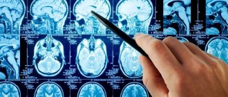
When a patient with signs of a stroke comes to the Yusupov Hospital, specialists strive to accurately determine the state of blood flow in the brain vessels of the right hemisphere. The first priority in complex diagnostics are visualization methods: CT and MRI, which make it possible to establish the localization of the lesion, the type and subtype of stroke, and also determine which vessel pathology led to disruption of brain nutrition.
With ischemic stroke, a “mismatch” phenomenon occurs, which can be detected during perfusion CT. A feature of this condition is that with a decrease in blood volume and the speed of its movement, an ischemic penumbra zone appears. This area of the brain contains cells whose death can be prevented.
Comprehensive diagnosis of ischemic stroke of the right hemisphere at the Yusupov Hospital is carried out using high-precision methods:
- cerebral angiography is prescribed to patients if surgical intervention is necessary;
- transcranial Doppler sonography is carried out to study the state of blood flow in the brain;
- electroencephalography is used to establish areas of subcortical and cortical damage;
- Positron emission tomography and scintigraphy are performed to assess the nature of brain metabolism.
Causes
The causes of the disease most often lie in bad habits or the presence of chronic vascular diseases. As with many diseases, it is difficult to identify specific causes of transient ischemic attack. Therefore, it is customary to talk about risk factors - factors that increase the likelihood of developing TIA.
- Atherosclerosis of cerebral vessels.
- Arterial hypertension.
- Heart diseases - myocardial infarction, endocarditis, atrial fibrillation.
Risk factors also include some features of the patient’s lifestyle:
- Having a smoking addiction.
- Obesity.
- Sedentary lifestyle.
All of these factors negatively affect the blood vessels of the brain, contributing to loss of elasticity or narrowing. As a result, when the brain needs an influx of oxygen and energy, the vessels cannot provide sufficient nutrition to the cells. This is how ischemia develops.
Treatment
A patient who has suffered a stroke requires emergency care and hospitalization. Numerous studies of this disease confirm that treatment carried out in the first three days is most effective, since the activity of nerve cells surrounding the lesion is high.
Neurologists at the Yusupov Hospital begin treating the consequences of a stroke after a comprehensive examination of the patient. The treatment and rehabilitation programs developed by our specialists are based on the principle of the individuality of each patient, however, if the blood supply to the brain is impaired, the following measures are mandatory:
- monitoring of cardiac activity and blood pressure;
- prevention of early contractures, pneumonia;
- ensuring the required level of blood oxygen saturation and removing excess carbon dioxide;
- maintaining homeostasis - equilibrium of the internal environment;
- monitoring the condition of the intestines and bladder;
- swallowing control;
- skin care and prevention of bedsores.
Treatment of patients after pneumonia involves the use of drugs and non-drug methods. To take into account contraindications, cardiologists, neurosurgeons and resuscitators take part in drawing up the treatment program.
Drug therapy after a stroke involves the use of the following drugs:
- antihypertensive drugs for high blood pressure;
- osmotic diuretics and dexazone are used for cerebral edema, as well as to prevent increased intracranial pressure;
- anticoagulants, which include heparin, help control blood clotting;
- antiplatelet agents (trental, beta blockers, calcium antagonists) are used to identify signs of insufficient blood supply;
- drugs with a neuroprotective effect are used to restore neurons.
The Yusupov Hospital creates comfortable conditions for every patient. After a stroke, patients need professional care, which is provided by the staff of the neurology clinic:
- introducing fluids and nutrients into the body;
- turning the patient from one side to the other every 2 hours;
- performing cleansing enemas once every two days;
- wiping the skin every 8 hours with camphor alcohol;
- cleaning the nasopharynx and oropharynx using suction and then rinsing them.
Treatment of arterial stroke
If the symptoms described above appear, you must immediately call an ambulance. The time from the onset of acute cerebrovascular accident to the development of irreversible processes of necrotization of the nervous tissue of the brain is 2-2.5 hours. The treatment approach varies for different types of stroke. In hemorrhagic stroke, it is important to promptly begin brain decompression therapy to avoid herniation of the brain stem into the foramen magnum, which is fatal. For this use:
- Antihypertensive drugs that lower blood pressure;
- Diuretics that cause diuresis and reduce the volume of circulating blood. If there is a large amount of bleeding and the hematoma grows, surgical intervention with craniotomy is required.
For ischemic stroke, the tactics are completely different. The patient is prescribed thrombolytic therapy aimed at eliminating thrombosis. In some cases, it is possible to use surgical intervention under X-ray control. Embolectomy and stenting of the affected arteries are performed, and blood flow is restored. The Ural Center for Resuscitation Neurorehabilitation offers high-quality services for the treatment and rehabilitation of even the most severe forms of stroke. Only correct diagnosis, comprehensive treatment and full rehabilitation can avoid serious consequences and complications after a stroke.
Rehabilitation
The implementation of the rehabilitation program begins after the patient’s condition improves. In the rehabilitation clinic, patients interact with speech therapists who offer exercises to gradually restore written and spoken language skills. Physiotherapeutic procedures and massages are carried out by professional rehabilitation therapists. Exercise therapy instructors at the Yusupov Hospital restore motor function through therapeutic exercises, physical education and kinesitherapy.
Stroke rehabilitation programs may include the following:
- magnetic therapy;
- laser therapy;
- acupuncture;
- kinesio taping;
- Voita therapy;
- massage sessions;
- treatment by position and movement.
The rehabilitation clinic of the Yusupov Hospital has modern technical equipment, which, combined with the knowledge of specialists, ensures effective restoration of functions after an ischemic stroke on the right side of the brain.
Treatment of cerebral vessel atherothrombosis
First aid for the patient consists of urgent surgical intervention. A carotid endarterectomy is performed, during which the damaged area is removed. Next, drug treatment is prescribed:
- Using special medications, existing blood clots are dissolved.
- Drugs are prescribed that correct pathological disorders of the cardiovascular system and reduce blood clotting;
- If thrombosis continues to progress, then direct and indirect anticoagulants are prescribed;
- To further protect and stabilize the functioning of the nervous system, neuromodulators and neurotrophic drugs are used;
- General strengthening therapy is carried out, accelerating energy metabolism;
- To regenerate damaged tissues, antioxidants are prescribed
Clinical Brain Institute Rating: 4/5 — 2 votes
Share article on social networks
Forecast
Right hemisphere ischemic stroke is a disease associated with high mortality. However, the achievements of modern medicine allow neurologists to fight for the life of each patient and improve its quality. The prognosis for ischemic right-sided stroke depends on factors such as the timeliness and correctness of first aid, the age of the patient, treatment and rehabilitation tactics, and the patient’s readiness.
Restoring functions lost due to a stroke lasts up to a year. However, activities carried out in the first trimester are most effective. There are cases where specialists are unable to restore speech in left-handed people with a right-sided stroke, while other functions are completely restored.
The patient and his loved ones must be prepared for the fact that rehabilitation measures are delayed for several years. During this period, it is important to follow your doctor’s orders, do home exercises, and attend procedures. At the Yusupov Hospital, relatives are trained in the basics of care and interaction with the patient.
The rehabilitation programs implemented at the Yusupov Hospital are highly effective, this conclusion can be drawn from research conducted by neurologists. Some patients who underwent treatment and rehabilitation were able not only to return to a full life, but also to continue working. The achievement of other people after rehabilitation was that they were able to take care of themselves and communicate with others.
Treatment and rehabilitation after ischemic stroke of the right hemisphere is carried out in accordance with international standards. The medical staff provides a wide range of services and creates conditions for the recovery of patients. To schedule a consultation or get answers to your questions, you can contact the Yusupov Hospital by phone.


