One of the types of ischemic stroke is lacunar, localized in the pontomesencephalic region or cerebral hemispheres. This is the most common form of pathology diagnosed at the Yusupov Hospital in many patients with ischemic stroke. Early diagnosis will avoid complications and can be performed in a modern hospital clinic equipped with high-precision medical equipment. Yusupov Hospital, rightfully considered a leading clinic for the treatment of strokes.
Overview of Lacunar Ischemic Stroke
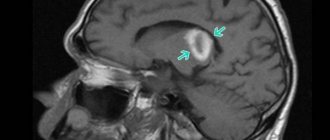
Lacunar ischemic stroke leads to pathological disorders in the deep layers of the brain and the formation of so-called lacunae (cavities), the diameter of which ranges from one to fifteen millimeters. Sometimes the lacunae merge, resulting in the formation of large cavities filled with blood or plasma and fibrin.
With this pathology, the patient’s consciousness, vision, speech, and other functions of the cerebral cortex are practically not impaired. In addition, symptoms of brain stem damage are also completely absent. Thanks to these distinctive features, lacunar stroke can be differentiated from other types of this pathology.
In the absence of qualified neurological assistance, this type of brain catastrophe threatens the development of severe complications, including death. According to statistics, after patients have suffered a lacunar ischemic stroke of the brain, the survival prognosis is: in the first month - 70-80%, within a year - about 50%. Therefore, early diagnosis of pathology plays a vital role.
Symptoms
The disease may not manifest itself in any way throughout life. The occurrence of symptoms is directly related to the size of the formation and its location; a cyst on the right, a cyst on the left, in the occipital, temporal, parietal region can be diagnosed. If the lacuna is small and does not put pressure on nearby tissues, a person will never guess about the pathology.
The severity of symptoms depends on which areas are under pressure:
- A cyst of the basal ganglia leads to a lack of coordination;
- Damage to the parietal lobe – disturbances in written and oral speech, deterioration of coordination;
- The consequence of damage to the occipital region is visual impairment;
- Pressure in the temporal region leads to loss of taste and smell;
- Damage to the insular zone leads to a loss of critical assessment of one's condition;
- Damage to the frontal lobe causes disturbances in facial expressions and epileptic seizures.
Consequences
A number of complications with an increase in the size of the lacuna:
- Cephalgia, the strength of which changes with changes in body position;
- Nausea;
- Vomit;
- Presence of seizures;
- Hallucinations.
Clinical manifestations of the disease include high cranial pressure due to tumor growth and disturbances in the circulatory system of the brain. A feeling of heaviness, headache, and oppressive nature are the main symptoms of the disease.
Causes of lacunar ischemic stroke
The main cause of lacunar stroke is considered to be arterial hypertension, which leads to brain damage and depends on blood pressure, the degree of damage to the arterial walls and their condition. In order to prevent the development of a cerebral catastrophe, doctors at the Yusupov Hospital monitor surges that occur during the day, since lacunar stroke occurs against the background of sharp changes in arterial pressure.
The risk group consists of persons suffering from the following pathologies:
- hyaline dystrophy AG;
- atherosclerosis;
- infectious inflammation of arterioles localized in the brain (previously suffered);
- diabetes mellitus
Along with the above, lacunar ischemic stroke of the brain can be caused by vasculitis, the form of which can be either specific or nonspecific.
Consequences
The lack of quality therapy leads to the worsening of old symptoms and the appearance of new ones. A person loses coordination, his vision decreases, up to complete loss. Facial expressions and speech deteriorate, and it becomes difficult to walk. A person can be paralyzed.
Infants are at risk of developing hydrocephalus. Liquor fluid accumulates in the ventricles, and the brain contracts. With proper diagnosis and treatment, you can get rid of symptoms and not worry about the appearance of new formations. The main thing is a correct approach to the problem and prevention, which usually lasts a little less than 2 years.
A lacunar cyst is a benign tumor that does not develop into a malignant one. If the disease is not treated, the formation will increase in volume and affect parts of the cerebral cortex, impairing the functioning of the body. There is no need to panic; in most cases, the cyst does not interfere with the functioning of organs and systems, and the person lives peacefully, unaware of the pathology.
Diagnosis of lacunar ischemic stroke
In order to determine lacunar ischemic stroke, the Yusupov Hospital uses modern diagnostic methods such as computed tomography and magnetic resonance imaging, which are the most informative in this situation. Using these studies, the location, number and volume of formed lacunae are revealed. With lacunae of small diameter, fixation of lesions is difficult. To make a final diagnosis, along with the results of the studies, the neurologist takes into account the patient’s medical history, especially if he is diagnosed with diabetes mellitus, arterial hypertension or alcoholism.
Types of cysts
Lacunar cysts are divided into congenital and acquired. The first type is classified as a primary type of disease, the second - as a secondary one. A congenital cyst develops in a baby in the womb. An acquired lacunar cyst appears due to external factors.
The primary type of the disease, as mentioned above, develops even before birth. This is influenced by the behavior of the mother who abuses bad habits. Alcohol, smoking, and drugs negatively affect the development of the fetus. Such habits cause the child’s genes to mutate and impair fetal nutrition and blood supply. In addition to the appearance of a lacunar cyst, other congenital diseases are possible in a child.
In addition to the bad habits of the mother, the child is affected by diseases suffered by the woman during pregnancy. They affect the further development of the central nervous system of the fetus. Cyst formation occurs at three stages of pregnancy, but often the disease appears in the first trimester.
The secondary type of disease occurs due to acquired diseases. The range of problems is significant: meningitis, head injuries. A lacunar cyst can form due to diabetes, hypertension, or a connective tissue disorder. Its formation is affected by weakened immunity. A mother's weak immune system has a negative impact on the child.
Treatment of lacunar ischemic stroke
Treatment of lacunar stroke at the Neurology Center of the Yusupov Hospital is based, first of all, on the use of drugs whose action is aimed at improving cerebral circulation, as well as having a neuroprotective effect. The development of collateral blood flow is promoted by drugs such as cinnarizine and Cavinton.
The Yusupov Hospital attaches great importance to the treatment of the underlying disease that caused vascular damage. Blood pressure is constantly monitored. If its levels are high, the patient is prescribed antihypertensive drugs. High cholesterol levels, determined by laboratory tests, are corrected by taking lipid-lowering drugs - statins, the action of which is aimed at blocking enzymes that help the synthesis of cholesterol compounds in the liver.
Specialists at the Neurology Clinic of the Yusupov Hospital constantly monitor the function of the patient’s cardiovascular and respiratory systems. If necessary, the patient is prescribed medications that correct the water-electrolyte balance in the body and reduce swelling of brain tissue.
To prevent relapse, the patient is prescribed antiplatelet agents. For arterial thromboembolism, warfarin is used. In some cases, a course of anticonvulsant drugs is recommended.
To prevent dementia, patients are prescribed a course of drugs Neuromidin or Gliatilin. In the presence of pseudobulbar syndrome, fluoxetine is used.
For patients with lacunar ischemic stroke, doctors at the Yusupov Hospital use an individual approach, resulting in high treatment results.
Experts' prognosis for lacunar ischemic stroke
If a patient is diagnosed with a single lacunar stroke of the brain, the prognosis is favorable. As a rule, after rehabilitation, the patient experiences restoration of all functions, although the presence of sensory residual and motor symptoms may sometimes be observed.
During a relapse, a lacunar state of the brain may develop, and the risk of this complication is very high: according to statistics, after a repeated lesion, this occurs in almost 70% of cases.
Despite the restoration of all impaired functions, lacunar ischemic stroke negatively affects the mental state of the patient, in which gradual changes occur. There is the appearance of memory loss, disorientation and difficulty communicating, tearfulness, frequent hysterics, a feeling of helplessness and a state of passion.
Clinical picture
Primary cysts most often do not manifest themselves in any way and do not interfere with a person’s life. As a rule, this pathology is not treated.
Symptoms of a secondary formation depend on its size and etiology. Small brain cysts do not have pathological symptoms; they are characteristic of large specimens. However, experts have identified several common signs of the disease:
- Severe pain in the head that is not relieved by painkillers.
- Dizziness, in some cases there is loss of consciousness.
- Bursting and pulsating sensations in the head area.
- Periodic nausea. Sometimes it ends in vomiting, which, in turn, does not bring relief.
- Disturbance of sleep and biological rhythms of patients.
- Cramps, tremor of fingers.
- Neurological symptoms.
- In children under one year of age, bulging and pulsation of the large fontanel and frequent regurgitation, in some cases vomiting, are observed.
- Coordination of movements worsens, gait changes.
- Partial or complete paresis and paralysis of the upper and lower extremities occur.
- Some areas of the skin may lose sensitivity.
- Pathological changes in hearing, vision, and speech are possible.
It is also necessary to note individual symptoms of cerebellar cysts depending on their type:
- hallucinations are characteristic of a retrocerebellar cyst;
- when the right half of the cerebellum is damaged, a person becomes quickly fatigued, both physical and mental;
- paralysis is more often observed with the development of pathology in the left half of the organ;
- hydrocephalus is a sign of cyst-like expansion of the subarachnoid space.
Rehabilitation after lacunar ischemic stroke
Rehabilitation at the Yusupov Hospital involves a whole range of measures: medical, social and psychological. They are aimed at restoring functions lost after a stroke. The hospital’s highly qualified doctors: neurologists, physiotherapists, psychotherapists have extensive practical experience in the field of rehabilitation medicine; they have in their arsenal the world’s leading techniques, modern medical equipment and the latest drugs for treating the consequences of brain accidents, thanks to which they can achieve high results. The clinic provides services for transporting patients to the hospital. Call and the coordinating doctor will answer all your questions.
Paranasal sinus cyst - symptoms and treatment
Treatment depends on the type of cyst, its location and size. If it is less than 1 cm and there is no discharge, then the doctor suggests conservative methods: regular examination, tablets, sprays and drops. If the cyst is large or there is purulent discharge, then surgery will be required.
Drug treatment of cysts
The method allows you to eliminate unpleasant symptoms and causes of cyst formation: swelling, inflammation and blockage of the sinus anastomosis. Drug treatment is effective and can alleviate the patient's condition in the early stages of the disease. However, it will not help when the fibrous membrane of the cyst has become too dense.
Drugs used:
- vasoconstrictor drops - quickly reduce swelling in the anastomosis area and normalize ventilation of the sinuses of the nasal cavity; medications in this group are addictive and can be safely used for no more than 5 days in a row;
- antihistamines - will help if the cyst is caused by allergies;
- mucolytics - normalize the outflow of mucus;
- antiseptics - help fight inflammation and cleanse the surface of the nasal mucosa;
- topical glucocorticosteroids are most effective in the treatment of allergic rhinosinusopathy, prolonged sinusitis, hyperplasia of the mucous membrane in the area of the anastomosis and ostiomeatal complex;
- painkillers - reduce pain caused by the pressure of the cyst on surrounding tissues [6];
- probiotics - normalize the microflora of the nasal cavity and nasopharynx, which is important in the treatment of chronic inflammation of the upper respiratory tract.
Surgery
Indications:
- cyst more than 8 mm;
- ineffectiveness of conservative treatment;
- signs of suppuration inside the cyst [7].
The most popular method of treating cysts is puncture or puncture. The surgeon pierces the wall of the cyst with a thin needle and pumps out the purulent discharge [1]. However, this method only has a temporary effect. Piercing the cyst, as well as spontaneous leakage of its contents, relieves pain for a while. But gradually the cyst grows together again and begins to accumulate purulent fluid. To remove it completely, more serious surgery will be required [8].
Maxillary sinusotomy according to Caldwell-Luke
It is performed through an incision under the upper lip in the mouth. This is a classic method for removing sinus cysts, but now it is practically not used due to its high morbidity: after the operation, scars, adhesions can form and the functioning of the sinuses can be disrupted.
Microsinusrotomy
A minimally invasive surgical technique that is performed through the anterior wall of the maxillary sinus. A 4–8 mm hole is formed in the bone, and the cyst is removed with special instruments. Performed under general anesthesia. The disadvantage is the risk of damage to the branches of the trigeminal nerve and the formation of persistent facial neuralgia.
Functional Endoscopic Sinus Surgery (FESS surgery)
This method is considered the most gentle and effective: during the procedure, unnecessary incisions and punctures are not made on the patient’s face. It is performed through the nasal cavity under endoscopic control. Allows you to reach all paranasal sinuses, including the sphenoid, frontal sinuses and cells of the ethmoid labyrinth. The operation is performed under local and general anesthesia [17].
Additional benefits:
- can be used to treat children;
- reduce the likelihood of cyst recurrence;
- you need to stay in the hospital for no more than two days;
- the incision heals quickly;
- the operation leaves no scars;
- there is no risk that adhesions will appear [18].
Relative disadvantages: trained specialists and special equipment are required, which makes the operation expensive.