Demyelinating disease is a pathological process of destruction of the myelin sheath that affects neurons in the brain and spinal cord. At the same time, the conductivity of impulses in the nervous system deteriorates. The disease is characterized by the destruction of brain myelin. This dangerous condition affects the functioning of the entire body. The disease occurs with equal frequency in both adults and children. Modern medicine does not have the means to completely cure this disease. It can only be weakened and the flow slowed down.
Demyelination
Demyelinating brain disease according to ICD-10 has codes G35, G36 and G77. The process caused by damage to the nervous tissue negatively affects the functioning of the entire organism as a whole. Certain nerve endings are covered with a myelin sheath, which performs important functions in the body. For example, myelin ensures the rapid transmission of electrical impulses and, accordingly, if this process is disrupted, the entire system suffers. Myelin consists of lipids and protein compounds in a 70/30 ratio.
Expert opinion
Author: Daria Olegovna Gromova
Neurologist
The most common demyelinating brain disease is multiple sclerosis. Its symptoms are very diverse, so the patient rarely immediately comes to a neurologist. Decreased vision, numbness of the limbs, problems with motor skills - different doctors give different recommendations, although it is enough to simply send the patient for an MRI and laboratory tests that will confirm fears or rule out brain diseases.
Demyelinating disease is not only multiple sclerosis, it is also neuromyelitis optica and acute disseminated encephalomyelitis. These diseases have no cure, but their progression can be slowed down. In general, doctors give a favorable prognosis when treating these pathologies. The diagnosis of “multiple sclerosis” is now made more often, but the disease itself is milder than 30-40 years ago.
Thanks to modern methods of therapy and research, a benign course is observed in approximately half of the cases - patients fully retain their ability to work, start families, and give birth to children. There are some restrictions, for example, visiting baths and saunas, prolonged exposure to the sun is prohibited, but otherwise the lifestyle remains the same, no special diets are needed.
Development mechanisms
The nervous system consists of central and peripheral sections. The regulation mechanism between them works as follows: impulses from the receptors of the peripheral system are transmitted to the nerve centers of the spinal cord, and from there to the brain. The disruption of this complex mechanism causes demyelination.
Nerve fibers are covered with a myelin sheath. As a result of the pathological process under consideration, this membrane is destroyed and fibrous tissue is formed in its place. She, in turn, cannot conduct nerve impulses. In the absence of nerve impulses, the functioning of all organs is disrupted, since the brain cannot issue commands.
What is demyelination
The demyelinating process that occurs in the brain is a disorder that leads to a pathological change in the structure of the nervous tissue, which often provokes the appearance of neurological symptoms and causes disability and death. The white shell is a multilayer cell membrane. Biocurrents are not able to overcome the myelin sheath.
Electrical impulses are transmitted to the areas of numerous nodes of Ranvier, where myelin is absent. The interceptions are located at regular intervals of approximately 1 µm in length. In the peripheral system, the protective membrane is formed from lemmocytes (Schwann cells). Demyelination of nerve fibers is a lesion of nervous tissue that leads to a large number of central nervous system diseases.
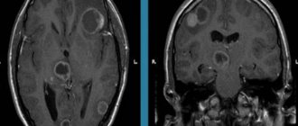
Foci of demyelination are structural formations in the brain or spinal cord that lack myelin, which leads to disruption of the transmission of nerve impulses and disruption of the nervous system as a whole. The diameter of the lesions varies significantly; it can be several millimeters or reach several centimeters.
Causes
Demyelinating diseases of the brain are characterized by damage to the peripheral and central nervous systems. They most often occur against the background of a genetic predisposition. It also happens that a combination of certain genes provokes disturbances in the functioning of the immune system. There are other causes of demyelinating diseases:
- state of chronic or acute intoxication;
- solar radiation;
- ionizing radiation;
- autoimmune processes in the body and genetic pathologies;
- complication of bacterial and viral infection;
- poor nutrition.
The Caucasian race is considered to be the most susceptible to this pathology, especially those representatives who live in northern latitudes. This type of disease can be provoked by a head or spinal injury, depression, or bad habits. Some types of vaccines can also trigger the development of such pathologies. This applies to vaccinations against measles, smallpox, diphtheria, influenza, whooping cough, and hepatitis B.
Prevention
To prevent the development of demyelinating diseases, experts recommend promptly seeking medical help to quickly get rid of the main etiological provocateurs. Persons belonging to the high-risk community (persons who have suffered central nervous system disorders or brain pathologies) should undergo regular examinations by their attending physician and be especially attentive to their well-being. Self-medication in such situations is absolutely not allowed!
On our website we have collected more than 250 medical centers with reliable addresses and contact phone numbers, which can be sorted in the selection system according to parameters that are important to you. In the special catalog you will find current promotions and discounts. Managers of mrt-mozga.ru will book you in at a convenient time!
Classification
Demyelinating disease of the nervous system is classified into different types, which are based on the destruction of the myelin sheath. In this regard, the pathology under consideration is divided accordingly into multiple sclerosis, Marburg disease, Devic disease, progressive multifocal leukoencephalopathy and Guillain-Barré syndrome.
Multiple sclerosis
Multiple sclerosis is characterized as a severe chronic and immunodegenerative disease of the central nervous system, prone to progression. In most cases, the disease occurs at a young age and almost always leads to disability. This demyelinating disease of the central nervous system is assigned code G35 according to ICD-10.
Currently, the causes of the development of multiple sclerosis are not fully understood. Most scientists are inclined to the multifactorial theory of the development of this disease, when genetic predisposition and external factors are combined. The latter include:
- infectious diseases;
- state of chronic intoxication;
- taking certain medications;
- change of place of residence with a sharp change in climate;
- lack of fiber and high-calorie diet.
The relationship between the symptoms of multiple sclerosis and the stage of the disease is not always clear. The pathology can have a wave-like course. Exacerbations and remissions can be repeated at varying intervals. A feature of multiple sclerosis is that each new exacerbation has a more severe course compared to the previous one.
The progression of multiple sclerosis is characterized by the development of the following symptoms:
- sensory disturbances (goosebumps, numbness, tingling, burning, itching);
- visual impairment (impaired color rendering, decreased visual acuity, blurred picture);
- trembling of the limbs or torso;
- headache;
- speech disorders and difficulty swallowing;
- muscle spasms;
- gait changes;
- cognitive impairment;
- heat intolerance;
- dizziness;
- chronic fatigue;
- libido disturbance;
- anxiety and depression;
- stool instability;
- insomnia;
- autonomic disorders.
There are also secondary symptoms of multiple sclerosis. They imply complications of the disease. Foci of demyelination in the brain are determined using magnetic resonance imaging, including the introduction of a contrast agent.
Treatment of multiple sclerosis is carried out using methods such as:
- plasmapheresis;
- taking cytostatics;
- prescription of immunosuppressants;
- use of immunomodulators;
- taking beta interferons;
- hormonal therapy.
- symptomatic therapy with the prescription of antioxidants, nootropic drugs and vitamins.
During the period of remission, patients are prescribed sanatorium treatment, massage, and physical therapy. In this case, all thermal procedures should be excluded. To alleviate the symptoms of the disease, medications are prescribed: those that reduce muscle tone, eliminate tremors, normalize urination, stabilize the emotional background, and anticonvulsants.
Multiple sclerosis is classified as an incurable disease. Therefore, these treatment methods are aimed at reducing symptoms and improving the patient’s quality of life. The life expectancy of patients with this disease depends on the nature of the pathology.
Demyelinating diseases of the central nervous system (multiple sclerosis, multiple encephalomyelitis)
Demyelinating diseases of the brain and spinal cord are pathological processes that lead to the destruction of the myelin sheath of neurons and disruption of the transmission of impulses between nerve cells of the brain. It is believed that the etiology of diseases is based on the interaction of the body's hereditary predisposition and certain environmental factors. Impaired impulse transmission leads to a pathological state of the central nervous system.
There are the following types of demyelinating diseases:
- Multiple sclerosis is a demyelinating disease of the central nervous system. The demyelinating disease multiple sclerosis is the most common pathology. Multiple sclerosis is characterized by a variety of symptoms. The first symptoms appear at the age of 20-30 years, women are more often affected. Multiple sclerosis is diagnosed by the first signs, which were first described by psychiatrist Charcot - involuntary oscillatory eye movements, trembling, scanned speech. Patients also experience urinary retention or very frequent urination, absence of abdominal reflexes, pallor of the temporal halves of the optic discs;
- ADEM, or acute disseminated encephalomyelitis. It begins acutely and is accompanied by severe cerebral disorders and manifestations of infection. The disease often occurs after exposure to a bacterial or viral infection and may develop spontaneously;
- diffuse disseminated sclerosis. It is characterized by damage to the spinal cord and brain and manifests itself in the form of convulsive syndrome, apraxia, and mental disorders. Death occurs within a period of 3 to 6-7 years from the moment the disease is diagnosed;
- Devic's disease, or acute neuromyelitis optica. The disease begins as an acute process, is severe, and progresses, affecting the optic nerves, which causes complete or partial loss of vision. In most cases, death occurs;
- Balo's disease, or concentric sclerosis, periaxial concentric encephalitis. The onset of the disease is acute, accompanied by fever. The pathological process occurs with paralysis, visual disturbances, and epileptic seizures. The course of the disease is rapid - death occurs within a few months;
- leukodystrophies - this group contains diseases that are characterized by damage to the white matter of the brain. Leukodystrophies are hereditary diseases; as a result of a gene defect, the formation of the myelin sheath of the nerves is disrupted;
- Progressive multifocal leukoencephalopathy is characterized by decreased intelligence, epileptic seizures, the development of dementia and other disorders. The patient's life expectancy is no more than 1 year. The disease develops as a result of decreased immunity, activation of the JC virus (human polyomavirus 2), often found in patients with HIV infection, after bone marrow transplantation, in patients with malignant blood diseases (chronic lymphocytic leukemia, Hodgkin's disease);
- diffuse periaxial leukoencephalitis. A hereditary disease that most often affects boys. Causes visual, hearing, speech, and other disorders. Progresses quickly - life expectancy is just over a year;
- Osmotic demyelination syndrome is very rare and develops as a result of electrolyte imbalance and a number of other reasons. A rapid increase in sodium levels leads to the loss of water and various substances from brain cells, causing the destruction of the myelin sheaths of nerve cells in the brain. One of the posterior parts of the brain is affected - the Varoliev bridge, which is most sensitive to myelinolysis;
- myelopathy is a general term for lesions of the spinal cord, the causes of which are varied. This group includes: tabes dorsalis, Canavan disease, and other diseases. Canavan disease is a genetic, neurodegenerative autosomal recessive disease that affects children and causes damage to nerve cells in the brain. The disease is most often diagnosed in Ashkenazi Jews living in Eastern Europe. Tabes dorsalis (locomotor ataxia) is a late form of neurosyphilis. The disease is characterized by damage to the posterior columns of the spinal cord and spinal nerve roots. The disease has three stages of development with a gradual increase in symptoms of damage to nerve cells. Coordination when walking is impaired, the patient easily loses balance, bladder function is often disrupted, pain appears in the lower limb or lower abdomen, and visual acuity decreases. The most severe third stage is characterized by loss of sensitivity of muscles and joints, areflexia of the tendons of the legs, development of astereognosis, the patient cannot move;
- Guillain-Barré syndrome - occurs at any age, refers to a rare pathological condition that is characterized by damage to the peripheral nerves of the body by its own immune system. In severe cases, complete paralysis occurs. In most cases, patients recover completely with adequate treatment;
- neural amyotrophy of Charcot-Marie-Tooth. A chronic hereditary disease, which is characterized by progression, affects the peripheral nervous system. In most cases, the myelin sheath of nerve fibers is destroyed; there are forms of the disease in which pathology of the axial cylinders in the center of the nerve fiber is detected. As a result of damage to the peripheral nerves, tendon reflexes fade and atrophy of the muscles of the lower and then upper extremities occurs. The disease belongs to progressive chronic hereditary polyneuropathies. This group includes: Refsum's disease, Roussy-Lévy syndrome, Dejerine-Sotta hypertrophic neuropathy and other rare diseases.
Demyelinating diseases of the nervous system: classification
Demyelinating diseases (DD) are divided into primary (acute disseminated encephalomyelitis, clinical forms - polyencephalitis, encephalomyelopolyradiculoneuritis, optomyelitis, disseminated myelitis, optoencephalomyelitis) and secondary (vaccinal - develop during vaccination with DPT, DTP, atirabiic vaccine; parainfectious - encephalomyelitis with influenza, coc Lushe, Corey , chicken pox, other diseases).
Subacute forms of the disease manifest themselves in the form of diseases:
- multiple sclerosis. Clinical forms – cerebrospinal and spinal, optical, cerebral, brainstem, cerebellar;
- chronic forms of ID – Dawson, Doering, Pette, Van Bogart encephalitis, Schilder’s periaxial diffuse leukoencephalitis;
- Disorders affecting predominantly peripheral nerves - infectious polyradiculoneuropathy, infectious-allergic primary Guillain-Barré polyradiculoneuritis, toxic polyneuropathy, diabetic and dismetabolic polyneuropathy.
Treatment of demyelinating diseases of the nervous system
Treatment of demyelinating disease depends on the type of disease and its severity, and is a long-term and complex process. In addition to drug treatment (pulse therapy at the onset of the disease and exacerbation), the patient is prescribed a diet, recommended to follow a strict daily routine, sleep and wakefulness, regularly undergo massage, and engage in physical therapy. Each patient with demyelinating disease requires an individual approach; maintenance therapy for patients with demyelinating disease takes many years.
« Back to previous page
Marburg disease
Hemorrhagic fever or Marburg disease is an acute infectious pathology caused by the Marburg virus. It enters the body through damaged skin and mucous membranes of the eyes and mouth.
Infection occurs through airborne droplets and sexual contact. In addition, you can become infected through the blood and other secretions of the patient. After a person recovers from this disease, he develops a stable and long-lasting immunity. Re-infection was not encountered in practice. A person who has recovered from Marburg hemorrhagic fever experiences the formation of necrosis and foci of hemorrhage in the liver, myocardium, lungs, adrenal glands, kidneys, spleen and other organs.
The symptoms of the disease depend on the stage of the pathological process. The incubation period lasts from 2 to 16 days. The disease has an acute onset and is characterized by an increase in body temperature to high levels. Along with fever, chills may appear. Signs of intoxication increase, such as weakness, headache, pain in muscles and joints, intoxication and dehydration. 2-3 days after this, gastrointestinal dysfunction and hemorrhagic syndrome appear. A stabbing pain appears in the chest area, which intensifies during breathing. In addition, the patient may develop a dry cough and substernal stabbing pain. The pain may spread to the throat area. The mucous membrane of the pharynx becomes very red. Almost all patients experienced diarrhea, which lasted almost 7 days. A characteristic sign of this disease is the appearance of a measles-like rash on the body.
All symptoms intensify by the end of the first week. Bleeding from the nose, gastrointestinal tract, and genital tract may also be observed. By the beginning of week 2, all signs of intoxication reach their maximum. In this case, convulsions and loss of consciousness are possible. According to the blood test, specific changes occur: thrombocytopenia, poikilocytosis, anisocytosis, granularity of erythrocytes.
If a person is suspected of having Marburg disease, he is urgently hospitalized in the infectious diseases department and must be kept in an isolated box. The recovery period may take up to 21-28 days.
Devic's disease
Neuromyelitis optica or Devic's disease has a chronic pattern similar to multiple sclerosis. This is an autoimmune disease, the causes of which are still unclear. One of the reasons for its development is an increase in the permeability of the barrier between the meninges and the vessel.
Some autoimmune diseases can trigger the progression of Devic's disease:
- rheumatoid arthritis;
- systemic lupus erythematosus;
- Sjögren's syndrome;
- dermatomyositis;
- thrombocytopenic purpura.
The disease has specific symptoms. Clinical manifestations are caused by disruption of conductive impulses. In addition, the optic nerve and spinal cord tissue are affected. In most cases, the disease manifests itself as visual impairment:
- a veil before the eyes;
- pain in the eye sockets;
- blurred vision.
As the disease progresses and there is no adequate treatment, the patient runs the risk of completely losing his vision. In some cases, regression of symptoms with partial restoration of eye function is possible. Sometimes it happens that myelitis precedes neuritis.
Neuromyelitis optica has two course options: a progressive increase in symptoms with simultaneous damage to the central nervous system. In rare cases, a monophasic course of the disease occurs. It is characterized by steady progress and worsening symptoms. In this case, the risk of death is increased. With the right treatment, the pathological process slows down, but complete recovery is not guaranteed.
The second option, the most common, is characterized by an alternating change of remission and exacerbation and is designated by the concept of “recurrent course.” It is also accompanied by visual disturbances and spinal cord dysfunction. During the period of remission, a person feels healthy.
To identify Devic's disease, a set of measures is carried out. In addition to standard diagnostic procedures, lumbar puncture with cerebrospinal fluid analysis, ophthalmoscopy and MRI of the spine and brain are performed.
Treatment of Devic's disease is long and difficult. The main goal is to slow the progression of the disease and improve the patient’s quality of life. As part of drug therapy, glucocorticosteroids, muscle relaxants, antidepressants, and centrally acting painkillers are used. In severe cases of the disease, the patient may face complications such as paralysis of the legs, blindness, or persistent dysfunction of the pelvic organs. With timely and correct treatment, a complete recovery is guaranteed.
Multiple lesions
Such lesions are much less common compared to single gliosis of the brain. This pathology can develop against the background of various diseases of the blood vessels, the central nervous system (in particular, white matter), as well as connective tissues.
Typically, the cause of such lesions is atherosclerosis, as well as previous strokes and heart attacks. Also, multiple lesions can occur due to traumatic brain injury.
It is important to note that with brain trauma, a process of necrosis occurs, and in different areas and in different areas. As a result, it is the subcortical foci of tissue death that are much more susceptible to gliotic degeneration.
When receiving minor head injuries, the size of the lesions is no more than a few millimeters. Thanks to this, the functions of human thinking remain practically unchanged.
If a patient has even the smallest structural changes, they can easily be detected using an MRI examination.
Progressive multifocal leukoencephalopathy
People with immune deficiency may experience progressive multifocal multifocal leukoencephalopathy. This is an infectious disease caused by the penetration of the JC virus, which belongs to the Polyomavirus family, into the body. A feature of the pathology is that asymmetric and multifocal brain damage occurs. The virus affects the membranes of nerve endings, which are made of myelin. Therefore, this disease belongs to the group of demyelinating pathologies.
Almost 85% of patients with this diagnosis are AIDS or HIV infected. The risk group includes patients with malignant tumors.
Main symptoms of the disease:
- mood swings;
- visual disturbances;
- paresthesia and paralysis;
- memory impairment.
Unit formations
Such formations are more common in different age groups.
MRI of the brain can detect foci of gliosis in both infants and elderly patients.
In the first case, the formation of altered lesions may be associated with the consequences of postpartum trauma; in the second, it may be the result of degeneration of brain tissue due to age-related changes.
This process is a natural age-related change that is visible in 60% of cases when scanning white matter in people over 85 years of age.
As a rule, such lesions are detected by chance, since they do not cause discomfort. However, when lesions form in the left frontal lobe, they often provoke the appearance of hallucinations.
This phenomenon is due to the fact that in this part there are the most important centers responsible for deep feelings and sensations.
The main feature of such a lesion is that it is not predisposed to proliferation and carries a significant risk in the development and exacerbation of chronic diseases.
Guillain-Barre syndrome
An acute inflammatory disease characterized by “demyelinating polyradiculoneuropathy.” It is based on autoimmune processes. The disease manifests itself as sensory disturbances, muscle weakness and pain. It is characterized by hypotension and disorder of tendon reflexes. Respiratory failure may also occur.
All patients with this diagnosis should be admitted to the intensive care unit. Since there is a risk of developing respiratory failure and a ventilator may be required, the department must have intensive care. Patients also need proper care with the prevention of bedsores and thromboembolism. It is also necessary to stop the autoimmune process. For this purpose, plasmapheresis and pulse therapy with immunoglobulins are used. Full recovery of patients with this diagnosis should be expected within 6-12 months. Deaths occur due to pneumonia, respiratory failure and pulmonary embolism.
Symptoms
Demyelination always manifests itself as a neurological deficit. This sign indicates the beginning of the process of myelin destruction. The immune system is also involved. Brain tissue - spinal and brain - atrophies, and expansion of the ventricles is observed.
Manifestations of demyelination depend on the type of disease, causative factors and location of the lesion. Symptoms may be absent when the damage to the brain substance is minor, up to 20%. This is due to a compensatory function: healthy brain tissue performs the tasks of the affected areas. Neurological symptoms rarely appear - only when more than 50% of the nervous tissue is damaged.
The following are common signs of demyelinating brain diseases:
- paralysis;
- limited muscle mobility;
- tonic spasms of the limbs;
- dysfunction of the intestines and bladder;
- pseudobulbar syndrome (impaired pronunciation of sounds, difficulty swallowing, change in voice);
- impaired fine motor skills of the hands;
- skin numbness and tingling;
- visual dysfunction (decreased visual acuity, blurred images, fluctuations in the eyeballs, color distortions).
Neuropsychological disorders characteristic of the pathology in question are caused by memory deterioration and a decrease in mental activity, as well as changes in behavior and personal qualities. This is manifested by the development of neuroses, depression, dementia of organic origin, emotional swings, severe weakness and decreased performance.
How does it manifest itself?
Depending on the location of the inflammation, different symptoms and MR signs appear. All abnormal problems can be divided into several conditional categories:
- Malfunctions in the musculoskeletal system - the formation of paresis, paralysis, abnormal reflexes.
- The likelihood of damage to one of the parts of the brain (cerebellum) is difficulties in orientation in space and coordination of movements, spastic paralysis.
- Symptoms of damage to the base of the brain are disorders of the speech apparatus, swallowing ability, and the inability to carry out actions with the eye orbits.
- Unstable operation of the sensitive features of the surface layer of the skin.
- Deformations in the genitourinary system and internal objects of the small pelvis - painful emptying, incontinence.
- Ophthalmological disorders - decreased vision, even blindness.
- Cognitive imbalance - memory impairment, low speed of thought process and attention.
Diseases included in the demyelinating group are capable of creating a greater neurological defect each time, which causes changes in the patient's behavior, poor health, low level of performance, etc. At the last stage, the patient can expect the most severe consequences, for example, heart failure, shutdown of the breathing apparatus.
If the above-described complaints occur, you must urgently seek medical help. Specialists will be able to provide treatment and relieve the symptoms of the disease. The search service mrt-mozga.ru will help you make an appointment with local doctors for a consultation or body examination. The company provides services for the quick selection of clinical centers performing MRI, CT, ultrasound and other types of diagnostics. On the website you can apply for an appointment with a doctor in the capital in a few clicks, without queuing! Using the services of the search portal, you can get advantageous discounts and promotional offers.
Diagnostics
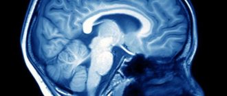
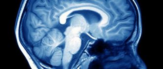
An early stage of the pathology with the absence of characteristic symptoms is accidentally discovered during a diagnostic examination for another reason. To confirm the diagnosis, neuroimaging is performed, and the neurologist determines the degree of impairment of the conductive function of the brain. The main diagnostic method is magnetic resonance imaging. In the photographs you can clearly see areas of the affected tissue. If you do angiography, you can determine the extent of vascular damage.


