The most common complaint that doctors hear from their patients is headache. Both adults and children complain about it. It is impossible to ignore this. Especially if there are other symptoms. Parents should pay special attention to the child’s headaches and the baby’s behavior, because he cannot say that he is in pain. Perhaps these are the consequences of a difficult birth or congenital anomalies, which can be determined at an early age. Maybe these are liquorodynamic disturbances. What is it, what are the characteristic signs of this disease in children and adults and how to treat it, we will consider further.
What does liquorodynamic disturbances mean?
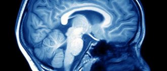
Liquor is cerebrospinal fluid that constantly circulates in the ventricles, cerebrospinal fluid ducts and in the subarachnoid space of the brain and spinal cord. Liquor plays an important role in metabolic processes in the central nervous system, in maintaining homeostasis in brain tissue, and also creates a certain mechanical protection for the brain.
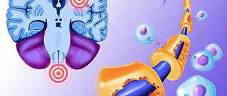
Liquorodynamic disorders are conditions in which the circulation of cerebrospinal fluid, its secretion and reabsorption are impaired. These processes are regulated by glands located in the choroid plexuses of the brain ventricles that produce fluid.
In the normal state of the body, the composition of the cerebrospinal fluid and its pressure are stable.
Basic information about pathology
A special cerebrospinal fluid, or cerebrospinal fluid, is produced in the choroid plexuses of the brain, called the ventricles, and fills the subarachnoid space. Excess cerebrospinal fluid is released into the bloodstream through the arachnoid membrane. When problems with absorption, circulation occur, or excessive secretion of cerebrospinal fluid occurs, hydrocephalus develops. It can be congenital or acquired as a result of certain diseases. In addition, the following forms of the disease are distinguished:
- internal hydrocephalus - overflow of one or more ventricles inside the brain with cerebrospinal fluid;
- external hydrocephalus - fluid injection between the brain and the enveloping subarachnoid membrane.
In its acute form, the disease develops over several days. Subacute course involves the development of decompensation within a month from the onset of the first symptoms, chronic - longer than six months. In addition, the classification of hydrocephalus includes stable and progressive forms of pathology, characterized, respectively, by stability or a gradual increase in pressure inside the skull.
What is the mechanism of violations
Let's consider how liquorodynamic disorders of the brain can develop:
- The rate of production and release of cerebrospinal fluid by the choroid plexuses increases.
- The rate of absorption of cerebrospinal fluid from the subarachnoid space slows down due to the blocking of the narrowing of the cerebrospinal fluid vessels due to previous subarachnoid hemorrhages or inflammatory diseases of the meninges.
- The rate of CSF production decreases during the normal absorption process.
The rate of absorption, production and release of cerebrospinal fluid is influenced by:
- On the state of cerebral hemodynamics.
- State of the blood-brain barrier.
The inflammatory process in the brain increases its volume and increases intracranial pressure. The result is poor circulation and blockage of the vessels through which the cerebrospinal fluid moves. Due to the accumulation of fluid in the cavities, partial death of intracranial tissue may begin, and this will lead to the development of hydrocephalus.
Symptoms of cerebrospinal fluid dynamics disorders
Intracranial hypertension syndrome
Intracranial hypertension syndrome develops as a result of a persistent increase in intracranial pressure above 200 mm of water column. Synonyms for intracranial hypertension syndrome are hypertensive syndrome, hypertensive syndrome.
Symptoms of intracranial hypertension syndrome, hypertension syndrome, signs of fluid-fluorodynamic disorders, symptoms of hypertensive cerebral syndrome
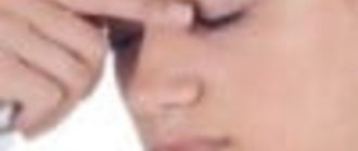
The clinical picture of hypertension syndrome is determined by the rate of increase in intracranial pressure. A characteristic symptom of this syndrome is a bursting headache (bursting headache), which occurs due to irritation of the receptors of the dura mater and intracranial vessels. In the initial period, the headache can be paroxysmal, usually manifests itself in the morning, and intensifies after physical activity. Later, the headache becomes constant and periodically intensifies. In young children, specialists from private medical practice in Saratov judge the presence of headaches by restless behavior and periodic crying. At the height of the headache, nausea and vomiting are often observed, which bring short-term relief. Autonomic reactions are noted in the form of increased sweating, changes and fluctuations in the child’s body temperature. At subsequent stages of development of intracranial hypertension, congestion in the fundus of the eye, disorders of higher nervous activity, delayed speech development, and delayed mental and motor (psychomotor) development in young children appear. Also observed are rapid heartbeat (tachycardia), disturbance of consciousness, and generalized clonic-tonic (tonic-clonic) convulsions. The final terminal stage of the disease is characterized by profound disturbances of consciousness (coma, stupor), the transition of tachycardia to bradycardia, and respiratory rhythm disorder. Manifestations of hypertension syndrome develop against the background of symptoms of the underlying disease. In young children, as intracranial pressure increases, there is an increase in the size of the head, fontanels, divergence of the sutures on the skull, and an increase in the venous pattern on the scalp. In older children, changes in the bones of the skull are manifested by increased vascular grooves, the appearance of digital impressions, and porosity of the dorsum sella.
Intracranial hypotension syndrome
Intracranial hypotension syndrome develops with a persistent decrease in cerebrospinal fluid pressure below 100 mm H2O. Art. Synonyms for intracranial hypotension syndrome are hypotensive syndrome, hypotensive syndrome.
Primary intracranial hypotension is quite rare. More often it develops as a result of liquorrhea (loss of cerebrospinal fluid), which can be observed after therapeutic and diagnostic interventions on the cerebrospinal fluid tract, as well as with arterial hypotension or an overdose of dehydrating drugs. The main symptom of intracranial hypotension is a headache, often of a squeezing nature (squeezing pain), which is relieved by lying down with the head down or by pressing the jugular veins. With intracranial hypotension syndrome, general weakness, dizziness, nausea, vomiting, and tachycardia are also observed. With a pronounced decrease in intracranial pressure, disorders of consciousness are observed from mild to stupor and coma.
CSF dystonia syndrome
CSF dystonia syndrome is characterized by instability of intracranial pressure and frequent changes. The symptoms of cerebrospinal fluid dystonia are the same as those of hypertension syndrome and hypotension syndrome.
Occlusive syndromes, Brunx attack
Occlusive syndromes develop as a result of blockade of the cerebrospinal fluid pathways at any level of the ventricular system. Clinically, occlusive syndromes are characterized by intracranial hypertension in combination with symptoms of damage to parts of the brain lying above the level of occlusion and the underlying disease. They are accompanied by Bruns' attacks. Bruns' attack has a number of symptoms - vomiting, rapidly increasing headache, impaired consciousness, forced head position, signs of brain stem dislocation.
Obstruction to the outflow of cerebrospinal fluid can occur at any level of the ventricular system.
Classification of violations
Liquorodynamic disorders are classified in the following areas:
- How does the pathological process proceed:
- Chronic course.
- Acute phase.
2. Stages of development:
- Progressive. Intracranial pressure increases and pathological processes progress.
- Compensated. Intracranial pressure is stable, but the ventricles of the brain remain dilated.
- Subcompensated. Great danger of crises. Unstable condition. Blood pressure can rise sharply at any moment.
3. In which cavity of the brain is the cerebrospinal fluid located:
- Intraventricular. Fluid accumulates in the ventricular system of the brain due to obstruction of the cerebrospinal fluid system.
- Subarachnoid. Liquorodynamic disturbances of the external type can lead to destructive lesions of brain tissue.
- Mixed.
4. Depending on the pressure of the cerebrospinal fluid:
- Hypertension. Characterized by high intracranial pressure. The outflow of cerebrospinal fluid is impaired.
- Normotensive stage. Intracranial pressure is normal, but the ventricular cavity is enlarged. This condition is most common in childhood.
- Hypotension. After surgery, excessive outflow of cerebrospinal fluid from the ventricular cavities.
Causes
There are congenital and acquired etiological factors for the development of disorders of cerebrospinal fluid dynamics of the brain.
Congenital causes:
- pathologies of intrauterine development in the form of chromosomal aberrations at the genetic level;
- dysfunction of neuronal connections between the right and left hemispheres of the brain;
- congenital formations that imitate the cavities of the ventricles of the brain;
- syndrome of compression of the medulla oblongata and dysfunction of the cerebellum (Arnold-Chiari anomaly);
- disruption of the outflow of cerebrospinal fluid caused by insufficient functional development of the cerebellar vermis or tumor (Walker-Dandy anomaly).
Acquired reasons:
- traumatic damage to the structures of the central and peripheral nervous system;
- tumors of various origins that disrupt the outflow of cerebrospinal fluid into the central nervous system and PNS;
- infectious and inflammatory diseases in the cells of the brain and spinal cord (meningitis, encephalitis, measles, rubella and others).
- Thrombosis and embolization of vascular walls of the brain.
Causes congenital
There are congenital anomalies that can contribute to the development of liquorodynamic disorders:
- Genetic disorders in intrauterine development of the fetus.
- Agenesis of the corpus callosum.
- Dandy-Walker syndrome.
- Arnold-Chiari syndrome.
- Encephalocele.
- Stenosis of the cerebral aqueduct, primary or secondary.
- Porencephalic cysts.
Acquired reasons
Liquorodynamic disorders can begin to develop for acquired reasons:
- Spinal cord and brain injuries.
- Various infectious diseases and parasitic infections that affect the nervous system.
- Neoplasms inside the skull that block the cerebrospinal fluid pathways.
- Thrombosis.
- Intrauterine hypoxia in the first two days after birth.
- Papillomas of the choroid plexus.
Cephalgia (headache)
Cranial therapy (craniosacral therapy, manual and osteopathic techniques)
Craniosacral therapy is one of the gentlest and at the same time most profound methods of modern therapy. It affects the physical and psychological causes of the disease, which are often beyond the reach of other methods. In the internal space of the spine and skull, cerebrospinal fluid, or cerebrospinal fluid, is renewed and circulates. It washes the substance of the spinal cord and brain. As a result of the movement of the cerebrospinal fluid, its pressure within the dura mater changes cyclically, and a number of bones, tissues and internal organs cyclically gently expand and contract.
The therapist’s influences during the session are very light and precise, taking place in the area of the skull, sacrum, and diaphragms. As a result, the natural rhythm of cerebrospinal fluid production is restored, the body is freed from restrictions on physiological mobility, the circulation of cerebrospinal fluid is improved, blood pressure and the functioning of the central and peripheral nervous systems are normalized. An anti-stress effect is achieved and rehabilitation processes are launched.
CST protocols can be integrated into any bodily practice, or used as an independent type of preventive and health-improving effect on the body. Applicable for children of any age, adults and the elderly.
Those who have completed a series of seminars: are given the opportunity to enter the Association of Manual Practitioners free of charge and receive a personal account on the website osteonika.ee in the “pro association” section, where there will be your photo, contacts and a list of seminars plus access to educational files (admission depends on the level of training in school).
Visceral therapy (manual and osteopathic techniques)
The course of visceral osteopathy and manual therapy is aimed at studying the causes leading to diseases of internal organs and organ systems (respiratory, digestive, urogenital, cardiovascular, nervous).
During the course, anatomical, physiological and biomechanical connections, their role in health and the development of pathological conditions are studied in detail. Techniques for palpation and visual diagnostics and the use of functional testing are given. Students gain valuable practical skills, and each therapeutic protocol is carefully worked out under the supervision of a supervisor.
The result of each seminar is several effective protocols that can be immediately used in your work.
Those who have completed a series of seminars: are given the opportunity to enter the Association of Manual Practitioners free of charge and receive a personal account on the website osteonika.ee in the “association-pros” section, where there will be your photo, contacts and a list of seminars plus access to educational files (depending on the level completed in School of Training).
Structural therapy (manual and osteopathic techniques)
Structural osteopathy (manual therapy) studies the functioning of the musculoskeletal system. From the point of view of biomechanics, pathologies are considered: vertebral subluxations, bone displacement, joint diseases; imbalance of tension in muscles, fascia and tendons, including the structures running within them. All disorders lead to local manifestations in the form of tissue swelling and pain due to irritation of nerve endings.
Structural osteopathy and manual therapy are effective in preventing diseases of the musculoskeletal system, for restoring the body after sports training, heavy physical activity; injuries and stress, also associated with physiological states of the body (including during pregnancy). In the case of chronic pathologies, there is a prolongation of the period of remission and a clear improvement in the general well-being of patients.
The result of each seminar is several effective protocols that can be immediately used in your work.
Those who have completed a series of seminars: are given the opportunity to enter the Association of Manual Practitioners free of charge and receive a personal account on the website osteonika.ee in the “association-pros” section, where there will be your photo, contacts and a list of seminars plus access to educational files (depending on the level completed in School of Training).
Clinical osteopathy and manual therapy
A series of seminars as part of training at the School or as a continuing education program for osteopaths and osteopractitioners, chiropractors and practitioners, rehabilitation therapists who have received a basic education in osteopathy (at least 700 academic hours).
The program of each thematic seminar includes a specific organ system:
- Anatomy and topography (normal)
- Normal physiology
- Innervation
- Pat. anatomy and physiology, pathomorphology
- Clinical manifestations of diseases
- History, manual osteopathic examination and diagnosis
- Critical laboratory values
- Radiological examination data
- Indications and contraindications for manual osteopathic treatment
Principles of drawing up proprietary protocols and individual methods of osteopathic and manual rehabilitation.
Those who have completed a series of seminars : are given the opportunity to enter the Association of Manual Practitioners free of charge and receive a personal account on the website osteonika.ee in the “association-pros” section, where there will be your photo, contacts and a list of seminars plus access to educational files (depending on the level completed in School of Training).
Aesthetic osteopathy
The technique of aesthetic osteopathy is physiological, universal and has no age criteria.
A session of aesthetic osteopathy is a complex and profound effect on the entire body, but the specialist pays maximum attention to the main processes that affect appearance. This is an increase or decrease in muscle tone; disruption of arterial, venous and lymph flow; changes in the positions of the bones of the facial, cranial skull and other bone structures of the skeleton; imbalance of the nervous and endocrine systems.
When there are deviations from the norm (due to chronic stress, injury or illness, negative or pent-up emotions), collagen and elastin fibers become deformed. The sad consequences are wrinkles, sagging skin, fat folds and changes in the contours of the face and body.
During the session, the patient is in a state of restorative rest, and the body’s natural restorative forces are activated. This is where the holographic principle comes into play: the whole body responds to working with the face, and recurrent pain in different regions of the body, the causes of which could not previously be identified, is eliminated.
Aesthetic osteopathy is physiological, universal and has no age criteria. There are very few specialists and teachers in this field, but you have a real opportunity to master this rare and effective technique.
Those who have completed seminars on aesthetic osteopathy: are given the opportunity to enter the Association of Manual Practitioners free of charge and receive a personal account on the website osteonika.ee in the “pro association” section, where there will be your photo, contacts and a list of seminars plus access to educational files (depending on the level completed at the School of Training).
Symptoms of disorders in infants
Liquorodynamic disorders in children under one year of age have the following symptoms:
- Frequent and profuse regurgitation.
- Unexpected crying for no apparent reason.
- Slow overgrowth of the fontanel.
- Monotonous crying.
- The child is lethargic and sleepy.
- Sleep is disturbed.
- Seams coming apart.
Over time, the disease progresses more and more, and signs of liquorodynamic disorders become more pronounced:
- Tremor of the chin.
- Twitching of limbs.
- Involuntary shudders.
- Life support functions are disrupted.
- Disturbances in the functioning of internal organs for no apparent reason.
- Possible squint.
Visually, you can notice the vascular network in the area of the nose, neck, and chest. When crying or tense muscles, it becomes more pronounced.
The neurologist may also note the following signs:
- Hemiplegia.
- Extensor hypertonicity.
- Meningeal signs.
- Paralysis and paresis.
- Paraplegia.
- Graefe's symptom.
- Nystagmus is horizontal.
- Delay in psychomotor development.
You should visit your pediatrician regularly. At the appointment, the doctor measures the volume of the head, and if pathology develops, changes will be noticeable. So, there may be such deviations in the development of the skull:
- The head grows quickly.
- It has an unnaturally elongated shape.
- The large and small fontanelles swell and pulsate.
- Sutures are coming apart due to high intracranial pressure.
All these are signs that a syndrome of liquorodynamic disorders is developing in an infant. Hydrocephalus progresses.
I would like to note that it is difficult to determine liquorodynamic crises in infants.
Medical tactics
After confirming the diagnosis and determining the cause of development, complex therapy for the disease is recommended.
If the causes are vascular changes in the form of thrombotic complications, anticoagulant and restorative vitamin therapy is prescribed. If impaired circulation of cerebrospinal fluid is caused by infectious agents, antibacterial and anti-inflammatory therapy is prescribed.
If the outflow and secretion of cerebrospinal fluid in the head is impaired due to dynamically progressing neoplasms, chemotherapy is resorted to. If there is a favorable prognosis during surgery, the tumor is surgically removed.
For the syndrome of liquor-dynamic disturbances in infants, osmotic and loop diuretics and glucocorticosteroids are prescribed.
These drugs have a large list of side effects. But they are used in neonatological practice when the expected benefits of these drugs are higher than the possible risks.
Surgical treatment is indicated for CSF crisis, cerebral vascular occlusion, and hydrocephalus.
Signs of liquorodynamic disorders in children after one year
After one year, a child’s skull is already formed. The fontanelles have completely closed and the sutures have ossified. If there are liquorodynamic disturbances in a child, signs of increased intracranial pressure appear.
There may be such complaints:
- Headache.
- Apathy.
- Worry for no reason.
- Nausea.
- Vomiting, after which there is no relief.
The following signs are also characteristic:
- Gait and speech are impaired.
- There are disturbances in the coordination of movements.
- Vision decreases.
- Horizontal nystagmus.
- In advanced cases, “bobble doll head”.
And also, if liquorodynamic disorders of the brain progress, the following deviations will be noticeable:
- The child speaks poorly.
- They use standard, memorized phrases without understanding their meaning.
- Always in a good mood.
- Delayed sexual development.
- Convulsive syndrome develops.
- Obesity.
- Disturbances in the functioning of the endocrine system.
- Lag in the educational process.
Classification
Taking into account the morphology, pathogenesis, cerebrospinal fluid pressure, clinical picture and course, there are several classifications of cerebrospinal fluid dynamics disorders. Most of them are used only in scientific and experimental environments. The following characteristics of the disease exist :
- According to the course of the pathological process:
- acute phase.
- chronic phase.
- According to the stages of pathology development:
- progressive, characterized by the development of pathological changes and increasing intracranial pressure. The patient requires medical or surgical treatment;
- compensated, as a rule, during this period the intracranial pressure stabilizes, but the ventricular system of the brain remains dilated. This condition occurs during treatment;
- subcompensated - the condition is extremely unstable; when exposed to the slightest provocation, the pressure of the cerebrospinal fluid increases significantly. The largest number of crises occur at this stage.
- According to the localization of cerebrospinal fluid in the cavities of the brain:
- internal or intraventricular, occurs due to obstruction of the cerebrospinal fluid system and accumulation of cerebrospinal fluid in the ventricular system of the brain;
- external or subarachnoid, in which the sinuses, which leads to destructive lesions of brain tissue;
- mixed, more diverse in clinical symptoms.
- According to the level of cerebrospinal fluid pressure:
- hypertensive, occurs when the outflow of cerebrospinal fluid from the ventricular system of the brain is disrupted, intracranial pressure is high;
- normotensive - intracranial pressure is normal, enlargement of cavities occurs due to concomitant diseases (infections, injuries, tumors). This type of liquorodynamic disturbances is typical for children in whom compensatory processes are more developed;
- hypotensive – a consequence of complications of surgical treatment, when after surgical intervention there is an excessive outflow of cerebrospinal fluid from the cavities of the ventricles, as a result of which they almost completely collapse.
Features of clinical symptoms in children of the first year of life
In children under one year of age with cerebrospinal fluid circulation disorders, parents note frequent and profuse regurgitation, very slow overgrowth of fontanelles, suture dehiscence, spontaneous crying for no reason, after which the children become lethargic and drowsy. As the disease develops, the symptoms also increase, including twitching of the limbs, tremor of the chin, and involuntary shuddering of the baby.
It is very important for children under one year old to regularly visit the pediatrician . During the examination, the doctor, during the examination, measures the circumference of the child's head. Normally, the volume of the head increases by 6-7 cm in the first 3 months, and by 0.5-1 cm per month from the fourth month to one year. In the presence of pathological changes, the head grows very quickly and takes on an unnaturally elongated shape, usually in anteroposterior dimensions. The large and small fontanelles in such infants do not close, but rather swell and pulsate. Due to increased intracranial pressure and the plasticity of the connective tissue, the sutures separate. Thanks to this, children remain in the compensation stage for a long time. Increasing hydrocephalus is a clear sign of impaired cerebrospinal fluid dynamics.
When examined by a neurologist, focal neurological symptoms are noted: paralysis and paresis, para- and hemiplegia, extensor hypertonicity, nystagmus, Graefe's symptom, meningeal signs.
Young patients are characterized by monotonous crying and their sleep is often disturbed. In the area of the bridge of the nose, neck, and upper chest in sick children, a vascular network is pronounced, which becomes visible when the child tenses (crying, trying to raise his head, sit down). In young patients, it is difficult to identify liquorodynamic crises.
Children under one year of age may experience disorders of internal organs without objective reasons. Vital functions are impaired. Over time, such children begin to lag significantly behind in psychomotor development. Sometimes a mother’s attention is attracted by the child’s growing squint. An experienced ophthalmologist may already suspect a syndrome of liquorodynamic disorders based on changes in the fundus of the eye.
Clinic for children after one year
After the child’s skull has fully formed and the fontanelles have closed, the sutures have ossified, and symptoms of increased intracranial pressure begin to predominate. Children complain of headaches, apathy, followed by anxiety, impaired coordination of movements, gait, and speech.
Liquorodynamic headache occurs in attacks, often in the morning, and may be accompanied by nausea and vomiting. There is no relief after vomiting. Such children have poor vision, horizontal nystagmus is noted, and due to muscle paralysis they cannot look up. In severe cases, the “bobble doll head” symptom is observed. Increased symptoms are called “liquid dynamic crises.”
If the disease occurs at an early age, children speak poorly or do not speak at all . Changes in mental development from minimal to extreme idiocy. Such children use standard memorized phrases in their speech, often without understanding their meaning. They are always in a good mood. Children also have disorders of the endocrine system, most often they manifest themselves in the form of obesity and delayed sexual development. Over time, convulsive syndrome increases in young patients.
Clinical picture in the working population
In adults, the disease most often occurs due to injuries, tumors, infections and manifests itself in the form of high intracranial pressure. Such patients suffer from CSF headaches, dizziness, cardiac disorders and symptoms of the underlying disease. Liquorodynamic crises occur as a result of nervous shock or exacerbation of the underlying disease.
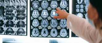
Diagnostics
In children under one year of age, the diagnosis of liquorodynamic disorders consists of collecting a pregnancy history, parental complaints, examining the child by specialists (neurologist, ophthalmologist), fundus examinations, and instrumental studies, including ultrasound, magnetic resonance imaging, neurosonography, computed tomography.
In children over one year of age, complaints are also collected for diagnosis, examined by specialists, and MRI and CT are used from instrumental studies.
In adults, the underlying disease plays a significant role in diagnosis.
ICD-10 classification
G.91. Acquired hydrocephalus.
G.94. Hydrocephalus in infectious and parasitic diseases and tumor processes.
Diagnosis of the disease in adults
If you experience headaches and the symptoms described above, you should consult a neurologist. To clarify the diagnosis and prescribe treatment, the following studies may be prescribed:
- Computed tomography.
- Angiography.
- Pneumoencephalography.
- ECHO of the brain.
- NMRI.
If there is a suspicion of a syndrome of cerebrospinal fluid dynamics disorders, a lumbar puncture may be prescribed with a change in cerebrospinal fluid pressure.
When diagnosing adults, much attention is paid to the underlying disease.
Signs of the disease in adults
As a rule, hydrocephalus of the brain in an adult or a teenager is directly related to increased intracranial pressure, which causes the patient to feel:
- severe headache that does not improve with analgesics;
- feeling of internal pressure on the eyeballs;
- nausea, sometimes leading to vomiting;
- impaired coordination, due to which the gait becomes unstable, hand movements become sharp and sweeping, handwriting changes, etc.;
- tinnitus, impaired visual acuity, loss of certain areas of space from the field of vision.
Sometimes changes affect the patient’s emotional sphere, causing rapid changes in mood for no apparent reason, neurasthenic manifestations and unmotivated aggression.
Treatment of liquorodynamic disorders
The earlier the disease is detected, the greater the chance of restoring lost brain functions. The type of treatment is selected based on the presence of pathological changes in the course of the disease, as well as the age of the patient.
In the presence of increased intracranial pressure, diuretics are usually prescribed: Furosemide, Diacarb. Antibacterial agents are used in the treatment of infectious processes. Normalization of intracranial pressure and its treatment is the main task.
To relieve swelling and inflammation, glucocorticoid drugs are used: Prednisolone, Dexamethasone.
Steroid medications are also used to reduce cerebral edema. It is necessary to eliminate the cause of the disease.
As soon as liquorodynamic disturbances are detected, treatment should be prescribed immediately. After undergoing complex therapy, positive results are noticeable. This is especially important during the period of child development. Speech improves, progress in psychomotor development is noticeable.
Surgical treatment is also possible. It may be prescribed in the following cases:
- Drug treatment is ineffective.
- Liquorodynamic crisis.
- Occlusive hydrocephalus.
Surgical treatment is considered for each case of the disease separately, taking into account age, characteristics of the body and the course of the disease. In most cases, surgery on the brain is avoided so as not to damage healthy brain tissue, and complex drug treatment is used.
It is known that if the syndrome of liquorodynamic disorders in a child is not treated, the mortality rate is 50% up to 3 years, 20-30% of children survive to adulthood. After surgery, mortality is 5-15% of sick children.
Mortality increases due to late diagnosis.
How are they treated?
Depending on the etiology and severity of the disease, conservative or surgical treatment of hydrocephalus may be chosen. Conservative therapy is most effective in cases of development of disorders due to inflammation, injury or minor hemorrhage and consists in eliminating the cause of the disease. To reduce intracranial pressure and alleviate the patient's condition, diuretic drugs are prescribed.
Surgical intervention is indicated for congenital pathologies to correct developmental defects, as well as in the presence of volumetric processes - large intracranial hematomas, tumors, abscesses, etc. In cases where the cause cannot be eliminated either by conservative treatment or surgical removal, the neurosurgeon prescribes shunt surgery and creates additional pathways for the outflow of excess cerebrospinal fluid.
Prevention of liquorodynamic disorders
Preventive measures include:
- Observation of pregnancy in the antenatal clinic. It is very important to register as early as possible.
- Timely detection of intrauterine infections and their treatment.
At 18-20 weeks, an ultrasound shows the development of the fetal brain and the state of the unborn child’s cerebrospinal fluid. At this time, it is possible to determine the presence or absence of pathologies.
- The right choice of delivery.
- Regular monitoring by a pediatrician. Measuring the circumference of the skull, if there is a need to conduct a fundus examination.
- If the fontanel does not close in a timely manner, it is necessary to conduct neurosonography and consult a neurosurgeon.
- Timely removal of tumors that block the cerebrospinal fluid pathways.
- Regular observation by a doctor and carrying out the necessary studies after suffering injuries to the brain and spinal cord.
- Timely treatment of infectious diseases.
- Prevention and therapy of chronic diseases.
- Quit smoking and alcohol.
- It is recommended to play sports and lead an active lifestyle.
It is easier to prevent any disease or take all measures to reduce the risk of developing pathology. If liquorodynamic disorders are diagnosed, then the earlier therapy is started, the greater the chance that the child will develop normally.
Treatment of intracranial pressure
Intracranial pressure needs to be normalized and treated. An integrated approach to the problem, even after the first courses of treatment, even in severely ill patients, gives positive dynamics, speech and psychomotor development in children improves. The earlier treatment is started, the faster the impaired functions are restored and intracranial pressure is normalized. At the first consultation, the doctor will tell you about the main factors and symptoms of the disease: what is hypertension syndrome in newborns, infants, infants, children, children, adults, what is mild, moderate, severe cerebral hypertensive syndrome , hypertensive syndrome , cerebrospinal fluid hypertensive syndrome , how the army and residual encephalopathy are related to hypertensive hydrocephalic syndrome, what intracranial pressure should be normal, how to measure, determine, check, and how to reduce, lower, relieve, treat intracranial pressure. On the website sarclinic.ru you can see a doctor online for free.
Sign up for a consultation. There are contraindications. Specialist consultation is required. Photo: Sergiyn | Dreamstime.com\Dreamstock.ru. The people depicted in the photo are models, do not suffer from the diseases described and/or all similarities are excluded.
Related posts:
Vegetative vascular dystonia in children, treatment of VSD in Saratov
Stuttering in children, stuttering treatment in Saratov, Russia
Dyslexia, treatment of dyslexia, correction, overcoming, elimination, alexia, treatment of alexia
Yactation, rocking, nodding movements of the head, torso, body
Enuresis, treatment of enuresis, how to treat nocturnal enuresis in Saratov, Russia