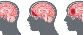
Causes of subarachnoid hemorrhage
In most cases, this condition is caused by non-traumatic factors:
- aneurysm rupture;
- rupture of a vessel in the brain against the background of arterial hypertension, atherosclerosis and other vascular diseases.
Subarachnoid hemorrhage in the brain can also be caused by traumatic factors:
- traumatic brain injury;
- brain contusion;
- fracture of the skull bones.
Main services of Dr. Zavalishin’s clinic:
- consultation with a neurosurgeon
- treatment of spinal hernia
- brain surgery
- spine surgery
Hemorrhage into the cerebellum
Hemorrhage into the cerebellum usually develops within several hours. With this localization of hemorrhage, loss of consciousness in the patient is rarely observed at first. Repeated vomiting is typical, the patient is unable to walk or stand. These clinical signs appear early and should raise suspicion of this diagnosis, which allows the neurosurgeon to promptly decide on surgical intervention. With hemorrhage in the cerebellum, the patient also experiences a headache in the occipital region and dizziness. A neurological examination of the patient reveals horizontal gaze paresis in the direction of the hemorrhage with a forced rotation of the eyeballs in the opposite direction and paresis of the abducens (VI) nerve on the affected side.
MRI of the brain of a patient with hemorrhage in the right hemisphere of the cerebellum (indicated by an arrow).
In the acute phase of hemorrhage, the patient may not have signs of damage to the cerebellum or they may be mild. Only sometimes, in patients with hemorrhage in the cerebellum, nystagmus or cerebellar ataxia in the extremities is detected during a neurological examination.
In the eyes of cerebellar hemorrhage, symptoms include spasm of the eyelid muscles (blepharospasm), involuntary closure of one eye, and squinting. Twitching of the eyeballs (ocular bobbing), usually regarded as a symptom of damage to the pons, may appear later when the patient’s consciousness is depressed to the level of coma. The vertical movements of the eyeballs are preserved, and the narrow pupils continue to react to light until the very late stages of the disease. On the affected side, weakness of the facial muscles and a decrease in the corneal (corneal) reflex are often noted.
There is no paralysis of the muscles of the limbs of the opposite half of the body (contralateral hemiplegia) and weakness of the facial muscles. Sometimes, at the onset of hemorrhage in the cerebellum, there is complete paralysis of the muscles of the arms and legs of both sides (tetraplegia) with preservation of consciousness. Sometimes the loss of voluntary movements in a patient manifests itself only in the form of spastic weakness of the muscles of the arms or legs (spastic paraparesis). Plantar reactions (reflexes) first have a flexion character, later - extension. Sometimes, a few hours after a hemorrhage in the cerebellum, the patient suddenly develops stupor and then coma as a result of compression of the brain stem, after which therapeutic measures aimed at reversing the development of the syndrome, and even surgical treatment, are rarely effective.
MRI of the brain of a patient with hemorrhage in the left hemisphere of the cerebellum (indicated by an arrow).
Neurological symptoms from the eyes are important for establishing the location of hemorrhages in the brain:
- with hemorrhages in the shell, the eyeballs deviate in the direction opposite to the side of paralysis
- with hemorrhage in the thalamus, the eyeballs deviate downward and pupillary reactions are lost
- with hemorrhage in the bridge, reflex turns of the eyes to the side are impaired; the pupils, although they react to light, are very weak
- with hemorrhage in the cerebellum, the eyeballs are turned in the direction opposite to the localization of the lesion, in the absence of paralysis
Headache is not considered a mandatory symptom of intracerebral hemorrhage in hypertension. Headache occurs in approximately 50% of patients, while vomiting occurs in almost all. The patient does not necessarily develop depression of consciousness to the point of coma. If the volume of the hematoma is small, the patient's consciousness can be maintained, even if blood has entered the ventricular system.
Epileptic seizures in patients as a result of hemorrhages in hypertension are rare - in less than 10% of cases. In most patients, the correct diagnosis is based on a combination of objective and subjective symptoms.
However, if the patient's consciousness is preserved, it is difficult to distinguish between cerebral infarction (ischemic stroke) and intracerebral hemorrhage. In such cases, magnetic resonance imaging (MRI) or computed tomography (CT) of the brain is indicated. Timely use of magnetic resonance imaging (MRI) and computed tomography of the brain (CT) makes it possible to accurately differentiate the type of lesion and establish their localization, especially in the most difficult to diagnose small hemorrhages in the brain against the background of hypertension in the patient.
Clinical picture of subarachnoid hemorrhage
This condition develops rapidly against the background of a person’s normal well-being and manifests itself:
- sharp acute headache, worsening with the slightest physical activity;
- nausea, vomiting;
- convulsions;
- psycho-emotional disorders (fear, agitation, drowsiness);
- increased body temperature (up to febrile and subfebrile levels);
- disorders of consciousness (from stupor to fainting, coma);
- symptoms of damage to the oculomotor nerve (gaze paresis, drooping eyelid), hemorrhage in the eyeball.
In half of the cases, this condition leads to death. The main complications and consequences of subarachnoid hemorrhage:
- spasm of cerebral vessels caused by ischemia;
- recurrence of an aneurysm episode (may occur early or several weeks after the first episode);
- hydrocephalus (in the initial stages or in the late period);
- pathologies caused by subarachnoid hemorrhage in the brain (myocardial infarction, pulmonary edema, bleeding from a stomach or duodenal ulcer).
Among the long-term consequences of subarachnoid hemorrhage in the brain: memory disorders, attention, psycho-emotional disorders (depression, insomnia, agitation). Also, a standard complaint of patients who have suffered this condition is headache. Somewhat less frequently, hormonal disorders develop in the hypothalamic and pituitary gland systems.
Hemorrhagic and ischemic stroke: treatment
Local and systemic risk factors are morphological and atherosclerotic changes in the main arteries of the head (MAG), atherosclerotic lesions of the mass arteries and vessels of the aortic arch, heart damage due to thrombus extraction of cerebral infarctions, fibromuscular dysplasia, ruptures of the walls of the MAG and cerebral arteries. And also, inflammation of the walls of the arteries, disorders in the cervical spine, pathologies of central and cerebral blood flow through the vessels, Vaquez disease, hemostasis disorders, some forms of leukemia, inhibition of the gas transport function of the blood, etc.
In addition, the occurrence of stroke is facilitated by various types of heart lesions - atrial fibrillation, cardiac arrhythmias, myocardial infarction, post-infarction aneurysms accompanied by thrombus formation, cardiac rheumatism, idiopathic cardiomyopathies, endocarditis, myocardiopathies, etc.
Hemorrhage in the brain occurs when a vessel ruptures or blood cells escape through the walls of capillaries and small veins into the surrounding tissue.
Causes of cerebral hemorrhage during hemorrhagic stroke:
- head injuries;
- a sharp increase in blood pressure;
- secondary hemorrhage in the brain stem;
- brain tumors, arteritis, coagulopathies.
Some genes play an important role in the development of hemorrhagic stroke (gene of the renin-angiotensin system, genes of the homeostasis system, etc.).
When cerebral circulation is impaired, pathobiochemical changes very quickly develop in the patient’s body, leading to irreversible damage to the nervous tissue of the brain due to necrosis of the brain area and programmed cell death.
Hemorrhage in the brain leads to the death of nerve tissue in damaged areas. The hematoma compresses the brain tissue, resulting in a sharp increase in intracranial pressure. The intensity of unnatural changes is directly proportional to the size of hemorrhage. Brain edema occurs within a few minutes after the development of local ischemia as a result of damage to the cell membrane and accumulation of fluid in the cell.
Brain edema causes an increase in intracranial pressure, which in turn leads to hemorrhagic transformation of the infarction and displacement of parts of the brain.
If death does not occur, cerebral edema gradually subsides over one to two weeks, and necrotic brain tissue undergoes resorption or liquefaction. Subsequently, a connective tissue scar and/or a cyst-like cavity is formed at the site of the infarction.
Diagnosis and treatment of subarachnoid cerebral hemorrhage
The doctor clarifies the victim’s medical history and conducts an external examination. The diagnosis is established on the basis of CT data, which makes it possible to determine the extent of the process, the presence of edema, and assess the condition of the cerebrospinal fluid system. If the results are negative, a lumbar puncture is performed.
MRI is a sensitive tool for diagnosing pathology after a few days. Vascular angiography is used to determine the source of bleeding in the brain.
Therapy for subarachnoid hemorrhage takes place in a neurological hospital and depends on the severity of the patient’s condition. It is aimed at solving the following problems:
- stabilization of the patient's condition;
- prevention of recurrent episode;
- normalization of homeostasis;
- treatment and prevention of ischemia, vascular spasm;
- therapy for the disease that caused the hemorrhage.
The aneurysm is clipped or endovascular occlusion is performed. Vasospasm and ischemia are treated with medication. In case of local spasm of a vessel, a vasodilator drug is injected directly into it or balloon angioplasty is performed.
In 50% of cases, subarachnoid hemorrhage leads to death. The majority develop significant impairments in the functional activity of the brain.
You can get help with this pathology at the Department of Neurosurgery of the City Clinical Hospital named after. A.K. Eramishantseva. The clinic has modern diagnostic equipment and advanced equipment for performing complex operations.
Hemorrhagic stroke
Hemorrhagic stroke is any (not associated with trauma) hemorrhage into the cranial cavity caused by diseases of the cerebral vessels:
- arterial hypertension of various origins,
- amyloid angiopathy,
- aneurysms and vascular malformations,
- blood diseases (erythremia, thrombophilia),
- vasculitis,
- systemic connective tissue diseases,
- hemorrhages during treatment with anticoagulants and fibrinolytic agents, as well as with the abuse of certain drugs (for example, amphetamine, cocaine).
The most common causes of hemorrhagic stroke are hypertension and amyloid angiopathy. The mechanism of hemorrhage in these diseases is associated with changes in the walls of blood vessels in the brain. Hemorrhagic stroke accounts for 8-15% of all strokes. In Russia, about 40,000 cerebral hemorrhages are recorded annually. Intracranial hemorrhages, depending on the location of the leaked blood, are divided into:
Intracerebral hemorrhage:
- acute sudden onset, against the background of high blood pressure, severe headache, dizziness, nausea and vomiting, rapid development of focal symptoms, impaired consciousness - from stupor to coma. The severity of the condition is due to cerebral edema, manifested by general cerebral symptoms and symptoms of dislocation.
The prognosis for hemorrhagic stroke is generally unfavorable. Mortality reaches 60-70%. The basis of prevention is drug treatment of hypertension (reduces the risk of stroke by 40-50%), eliminating risk factors for hypertension and stroke: smoking, drinking large doses of alcohol, diabetes, hypercholesterolemia.
Subarachnoid hemorrhage.
The frequency among other types of stroke does not exceed 5%. Subarachnoid hemorrhage can happen at any age, but it most often occurs between 40 and 60 years of age. The reason for the development may be:
primary vascular diseases of the central nervous system:
- arterial aneurysms of cerebral vessels (70 – 80%);
- vascular malformations;
- abnormalities of the cerebral vascular system (for example, dissecting aneurysms).
secondary vascular pathology of the central nervous system:
- arterial hypertension;
- vasculitis;
- blood diseases;
- disorders of the blood coagulation system when taking anticoagulants, antiplatelet agents, contraceptives and other drugs.
It develops acutely and is characterized by the occurrence of a sudden intense diffuse headache of the “blow” type, “spreading of hot liquid in the head,” nausea, vomiting, short-term loss of consciousness and rapid development of meningeal syndrome (the main distinguishing differential diagnostic feature) in the absence of focal neurological disorders. Prolonged loss of consciousness indicates severe hemorrhage, with blood escaping into the ventricular system of the brain. Subarachnoid hemorrhages occur most severely when cerebral aneurysms rupture.
Subdural hematomas
They account for approximately 2/5 of the total number of intracranial hemorrhages and occupy first place among the various types of hematomas. Most subdural hematomas are formed as a result of traumatic brain injury. Less commonly occur with vascular pathology of the brain (hypertension, aneurysms, malformations). Clinical manifestations are varied and consist of general cerebral, local and secondary brainstem symptoms. Typically, there is a so-called “bright” period —the time after injury when there are no symptoms of cerebral vascular damage. The duration of the “light” interval varies within a very wide range from several minutes and hours to several days (the so-called triphasic disorder of consciousness - primary loss of consciousness after injury, its recovery for a certain period and subsequent repeated switching off ).
The speed of detection and removal of a subdural hematoma is of great importance for the prognosis. The outcomes of surgical treatment are significantly better in patients operated on in the first 4-6 hours after injury. With rapid elimination of “brain compression,” ischemic disorders can be reversible.
