The spinal cord is the most important internal organ related to the structure of the central nervous system. The surface of the spinal cord has 3 membranes - arachnoid, hard and soft. The anatomy of the spinal cord is designed in such a way that the internal organ is the dominant system that supports the vital functions of the entire organism.
Structure of the spinal cord
Anatomical features
The spinal cord is located in the cavity of the spinal canal, which is formed by the processes of the vertebrae and their bodies. The beginning of the structure of the spinal cord is the foramen magnum of the brain. Next, the spinal cord is located in the canal, representing a 40-centimeter “cord” surrounded by three membranes.
The internal organ ends with a cluster of nerve fibers at the level of the first vertebrae in the lumbar region, called the cauda equina. This is where the narrowing begins, and then the internal organ is “stretched” into a terminal (terminal, terminal) filament, the diameter of which is 1 mm. The filum terminale extends to the coccygeal region, where it fuses with the periosteum.
The lower part of the trailing thread is tightly wrapped in ponytail fibers. When pain occurs in the coccyx area, doctors talk about a syndrome with the same name. The structure of the human spinal cord is such that the medulla itself is under constant protection - this is provided by the membranes and the spinal column itself.
The external structure is the shells and the space between them.
Blood supply
To ensure adequate nutrition of the brain, many large, medium and small blood vessels are connected to it. They originate from the aorta and vertebral arteries. The process involves the spinal arteries, anterior and posterior. The vertebral arteries supply the upper cervical segments.
Many additional vessels flow into the spinal arteries along the entire length of the spinal cord. These are radicular-spinal arteries, through which blood passes directly from the aorta. They are also divided into rear and front. The number of vessels may vary among different people, being an individual feature. Typically, a person has 6-8 radicular-spinal arteries. They have different diameters. The thickest ones feed the cervical and lumbar thickenings.
The inferior radicular-spinal artery (artery of Adamkiewicz) is the largest. Some people also have an additional artery (radicular-spinal) that arises from the sacral arteries. There are more radicular-spinal posterior arteries (15-20), but they are significantly narrower. They provide blood supply to the posterior third of the spinal cord along the entire cross section.
The vessels are connected to each other. These places are called anastomosis. They provide better nutrition to different parts of the spinal cord. The anastomosis protects it from possible blood clots. If a separate vessel is blocked by a blood clot, blood will still flow through the anastomosis to the desired area. This will save neurons from death.
In addition to the arteries, the spinal cord is generously supplied by veins, which are closely connected with the cranial plexuses. This is a whole system of vessels through which blood then flows from the spinal cord into the vena cava. To prevent blood from flowing back, there are many special valves in the vessels.
Histology.RU
Material taken from the site www.hystology.ru
Anatomically, the spinal cord consists of two symmetrical halves, delimited from each other by the ventral median fissure and the dorsal median septum. The centrally located gray matter of the spinal cord contains multipolar nerve cells that form the nuclei of the spinal cord. Its peripheral part, the white matter, is represented by a collection of nerve fibers that are part of the various pathways of the central nervous system.
Gray matter of the spinal cord . Anatomically, the gray matter of the spinal cord consists of two halves connected by a commissure. Each of them has dorsal and ventral horns. In the thoracic and lumbar segments of the spinal cord, the superolateral section of the ventral horns can be distinguished as its lateral horns. In the gray commissure, which connects the two halves of the gray matter, there is the central canal of the spinal cord.
Gray matter is formed by multipolar neurons, unmyelinated and myelinated nerve fibers and neuroglia. Groups of nerve cells of the same functional significance form the nuclei of gray matter.
Based on morphological characteristics, localization, participation in nerve conduction in the gray matter of the spinal cord, the following types of cells can be distinguished: radicular cells - cells whose neurites leave the spinal cord as part of its ventral roots; internal cells, the neurites of which form synapses on the cells of the gray matter of the spinal cord brain, and tuft cells. Their neurites form separate bundles in the white matter that conduct nerve impulses from certain nuclei of the spinal cord to its other segments or to certain parts of the brain, forming the pathways of the central nervous system.
In the gray matter of the spinal cord, areas are distinguished that differ in neuronal composition, the nature of nerve fibers and neuroglia. Thus, in the dorsal horns of the gray matter, the spongy layer, gelatinous substance, the proper nucleus of the dorsal horn, its dorsal nucleus, or Clark’s nucleus should be distinguished.
The spongy layer of the dorsal horns of the gray matter contains small tufted cells embedded in a broadly looped glial framework.
The gelatinous substance is formed mainly by glial elements, which contain small amounts of small tuft cells.
The dorsal horn contains the dorsal horn nucleus, the thoracic nucleus (Clark's nucleus), and a significant number of diffusely scattered small multipolar interneurons.
The nucleus of the dorsal horn contains tufted cells, the axons of which pass through the anterior white commissure
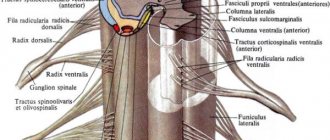
Rice. 176. Schematic section of the spinal cord and spinal ganglion:
1 - 2 - reflex pathways of conscious proprioceptive sensations and touch; 3 and 4 - reflex pathways of proprioceptive impulses; 5 - reflex pathways of temperature and pain sensitivity; b — posterior own bundle; 7 - lateral own beam; 8 - anterior own bundle; 9 - posterior and 10 - anterior spinocerebellar tracts; 11 - spinothalamic tract; 12 - delicate bun; 13 - wedge-shaped bundle; 14 - rubro-spinal tract; 15 - thalamo-spinal tract; 16 - vestibulo-spinal tract; 17 - reticulospinal tract; 18 - tecto-spinal tract; 19 - corticospinal (pyramidal) lateral tract; 20 - corticospinal pyramidal anterior tract; 21 - own nucleus of the posterior horn; 22 - thoracic nucleus (Clark's nucleus); 23, 24 — nuclei of the intermediate zone; 25 - lateral nucleus (sympathetic); 26 - nuclei of the anterior horn.
to the opposite side of the spinal cord into the lateral cord of the white matter, where they form the ventral spinocerebellar and spinothalamic tracts (Fig. 176).
The dorsal horns contain a significant number of small, multipolar associative and commissural neurons, the neurites of which form synapses on the cells of the gray matter of the spinal cord of the same side (associative cells) or the opposite (commissural).
The dorsal nucleus is formed by large cells, their axons enter the lateral cord of the white matter on the same side and, as part of the dorsal spinocerebellar tract, enter the cerebellum.
The nerve cells of the intermediate zone of the gray matter form two nuclei: the medial intermediate nucleus, the neurites of which join the fibers of the ventral spinocerebellar tract of the same side, and the lateral intermediate nucleus, which contains associative cells of the sympathetic nervous system. The axons of these cells leave the spinal cord through the ventral roots of the spinal cord and form the white connecting branches of the sympathetic trunk.
The nuclei of the ventral horns of the gray matter are formed by the largest nerve cells of the spinal cord (100 - 140 µm in diameter). Their neurites form the bulk of the fibers of the ventral roots. Through the mixed spinal nerves they enter the skeletal muscles and end in the motor nerve endings. In the ventral horns of the gray matter of the spinal cord, two groups of motor cells are distinguished: the medial one, which innervates the muscles of the trunk, and the lateral one, which is characteristic of the region of the cervical and lumbar enlargements of the spinal cord. The lateral nucleus of the ventral horns contains neurocytes that innervate the muscles of the limbs.
Nerve cells are scattered in the gray matter, their axons in the white matter are divided into longer ascending and shorter descending branches. These fibers form their own (main) bundles of white matter adjacent to its gray matter. They give rise to many collaterals, ending in synapses on the motor cells of the anterior horns of 4–5 adjacent segments of the spinal cord. The spinal cord has three pairs of its own bundles.
The white matter of the spinal cord consists of myelinated nerve fibers and a supporting neuroglial framework. Nerve fibers in the white matter make up pathways (fiber complexes) - links of certain reflex arcs. Individual pathways are characterized by the position and functional identity of the cells whose processes are their fibers, their synaptic connections and position in the white matter of the spinal cord.
Among the conducting pathways, one should highlight: 1) the pathways of the spinal cord’s own reflex apparatus, 2) the pathways connecting the spinal cord and brain, 3) ascending (afferent) and 4) descending efferent (see anatomy).
Reviews (0)
Add a review
Meninges
- Hard shell. It is located immediately behind the periosteum of the spine, but does not adhere closely to it. The epidural space is located between the periosteum and the dura mater. The tissue of the hard shell is connective; it contains vessels, lymphatic and circulatory. The epidural space is filled with fatty tissue. Venous plexuses are also located here.
- The arachnoid membrane is a network of thin plates of connective tissue, similar in structure to a spider’s web. The plates are composed of collagen and elastic fibers. Between the arachnoid and soft membrane there is a subarochnoid space with cerebrospinal fluid, which ensures the exchange and nutrition of neurons.
- Soft shell. This is a vascular environment that has serrated ligaments for fixation and provides communication and nutrition between the cerebrospinal fluid and the brain.
Conclusion
Thus, the loss of certain functions, for example, leg movements, makes it possible to determine which part was damaged. Injuries to this organ are among the most serious, and the damage is often irreparable. The main thing is to monitor the health of your spine and not overload it unless absolutely necessary.
The organ is located in the spinal canal, and its length is no more than 45 cm, which is less than the length of the spine itself. This is due to the fact that the brain grows only until the age of five, and the spine, as a rule, until the end of puberty.
Peculiarities
The internal structure of the spinal cord has thickenings in the lumbosacral and cervical regions. This structure is formed because in the corresponding parts of the spine there are a large number of exiting nerves that are directed to the lower or upper extremities.
- The cervical thickening is located at the level of the third and fourth cervical vertebrae and lasts until the second thoracic vertebra.
- The lumbosacral thickening is located from the level of the 9-10 thoracic vertebra and lasts until the 1st lumbar vertebra.
What diseases are possible?
Since this organ regulates the transmission of impulses to all systems and organs, the main sign of disruption of its activity is loss of sensitivity. Due to the fact that this organ is part of the central nervous system, diseases are associated with neurological characteristics. Typically, various spinal cord lesions cause the following symptoms:
- disturbances in the movement of limbs;
- pain syndrome of the cervical and lumbar regions;
- disorders of skin sensitivity;
- paralysis;
- urinary incontinence;
- loss of muscle sensitivity;
- increased temperature in affected areas;
- muscle pain.
You may be interested in: Spinal cord stroke: types, main signs, treatment methods
These symptoms can develop in different sequences, depending on the area in which the lesion is located. Depending on the causes of diseases, there are 3 groups:
- All kinds of developmental defects, including postpartum ones. The most common are congenital anomalies.
- Diseases that involve circulatory problems or various tumors. It happens that such pathological processes also cause hereditary diseases.
- All kinds of injuries (bruises, fractures) that disrupt the functioning of the spinal cord. These could be injuries resulting from car accidents, falls from a height, domestic injuries, or as a result of a bullet or knife wound.
Any spinal cord injury or disease that causes such consequences is very dangerous because it often deprives many people of the ability to walk and live a full life. You should consult a doctor as soon as possible to begin treatment on time if the above symptoms or the following disorders are observed after an injury or illness:
- loss of consciousness;
- blurred vision;
- frequent seizures;
- difficulty breathing.
Otherwise, the disease can progress and cause the following complications:
- chronic inflammatory processes;
- disruption of the gastrointestinal tract;
- disturbance in the functioning of the heart;
- circulatory disorders.
Therefore, you should seek help from a doctor in time to receive the correct treatment. After all, thanks to this you can save your sensitivity and protect yourself from pathological processes in the body that can lead to a wheelchair.
White and gray matter of the spinal cord
The cross-sectional diagram of the structure of the spinal cord is similar to the wings of a butterfly; this part of the internal organ is called gray matter. On the outside, gray matter is surrounded by white matter, but the cellular structure and functions of these substances differ significantly.
Gray matter consists of interneurons and motor neurons:
- interneurons provide close connections between the neurons themselves;
- motor neurons contribute to the transmission of motor reflexes.
The white matter contains axons - these are nerve processes that create fibers of descending and ascending pathways.
What does white matter consist of?
White matter is a special component of the central nervous system, represented by bundles of nerve fibers covered with a special myelin sheath, thanks to which the main purpose of this brain structure is fulfilled, which is to transmit information from the main functional centers of the nervous system to the underlying parts of the nervous system.
The myelin sheath allows electrical impulses to be transmitted over long distances at high speed without loss. It is a derivative of glial cells and, due to its special structure (the sheath is formed from a flat outgrowth of the glial body devoid of cytoplasm), wraps the nerve fiber along the periphery several times, interrupting only in the area of interceptions.
This characteristic feature allows you to increase the strength of the impulse sent by the gray matter several times. In addition, it performs an isolating function, allowing the signal strength to be maintained throughout the entire axon.
Regarding the chemical composition of white matter, myelin is mainly formed by lipids (organic compounds including fats and fat-like substances) and proteins, so white matter, at first glance, is a fat-like mass with corresponding characteristics.
The distribution of white matter in different parts of the central nervous system is heterogeneous in chemical composition: the spinal cord is “fatter” than the brain section of the nervous system. This is due to the fact that from the gray matter of this section, a greater amount of efferent information comes out to the peripheral nervous system.
Internal structure
Gray and white matter together form all the spinal cord pathways. They represent its internal composition. Gray matter is located in the center, and white matter is located throughout the periphery. Gray matter is formed as a result of the accumulation of short processes of neuronal cells and consists of 3 projections that form gray pillars. They are located along the entire length of this organ and in cross-section form:
- anterior horn, containing large motor neurons;
- the dorsal horn, formed with the help of small neurons that contribute to the formation of sensory columns;
- side horn
The gray matter of this organ of the nervous system also suggests the presence of kidney cells. They, located along the entire length of the gray matter, form tuft cells that conduct connections between all segments of the dorsal bridge.
The main part of the white matter is made up of long processes of neurons that have a myelin sheath, which gives the white tint to neurons. The white matter on both sides of the spinal cord is connected by the white commissure. The neurons of the white matter of the spinal cord are collected in special bundles; they are separated into 3 cords of the spinal cord using three grooves.
You may be interested in: Anterior roots of the spinal cord: structure, cross-sectional structure and main functions
In the cervical and thoracic sections of this organ there is a posterior cord, which is divided into thin and wedge-shaped. They continue in the initial part of the brain. In the sacral and coccygeal regions, these cords merge into one and are almost indistinguishable.
Of course, the white and gray matter together do not have a homogeneous structure, but they form an interconnection with each other, thanks to which nerve impulses are transmitted from the central nervous system to all peripheral nerves. Because of such a close connection with the brain, many doctors do not separate these two components of the human nervous system, as they consider them one whole. Therefore, it is very important to take care of preserving their functions, which are vital for every person.
A cross-section of the spinal cord can be seen with the naked eye that the spinal cord is composed of gray matter and surrounding white matter. The gray matter looks like the letter H or a butterfly with outstretched wings. Gray matter consists of nerve cell bodies and nerve cell processes called nerve fibers. White matter is formed only by nerve fibers. Along the spinal cord, gray matter forms gray columns symmetrically to the right and left of the central canal of the spinal cord. Anterior and posterior to the central canal, these gray columns are connected to each other by thin plates of gray matter, called the anterior and posterior commissures. In each column of gray matter, its front part is distinguished - the anterior column and its back part - the posterior column. At the level of the lower cervical, all thoracic and two upper lumbar segments (from CVIII to LI - LII) of the spinal cord, the gray matter on each side forms a lateral protrusion - the lateral column. In other parts of the spinal cord (above the VIII cervical and below the II lumbar segments) there are no lateral columns. In a cross-section of the spinal cord, the columns of gray matter on each side have the appearance of horns. There is a wider anterior horn (diagram, item 12) and a narrow posterior horn (diagram, item 9). The anterior and posterior horns correspond to the anterior and posterior columns. The lateral horn (diagram, item 10) corresponds to the lateral intermediate column (autonomous) of gray matter. When examining the spinal cord under a microscope, it is clear that large nerve root cells - motor (efferent) neurons - are located in the anterior horns. These neurons form five nuclei: two lateral (antero- and posterolateral), two medial (antero- and posteromedial) and a central nucleus. The dorsal horns of the spinal cord contain predominantly smaller neurons. The posterior, or sensory, roots of the spinal nerves are composed of central processes of pseudounipolar neurons located in the spinal (sensitive) ganglia. The gray matter of the dorsal horns of the spinal cord is heterogeneous. The bulk of the dorsal horn neurons form its own nucleus. In the white matter immediately adjacent to the apex of the posterior horn of the gray matter, a border zone is distinguished (diagram, paragraph 6). Anterior to the latter in the gray matter is the spongy zone (diagram, paragraph 7). It got its name due to the presence in this section of a large-loop glial network containing neurons. Even more anteriorly there is a gelatinous substance (diagram, item 8), consisting of small neurons. The processes of the nerve cells of the jellylike substance, the spongy zone and the tuft cells diffusely scattered throughout the gray matter communicate with the neurons of several neighboring segments. As a rule, these processes end with synapses on neurons located in the anterior horns of their segment, as well as on neurons of overlying and underlying segments. Directing from the posterior horns of the gray matter to the anterior horns, the processes of these cells are located along the periphery of the gray matter, forming a narrow border of white matter near it. These bundles of nerve fibers are called the anterior, lateral and posterior intrinsic bundles. The cells of all nuclei of the dorsal horns of the gray matter, as a rule, are interneurons. The processes of the nerve cells that make up the central and thoracic nuclei of the dorsal horns are directed in the white matter of the spinal cord to the brain. The intermediate zone of gray matter of the spinal cord is located between the anterior and posterior horns. Here, from the VIII cervical to the II lumbar segment, there is a protrusion of gray matter - the lateral horn (diagram, paragraph 10). In the medial part of the base of the lateral horn, the thoracic nucleus is noticeable, well outlined by a layer of white matter. It consists of large nerve cells and extends along the entire posterior column of gray matter in the form of a cellular cord (Clark's nucleus). This nucleus reaches its greatest diameter at the level from the XI thoracic to the I lumbar segment. The lateral horns contain the nerve centers of the sympathetic part of the autonomic nervous system in the form of several groups of small neurons combined into the lateral intermediate (gray) substance. The axons of these cells pass through the anterior horn and exit the spinal cord as part of the ventral roots. The intermediate zone contains the central intermediate (gray) matter. The processes of the cells of this nucleus participate in the formation of the spinocerebellar tract. At the level of the cervical segments of the spinal cord, between the anterior and posterior horns, and at the level of the upper thoracic segments, between the lateral and posterior horns, a reticular formation is located in the white matter adjacent to the gray matter. The reticular formation here looks like thin bars of gray matter intersecting in different directions and consists of nerve cells with a large number of processes. The gray matter of the spinal cord with the posterior and anterior roots of the spinal nerves and its own bundles of white matter bordering the gray matter forms its own, or segmental apparatus of the spinal cord. The main purpose of the segmental apparatus, as the phylogenetically oldest part of the spinal cord, is to carry out innate reactions (reflexes). White matter, as noted above, is localized outward from the gray matter. The grooves of the spinal cord divide the white matter into three cords symmetrically located on the right and left. The anterior funiculus is located between the anterior median fissure and the anterior lateral sulcus. In the white matter posterior to the anterior median fissure, the anterior white commissure is distinguished, which connects the anterior cords of the right and left sides. The posterior funiculus is located between the posterior median and posterior lateral sulci. The lateral funiculus is the area of white matter between the anterior and posterior lateral sulci. The white matter of the spinal cord is represented by processes of nerve cells. The combination of these processes in the spinal cord cords constitutes the spinal cord fascicle system. Their other names are: spinal cord tracts, or spinal cord pathways. They serve to transmit afferent information to regulators (nerve centers) and control signals from regulators to control objects - organs and organ systems.
Literature. Illustrations. References. Illustrations
Click here to access the site's library! Click here and receive access to the reference library!
- Acland RD Acland's DVD Atlas of Human Anatomy = Human Anatomy: DVD Atlas: The Upper Extremity, The Lower Extremity, The Trunk, The Head and Neck, Part 1, The Head and Neck, Part 2, and The Internal Organs, DVD Six Disc Set, Lippincott Williams & Wilkins, 2003, 653 MB. Video atlas, audio, 6 discs: upper limbs, lower limbs, torso, head and neck, internal organs, RealPlayer, *.avi format
. Access to this source = Access to the reference. URL: https://www.tryfonov.ru/tryfonov/serv_r.htm#0 quotation - Arslan O. Neuroanatomical Basis of Clinical Neurology = Neuroanatomical Basis of Clinical Neurology, The Parthenon Publishing Group, 2001, 368 p. Tutorial.
Wonderful illustrations . . Access to this source = Access to the reference. URL: https://www.tryfonov.ru/tryfonov/serv_r.htm#0 quotation - Baert AL, Van Goethem JWM, van den Hauwe L., Parizel PL Spinal Imaging: Diagnostic Imaging of the Spine and Spinal Cord = Diagnostic imaging of the spine and spinal cord, Springer, 2007, 602 p. Wonderful illustrations
. . Access to this source = Access to the reference. URL: https://www.tryfonov.ru/tryfonov/serv_r.htm#0 quotation - Cervero F., Jensen T.S., Eds. Pain: Handbook of Clinical Neurology = Pain: A Guide to Clinical Neurology, Elsevier, 2006, 944 p. Volume 81 of the Manual of Neurology
. Access to this source = Access to the reference. URL: https://www.tryfonov.ru/tryfonov/serv_r.htm#0 quotation - Cope T. Motor Neurobiology of the Spinal Cord = Neurobiology of motor functions of the spinal cord, CRC Press, 2001, 337 p. Collection of reviews
. . Access to this source = Access to the reference. URL: https://www.tryfonov.ru/tryfonov/serv_r.htm#0 quotation - DiLorenzo DJ, Bronzino JD Neuroengineering = Applied Neuroscience, CRC, 2007, 408 p. Collection of illustrated reviews
. Access to this source = Access to the reference. URL: https://www.tryfonov.ru/tryfonov/serv_r.htm#0 quotation - Goldberg S. Clinical Neuroanatomy Made Ridiculously Simple. 3rd ed., 2005, 88 p. Elements of clinical neuroanatomy. 3rd ed. 2005, 88 p. Tutorial
. Access to this source = Access to the reference. URL: https://www.tryfonov.ru/tryfonov/serv_r.htm#0 quotation - Hendelman W. Atlas of Funtional Neuroanatomy, 2nd ed., 2006, 15.5 MB. Wonderful illustrations
. . Access to this source = Access to the reference. URL: https://www.tryfonov.ru/tryfonov/serv_r.htm#0 quotation - Kimura J. Electrodiagnosis in Diseases of Nerve and Muscle: Principles and Practice = Electrodiagnosis of diseases of the nerves and muscles: principles and practice. 3rd ed., Oxford University Press, 2001, 1024 p. Well illustrated guide.
File in .CHM format . Access to this source = Access to the reference. URL: https://www.tryfonov.ru/tryfonov/donat.htm#0 quotation - Kimura J. Electrodiagnosis in Diseases of Nerve and Muscle: Principles and Practice = Electrodiagnosis of diseases of the nerves and muscles: principles and practice. 3rd ed., Oxford University Press, 2001, 1024 p. Well illustrated guide.
File in .pdf format . Access to this source = Access to the reference. URL: https://www.tryfonov.ru/tryfonov/donat.htm#0 quotation - Konrad P. The ABC of EMG. A Practical Introduction to Kinesiological Electromyography = Electromyography in kinesiology. Practical introduction. Noraxon, 2005, 60 p. Illustrated textbook
. Access to this source = Access to the reference. URL: https://www.tryfonov.ru/tryfonov/donat.htm#0 quotation - Lee HJ, DeLisa J,A. Manual of Nerve Conduction Study and Surface Anatomy for Needle Electromyography = Study of nerve conduction using surface electromyography. Anatomy of body surfaces. 4th ed. Lippincott Williams & Wilkins, 2005, 51 MB. Illustrated Guide
. Access to this source = Access to the reference. URL: https://www.tryfonov.ru/tryfonov/donat.htm#0 quotation - Leis AA, Trapani VC Atlas of Electromyography = Electromyography. Atlas. Oxford University Press, 2000, 218 p. Tutorial
. Access to this source = Access to the reference. URL: https://www.tryfonov.ru/tryfonov/donat.htm#0 quotation - Lin VW, Cardenas DD, Cutter NC, Hammond MC, Eds. Spinal Cord Medicine: Principles and Practice = Spinal Cord Medicine: Principles and Practice, 2nd ed., Demos Medical Publishing, 2002, 1069 p. Wonderful illustrations.
Beginning of chapters: History, anatomy, physiology, research methods . . Access to this source = Access to the reference. URL: https://www.tryfonov.ru/tryfonov/serv_r.htm#0 quotation - Mendoza J., Foundas AL Clinical Neuroanatomy: A Neurobehavioral Approach = Clinical neuroanatomy: a behavioral approach, Springer, 2007, 770 p. A beautifully illustrated guide
. . Access to this source = Access to the reference. URL: https://www.tryfonov.ru/tryfonov/serv_r.htm#0 quotation - Nieuwenhuys R., Voogd J., van Huijzen C. The Human Central Nervous System = Human Central Nervous System, 4th ed., Springer, 2007, 970 p. A beautifully illustrated guide
. . Access to this source = Access to the reference. URL: https://www.tryfonov.ru/tryfonov/serv_r.htm#0 quotation - Noback Ch.R., Strominger NL, Ruggiero DA, Demarest RJ The Human Nervous System: Structure and Function = The Human Nervous System: Structure and Function, 6th ed., Humana Press Inc., 2005, 475 p. Illustrated textbook for universities
. Access to this source = Access to the reference. URL: https://www.tryfonov.ru/tryfonov/serv_r.htm#0 quotation - Paxinos G., Mai JK, Eds. The Human Nervous System = Human Nervous System, Academic Press, 2003, 1366 p. Collection of illustrated reviews
. Access to this source = Access to the reference. URL: https://www.tryfonov.ru/tryfonov/serv_r.htm#0 quotation - Pierrot-Deseilligny E., Burke D. The Circuitry of the Human Spinal Cord: Its Role in Motor Control and Movement Disorders. Cambridge University Press, 2005, 664 p. Neronal circuits of the human spinal cord: their role in the control of movements in health and disease. 2005, 664 p. Illustrated Guide
. Access to this source = Access to the reference. URL: https://www.tryfonov.ru/tryfonov/serv_r.htm#0 quotation - Shimoji K., Willis WD Evoked Spinal Cord Potentials = Evoked Spinal Cord Potentials, Springer, 2006, 232 p. Illustrated Guide
. Access to this source = Access to the reference. URL: https://www.tryfonov.ru/tryfonov/serv_r.htm#0 quotation - Watson C., Paxinos G., Kayalioglu G., Eds. The Spinal Cord: A Christopher and Dana Reeve Foundation Text and Atlas = Spinal Cord. Text and atlas. Academic Press, 2008, 408 p. Illustrated textbook
. . Access to this source = Access to the reference. URL: https://www.tryfonov.ru/tryfonov/serv_r.htm#0 quotation - Waxman S.G., Ed. Clinical Neuroanatomy = Clinical neuroanatomy. 25th ed., McGraw-Hill Medical, 2007, 400 p. Illustrated textbook
. . Access to this source = Access to the reference. URL: https://www.tryfonov.ru/tryfonov/serv_r.htm#0 quotation - Waxman SG Clinical Neuroanatomy = Clinical Neuroanatomy, McGraw-Hill Medical, 2002, 400 p. Wonderful illustrations
. . Access to this source = Access to the reference. URL: https://www.tryfonov.ru/tryfonov/serv_r.htm#0 quotation - Weyreuther M., Heyde CE, Westphal M., Zierski J., Weber U. MRI Atlas: Orthopedics and Neurosurgery The Spine = MRI Atlas: Orthopedics and Neurosurgery of the Spine, Springer, 2006, 296 p. A beautifully illustrated clinical guide
. . Access to this source = Access to the reference. URL: https://www.tryfonov.ru/tryfonov/serv_r.htm#0 quotation
See: Neurology: Dictionary, Neurology: Internet Resources.
| LIBRARY = LIBRARY Human Physiology = Human Physiology, Human Anatomy = Human Anatomy, Human Biochemistry = Human Biochemistry, Human Psychology = Human Psychology, Medicine = Medicine, Mathematics = Mathematics, Chemistry = Chemistry, Physics = Physics, General Scientific Literature = General Science Lexis. Click here to access any source in the site's library! Click here and receive access to the any reference of the library! “I AM LEARNED. . . N E D O U C H A ?” T E S T V A S H E G O I N T E L L E C T A Premise : The effectiveness of the development of any branch of knowledge is determined by the degree of compliance with the methodology of knowledge - the knowable entity. |
Error? Click here and fix it! Search on the site E-mail of the author (author)
Functions of the organ
The structure and functions of the spinal cord are the most important system that supports the normal functioning of the human body.
The functionality of the spinal cord is divided into 2 parts:
- reflex - these are the simplest motor reflexes of the body, for example, when a hand is burned, a person begins to pull his hands away from the source of injury, or when he hits his knee with a hammer, a reflexive extension of the knee occurs;
- The conduction function is the transmission of nerve impulses from the brain region to the inner part of the spinal cord, as well as the transmission of nerve impulses from the brain to the internal organs of the human body.
With the help of conductive communication, almost every mental action is carried out - get up, walk, lie down, sit down, draw, pass, cut, etc. A person does not even think about most of the actions, performing them at a reflex level in everyday life.
Functions of the spinal cord: the main thing
The spinal cord is a highly complex system of nerve fibers that perform two important tasks in the life of the body:
- reflex,
- conductive.
Conductive function
What is the conducting function of the spinal cord? Any movement originates initially in your brain. Impulses come to it from the mucous membranes, skin or internal organs, after which it processes them and sends a signal to the spinal cord, and then to the peripheral nervous system. That, in turn, transmits signals along nerve endings that cause your muscles to contract.
When performing a certain movement, a person does not even think about which muscles need to be used at the moment - this function is automatically performed by the spinal cord.
Serious injuries, for example, rupture of an organ, lead to partial or complete loss of a person’s ability to move. In this case, the information simply does not reach the nerve endings that would cause the muscles to contract.
Here this organ acts as an intermediate link. The conductive function of the spinal cord is very important.
Reflex function
Each of you has probably accidentally touched a hot frying pan. Your nerve endings react to high temperature, which is an irritant. This information is sent directly to the spinal cord. In response to contact with a hot surface, an uncontrolled reflex function of the spinal cord is activated, causing the muscles to contract sharply. This contraction will cause you to immediately withdraw your hand and avoid a severe burn.
The reflex function of the spinal cord is not only the withdrawal of the hand upon contact with fire. The reflex also includes coughing during illness, closing the eyes during contact with ultraviolet light, and many other uncontrolled defensive reactions. At the same time, a certain segment is responsible for each reflex, and its damage causes the loss of a certain skill.
Segments
Spinal cord segment
-
this is a separate part of an organ
that is responsible for certain parts of the body, as well as for the functioning of all organs. There are 31 segments in total. To make it easier to understand the functions of each of the segments, which together make up the departments, you need to create a simple table.
Sections of the spinal cord and their functions:
| Departments | What are they responsible for? |
| Cervical | Movement of the diaphragm and elbow joints |
| Breasts | Skin sensitivity, muscle function, lungs, heart, digestive system |
| Lumbar | Work of the prostate, kidneys, bladder, adrenal glands, uterus, ureters |
| Sacral | Centers for defecation, urination and erection |
General information about gray matter
The human central nervous system (CNS) is one of the most complex structures of the body; it plays an extremely important role - it ensures the functional integrity of the body and its relationship with the outside world.
The central nervous system consists of the brain and spinal cord and their protective membranes, which, in turn, consist of gray and white matter. The gray substance (lat. substantia grisea) is responsible for most of the functions of higher nervous activity in humans. Thanks to it, a person perceives the external environment, hears, sees, speaks, and most importantly, a person can express an attitude, show sympathy or negative emotions, exhibit types of human behavior, empathy, etc.
The substance consists of approximately 86 billion neurons; of course, this number is extremely approximate, since modern medicine does not yet have the ability to count the exact number of nerve cells.
The white substance or white matter (lat. substantia alba) serves mainly to transmit signals and ensures the interconnection of both hemispheres, and also transfers information from the cerebral cortex to the nervous system.
Clusters of neurons form the nuclei of gray matter. Each nucleus has a corresponding responsibility and function: visual, auditory, circulation, breathing, movement, urination, etc.
The cerebral cortex consists of gray matter nuclei that form the corresponding centers. Substantia grisea is one of the main components of the spinal cord, and its nuclei are located in the cerebellar cortex and in the internal structures of the cerebrum (medulla oblongata, thalamus, hypothalamus, etc.).
Gray matter appears in the form of a membrane of the brain, under which there is white, however, in the spinal cord, substantia grisea is located in the inner part of the spinal system, enveloping a narrow central canal filled with cerebrospinal fluid, the substance forms the outline of the letter H, and it is already covered with white matter.
Organization of pathways
The pathways of the entire body are usually divided into:
- associative;
- afferent;
- efferent.
The task of associative pathways is to connect neurons between all segments. These connections are considered short.
Afferents provide sensitivity. These are ascending pathways that receive information from all receptors and send it to the brain. Efferent pathways transmit signals from the brain to neurons throughout the body. They belong to the descending paths.
How is gray and white matter distributed in the cerebral hemispheres?
To visually study the structure of the central nervous system, there are several methods that allow you to see the brain in cross-section. The most informative is the sagittal section, with the help of which the brain tissue is divided into 2 equal parts along the central line. At the same time, to study the location of gray and white matter in the thickness, a frontal section of the anterior section, and accordingly the cerebral hemispheres, is ideal, allowing one to isolate the hypothalamus, corpus callosum and fornix.
The white matter of the anterior section is located in the thickness of the large lobes, which are the springboard for the gray matter that makes up the cortex. It covers the entire surface of the hemispheres with a kind of cloak and belongs to the structures of higher nervous activity in humans.
Moreover, the thickness of the gray matter of the cortex is not the same throughout and varies from 1.5 to 4.5 mm, reaching its greatest development in the central gyrus. Despite this, it occupies about 44% of the volume of the forebrain, as it is located in the form of convolutions and grooves, which make it possible to increase the total area of this structure.
At the base of the white matter of the cerebral hemispheres, there are also separate accumulations of gray matter, which make up the basal ganglia. These formations are subcortical structures or central nodes of the base of the terminal section. Experts distinguish 4 types of such functional centers, which differ in form and purpose:
- caudate nucleus;
- lenticular nucleus;
- fence;
- amygdala.
All these structures are separated from each other by layers of white matter, which transmits information from them to the underlying parts of the brain through the black substance located in the middle section, and also connects the nuclei with the cortex and ensures their coordinated work.
