Reflex arc concept
Explaining in simple words what a reflex arc is is not that difficult.
This is a chain of neurons that, in a certain sequence, transmit nerve impulses from the source of irritation to the brain and central nervous system centers.
The neural arch is the basis for the full functioning of the entire human nervous system.
The unit of the arc is the reflex.
This is the body's response to a stimulus.
Structure and parts of the reflex arc
The neural arch consists of five main links:
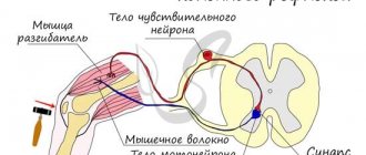
- Touch receptor
performs the functions of the link where the reflex arc begins. Essentially, this is the nerve ending of a neuron or cell, which is the first to receive the influence of a stimulus;
- The second link is afferent neuron
. Its task is to send information about the stimulus to the central nervous system, which was perceived by the receptor;
- The third link is nerve center
. Nerve cells located in the spinal cord or brain produce the desired reflex. The interneurons that make up the nerve center analyze, process and transmit impulses from the initial receptor to the next link - the efferent neuron;
- The fourth link is the same efferent neuron
. It is of two types depending on the reaction caused - motor, which addresses the muscles, and secretory - directed to the secretory formations;
- The last link of the arc, where it essentially ends, is working body
. These can be both muscles and secretory structures. The interaction of all links can be seen in a schematic diagram.
Interesting!
Normal manifestation of the reflex is possible only if all parts of the reflex arc are in working condition.
Types of reflexes
All reflex acts are usually divided into unconditional and conditional. Unconditional ones are transmitted hereditarily; they are characteristic of every biological species. Reflex arcs for unconditioned reflexes are formed before the birth of the organism and remain in this form until the end of its life (if there is no influence of negative factors and diseases).
Conditioned reflexes arise in the process of development and accumulation of certain skills. New temporary connections are developed depending on conditions. They are formed from unconditioned ones, with the participation of higher brain regions.
All reflexes are classified according to different criteria. According to their biological significance, they are divided into nutritional, sexual, defensive, orientation, locomotor (movement), postural-tonic (position). Thanks to these reflexes, a living organism is able to provide the main conditions for life.
In each reflex act, all parts of the central nervous system are involved to one degree or another, so any classification will be conditional.
Depending on the location of irritation receptors, reflexes are:
- exteroceptive (external surface of the body);
- viscero- or interoreceptive (internal organs and vessels);
- proprioceptive (skeletal muscles, joints, tendons).
Depending on the location of neurons, reflexes are:
- spinal (spinal cord);
- bulbar (medulla oblongata);
- mesencephalic (midbrain);
- diencephalic (diencephalon);
- cortical (cerebral cortex).
The reflex acts carried out by neurons of the higher parts of the central nervous system also involve fibers of the lower parts (intermediate, middle, medulla oblongata and spinal cord). In this case, the reflexes that are produced by the lower parts of the central nervous system necessarily reach the higher ones. For this reason, the presented classification should be considered conditional.
Depending on the response and the organs involved, reflexes are:
- motor, motor (muscles);
- secretory (glands);
- vasomotor (blood vessels).
However, this classification applies only to simple reflexes that combine certain functions within the body. When complex reflexes occur that irritate the neurons of the higher parts of the central nervous system, different organs are involved in the process. This changes the behavior of the organism and its relationship with the external environment.
The simplest spinal reflexes include flexion, which allows you to eliminate the stimulus. This also includes the scratching or rubbing reflex, knee and plantar reflexes. The simplest bulbar reflexes: sucking and corneal (closing of the eyelids when the cornea is irritated). Mesencephalic simple ones include the pupillary reflex (constriction of the pupil in bright light).
Types of reflex arcs
Biology distinguishes several types of nerve arches, which differ in structure, reception of stimulation and response. Let's look at the main ones.
Monosynaptic arc
In other words, a simple reflex arc, which consists of two neurons - afferent and efferent, interconnected by one synapse.
Such chains are practically never found in complexly developed organisms, since the simplest reflexes in them are polysynaptic.
Examples of monosynaptic reflexes include the following:
- knee;
- movement of the elbow joint;
- closing the mouth;
- Achilles tendon;
- abdominal reflex;
- irritation of the sole.
Polysynaptic
Complex reflex arcs include interneurons, receptors and effectors. Moreover, the last two elements are usually located in different organs.
Polysynaptic reflexes differ from monosynaptic reflexes in that the duration of the reflex is influenced by the strength of stimulation, and its severity is influenced by the intensity of stimulation.
Basic examples of polysynaptic reflexes:
- sucking;
- swallowing;
- sneezing;
- scratching reflex;
- pupillary;
- defensive (withdrawal of hand).
Somatic
The somatic nerve arch innervates the “soma,” i.e., organs that originate from somites:
- ligaments and tendons;
- skin;
- skeletal muscles.
The arc consists of a sensory, intercalary and motor neuron. It is responsible for conscious muscle movements and for reacting to pupillary, auditory and tactile stimuli.
For example, withdrawing your hand from a hot surface or sharp object, closing your eyes from bright light, moving your knee joint when the doctor checks reflexes in the area of the kneecap.
The somatic arc also carries out unconscious movements - walking, gesturing with hands, smiling.
Vegetative
The autonomic reflex arc does not have interneurons. It consists of a sensory neuron, which is located in the spinal nerve root, and a motor neuron, connected by a synapse. There are two such pairs in total.
Autonomic reflexes are responsible for metabolism, heat exchange, cardiovascular function, coughing, breathing, digestion, salivation, reproduction and growth. Autonomic reactions are not subject to consciousness.
The diagram of the somatic and autonomic reflex is shown in the figure. Labels allow you to understand where everything is located and how the relationship occurs.
Pupillary reflex
Let's consider the reflex arc using the example of the pupillary reflex. The path of the pupillary reflex passes along a complex reflex arc. It starts from the fibers of the rods and cones, which are part of the optic nerve. The fibers cross in the chiasm, passing into the optic tracts, stop in front of the geniculate bodies, partially twist and reach the pretectal region. From here, new neurons go to the oculomotor nerve. This is the third pair of cranial nerves, which is responsible for the movement of the eyeball, the light reaction of the pupils, and the raising of the eyelid.
The return path begins from the oculomotor nerve to the orbit and the ciliary ganglion. The second neuron of the link emerges from the ciliary ganglion, through the sclera into the perichoroidal space. A nerve plexus is formed here, the branches of which penetrate into the iris. The sphincter of the pupil has 70-80 radial neuron bundles entering it sectorally.
The signal for the muscle that dilates the pupil comes from the ciliospinal center of Budge, which is located in the spinal cord between the seventh cervical and second thoracic vertebrae. The first neuron goes through the sympathetic nerve and sympathetic cervical ganglia, the second starts from the superior ganglion, which enters the plexus of the internal carotid artery. The fiber that supplies the pupillary dilator nerves leaves the plexus in the cranial cavity and enters the optic nerve through the trigeminal ganglion. Through it, the fibers penetrate the eyeball.
The closedness of the circular work of the nerve centers makes it perfect. Thanks to the reflex function, the correction and regulation of human activity can occur voluntarily and involuntarily, protecting the body from changes and danger.
Sources used:
- Goldovskaya, I.L. Psychotropic therapy and the organ of vision / I.L. Goldovskaya. - M.: Medicine, 1987.
- Demirchoglyan, G.G. Physiology and pathology of the retina: Primary mechanisms of vision / G.G. Demircioglyan. — M.: Medicine
- Dormidontova, K.V. Some eye diseases and their prevention / K.V. Dormidontova. - Moscow
- FSBI "National Medical Research Center named after. V. A. Almazova" of the Ministry of Health of Russia
Principles of reflex activity
We have already discussed that the reflex arc is a multicomponent neural pathway. At the beginning of this path there are receptors that are constantly exposed to internal and external stimuli.
The receptors convert the resulting irritation into nerve impulses, which are transmitted through sensitive neurons to the nerve centers of the central nervous system located in the spinal cord or in the brain.
Interneurons located in the nerve centers receive this information and transmit it to the working organs by motor nerve cells.
Working organs can be any parts of the body that are touched by irritants. They receive an information impulse that tells them how to react to irritation - withdraw their hand, close their palm, sneeze, blink, swallow, raise their leg, bend their knee, digest food, close their eyes, scratch the back of their head, etc.
All reflexes occurring in the body, from a physiological point of view, can be divided into two large groups:
- Conditional
that appeared during life. These are salivation, reading, driving, and the flexion reflex. That is, everything that can be intentionally learned under the influence of certain conditions.
- Unconditional
, transmitted genetically. These include the blink reflex, chewing, swallowing, sucking, urination, coughing, blinking, and reproduction.
How the reflex works
A nervous process can provoke or increase the activity of an organ. When nervous tissue receives irritation, it goes into a special state. Excitation depends on differentiated concentrations of anions and cations (negatively and positively charged particles). They are located on both sides of the membrane of the nerve cell process. When excited, the electrical potential on the cell membrane changes.
When a reflex arc has two motor neurons in the spinal ganglion (nerve ganglion), the cell's dendrite will be longer (a branched process that receives information through synapses). It is directed towards the periphery, but remains part of the nervous tissue and processes.
The excitation speed of each fiber is 0.5-100 m/s. The activity of individual fibers is carried out in isolation, that is, the speed does not transfer from one to another.
Inhibition of excitation stops the functioning of the site of stimulation, slowing down and limiting movements and responses. Moreover, excitation and inhibition occur in parallel: while some centers fade away, others are excited. Thus, individual reflexes are delayed.
Inhibition and excitation are interconnected. Thanks to this mechanism, the coordinated operation of systems and organs is ensured. For example, the movements of the eyeball are carried out by alternating the work of muscles, because when looking in different directions, different muscle groups contract. When the center responsible for muscle tension on one side is excited, the center on the other slows down and relaxes.
In most cases, sensory neurons transmit information directly to the brain using a reflex arc and several interneurons. The brain not only processes sensory information, but also stores it for future use. In parallel, the brain sends impulses along the descending pathway, initiating a response from effectors (the target organ that performs the tasks of the central nervous system).