Organization of primary projection zones and specificity of associative fields of the human brain.
Primary projection fields correspond to those architectural areas in which the cortical sections of the analyzers are localized: the general sensitivity analyzer in the postcentral gyrus, the olfactory and auditory in the temporal lobe, and the visual in the occipital lobe. Simple, elementary functions are associated with these fields: general skin sensitivity, hearing, smell, vision.
Secondary projection fields are located around the primary ones. They are not directly related to specific pathways. Electrical stimulation of the secondary projection fields causes complex visual images and melodies in a person, in contrast to the elementary sensations (flash, sound) that arise in the case of stimulation of the primary fields. In the secondary projection fields, higher analysis and synthesis, more detailed processing of information, and awareness of it occur.
Secondary projection fields along with primary ones
form the central part of the analyzer, or its core.
The interaction of neurons in these zones is complex, ambiguous, and under conditions of normal brain activity it is based on a sequential change in excitatory and inhibitory processes in accordance with the nature of the final result. This provides dynamic localization properties.
Tertiary projection fields are directly related to higher mental functions. The functions of these zones are associated with the processes of learning and memory. They are unique to the human brain.
Associative field:
All sensory projection areas and the motor cortex occupy less than 20% of the surface of the cerebral cortex. The rest of the cortex constitutes the association region. Association areas of the human brain are most pronounced in the frontal, parietal and temporal lobes. It is believed that in associative areas the association of multisensory information occurs. As a result, complex elements of consciousness are formed. Each projection area of the cortex is surrounded by association areas.
In the parietal associative area of the cortex, subjective ideas about the surrounding space and our body are formed. Frontal associative fields have connections with the limbic part of the brain and are involved in organizing action programs during the implementation of complex motor behavioral acts.
Peculiarities:
· multisensory nature of neurons in the associative department. Moreover, what is received here is not primary, but rather processed information highlighting the biological significance of the signal. This allows you to formulate a program of targeted behavioral act.
· the ability to undergo plastic changes depending on the significance of incoming sensory information.
· long-term storage of traces of sensory influences. Destruction of the associative area of the cortex leads to severe impairments in learning and memory.
Telencephalon: structure, functions, symptoms of damage. Cytoarchitecture of the brain CBP.
Read:- B) Behavioral disturbances due to lesions of the frontal lobes of the brain. Clinical data
- E. - Overgrowth of the aqueduct of the brain.
- I-VII PAIRS OF CN: STRUCTURE, RESEARCH, SYMPTOMS AND SYNDROMES OF LESION.
- IX-XII PAIRS OF CN: STRUCTURE, RESEARCH, SYMPTOMS AND SYNDROMES OF LESION
- VI. Clinical and radiological symptoms and signs
- A) To assess the functional state of the thyroid gland, in other words, thyroid function, the following methods are currently used.
- Allergy in a child. Symptoms and treatment
- Anatomy of the brain
- APPENDICITIS IN A CHILD: CAUSES, SYMPTOMS, TREATMENT
- Arterial hypotension due to organic changes in brain structures.
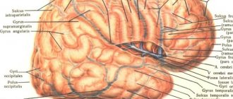
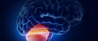
The cerebral hemispheres are the most massive part of the brain. They cover the cerebellum and brain stem. The cerebral hemispheres make up approximately 78% of the total brain mass. During the ontogenetic development of the organism, the cerebral hemispheres develop from the telencephalon of the neural tube, therefore this part of the brain is also called the telencephalon. The cerebral hemispheres are divided along the midline by a deep vertical fissure into the right and left hemispheres. In the depths of the middle part, both hemispheres are connected to each other by a large commissure - the corpus callosum. Each hemisphere has lobes : frontal, parietal, temporal, occipital and insula. The island, or the so-called closed lobule, is located in the depths of the lateral sulcus. The insula is separated from adjacent neighboring sections by a circular groove. The surface of the insula is divided by its longitudinal central groove into anterior and posterior parts. The lobes of the cerebral hemispheres are separated from one another by deep grooves. The most important are three deep grooves : central (Rolandic), separating the frontal lobe from the parietal lobe; lateral (Sylvian), separating the temporal lobe from the parietal, and parieto-occipital, separating the parietal lobe from the occipital lobe on the inner surface of the hemisphere. Each hemisphere has a superolateral (convex), inferior and internal surface. Each lobe of the hemisphere has cerebral convolutions separated from each other by grooves. The top of the hemisphere is covered with a cortex - a thin layer of gray matter, which consists of nerve cells. Under the cortex is the white matter of the hemispheres; it consists of processes of nerve cells - conductors. Functions: The frontal cortex of the cerebral hemispheres also takes an active part in the formation of thinking, the organization of purposeful activities, and long-term planning. The cortical part of the sensitive analyzer is localized in the parietal lobe. The temporal lobes play an important role in organizing complex mental processes, in particular memory. The function of the occipital lobe is associated with the perception and processing of visual information, the organization of complex processes of visual perception. A taste analyzer is projected in the island. Since the olfactory analyzer plays an important role in the regulation of emotions, its central section is classified as the limbic system. The corpus callosum connects the phylogenetically youngest parts of the hemispheres and plays an important role in the exchange of information between them. Symptoms of damage to the cerebral hemispheres For damage to the right side
cerebellum is characterized by three groups of symptoms: disturbances in body schema, changes in mental activity, parakinesis, or automated gesticulation.
Body schema disturbances manifest themselves in the form of autotopognosia, pseudopolymelia, and anosognosia. Various mental disorders are observed, which are classified as right hemisphere psychosyndrome: euphoria, decreased criticism of one’s own state, memory disorders, pseudo-reminiscences and confabulations. Acute damage to the right hemisphere (in the case of a stroke) is accompanied by parakinesis, or automated gestures (unconscious movements of “healthy” limbs). Due to damage to the left
hemisphere of the cerebrum, aphasia, agraphia, alexia, acalculia, and apraxia develop.
Cytoarchitecture of the KBP:
layer I is molecular, or zonal, the most superficial, poor in cells, its fibers have a direction mainly parallel to the surface of the cortex.
Layer II is the outer granular layer. Consists of a large number of densely located small granular nerve cells.
Layer III - small and medium pyramids, the widest. It consists of pyramidal cells, the sizes of which are unequal, which allows in most cortical fields to divide this layer into sublayers.
Layer IV is the inner granular layer. Consists of densely located small granular cells of round and angular shape. This layer is the most variable; in some fields it is divided into sublayers, and in some places it sharply thins and even disappears completely.
Layer V - large pyramids, or ganglion. Contains large pyramidal cells. In some areas of the brain, the layer is divided into sublayers; in the motor zone it consists of three sublayers, the middle of which contains Betz giant pyramidal cells, reaching a diameter of 120 microns.
Layer VI - polymorphic cells, or multiform. Consists mainly of triangular spindle-shaped cells.
Brodmann's cytoarchitectonic areas are sections of the cerebral cortex that differ in their cytoarchitectonics (structure at the cellular level). There are 52 Brodmann cytoarchitectonic areas.
Date added: 2015-09-27 | Views: 1527 | Copyright infringement
Previous3Next
PsyAndNeuro.ru
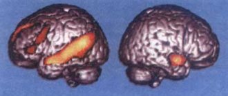
In January 2021, a study by Edmund T. Rolls et al. was published in the journal Translational Psychiatry, the authors of which focused on changes in functional connections in the brain over time, and then compared the findings in people with and without mental disorders. The authors set out to determine how these processes occur in people with long-term schizophrenia, compared with those who have suffered a first episode of the disease, as well as in comparison with those who suffer from attention deficit hyperactivity disorder (ADHD); and how different areas of the thalamus work in schizophrenia and ADHD. To do this, they used four databases.
Scientists selected 1017 people aged 22–35 from the Human Connectome Project (HCP). 123 long-term schizophrenic patients and 136 controls were recruited from Veteran General Hospital in Taiwan. 154 patients hospitalized for first episode psychosis and 112 controls were recruited from the Shanghai Mental Health Center. fMRI results of respondents with ADHD were taken from the ADHD-200 Consortium study. To clarify the functional and anatomical connections between brain regions, an automated anatomical atlas (The automated anatomical labeling atlas 3, AAL3) was used. Temporal variability of functional connections was calculated using a special formula.
Among 1017 HCP respondents, high temporal variability was found in functional connections formed by the amygdala, orbitofrontal cortex, caudal nuclei, cingulate gyrus, hippocampus, parahippocampal gyrus, lateral and medial geniculate nuclei of the thalamus. The smallest temporal variability was found in the motor and primary visual cortex. The authors explained this by saying that the visual cortex has stronger functional connections between its areas, which allows them to restructure simultaneously. In contrast, areas such as the hippocampus, parahippocampal gyrus, amygdala, and orbitofrontal cortex have functional connections with many other brain regions and change independently of each other. In addition, the visual cortex may have strong functional connections with the motor cortex.
The authors next compared respondents with chronic schizophrenia from the Veteran General Hospital with a control group. In the case of disease, the authors found high temporal variability in the primary visual cortex (inferior occipital gyrus, fusiform gyrus), temporal cortex, orbitofrontal cortex, thalamic nuclei (medial dorsal, ventral anterior and lateral nuclei), motor areas (putamen and globus pallidus ). According to data obtained from AAL3, regions of the brain with strong functional connections are weakened in chronic schizophrenia. These areas include the primary visual cortex (occipital region, calcarine sulcus, lingual gyrus, sphenoid gyrus) with functional connections formed both within it and with the temporal lobe, auditory cortex (Heschl’s gyrus, superior temporal gyrus). There are also changes in the thalamic nuclei. Strong connections were found in the medial dorsal, ventral anterior, lateral, and posterior nuclei. The connection in the area of the medial and lateral groups of thalamic nuclei is weakened.
The authors then studied first-episode psychotic respondents recruited from the Shanghai Mental Health Center. Temporal variability was greater between the temporal and visual cortices, in areas of the cingulate cortex and superior medial prefrontal cortex. There were no changes in internal functional connections in the area of the visual cortex. Functional connections in the temporal region, orbitofrontal cortex, hippocampus and parahippocampal gyrus were weak. Strong functional connections were found between the following nuclei of the thalamus: medial dorsal nucleus, ventral anterior and lateral nuclei, lateral and ventral posterior nuclei. The lateral and medial groups of thalamic nuclei demonstrated weak functional connectivity during the first episode of schizophrenia, as in the case of its chronic course.
The authors then examined fMRI results in respondents with ADHD. The area with low temporal variability was the primary visual cortex (sphenoid gyrus). Areas with a high ability to change functional connections over time were the longitudinal gyrus, posterior cingulate cortex, and ventromedial prefrontal cortex. The authors conclude that the visual cortex is quite resistant to changes in the brain that ADHD leads to. The stability of functional connections is preserved in the area of the medial and lateral groups of thalamic nuclei. Weaker functional connectivity was found in the medial dorsal, ventral anterior and lateral thalamic nuclei.
Thus, the authors found little variability over time in the functional connections of the visual cortex. Quite strong changes occurred in the orbitofrontal cortex, amygdala and hippocampus. Consequently, the functional connections of the visual cortex are stronger.
The importance of functional connections for the normal functioning of the brain is emphasized by the discovery of their changes in the case of diseases. Thus, in long-term schizophrenia, temporal variability is more pronounced than in the case of ADHD.
The hypothesis of the attractiveness of neural networks explains the relationship between the functional connections that arise between neurons, their stability or variability in connection with diseases. Excitation from active neurons passes to less active cells, and thereby ensures the constancy of the neural network. There are strong functional connections between neurons in the same brain region. And if a neuron or group of neurons forms a connection with cells in another area, the remaining cells take over their function in forming the network. In schizophrenia, there is a decrease in neuronal activity, especially in the prefrontal cortex. Instability is formed in the attractive neural network. This leads to a weakening of attention, and you have to use volitional efforts to maintain it. As a result, functional connections in sensory areas are weakened. Processes seem to shift towards one’s own thoughts, and the reaction to what is happening around decreases. The introduction of the concept of temporal variability demonstrates the dynamics of changes in the brain in schizophrenia and contributes to understanding the pathogenesis of certain symptoms, for example, how attention is impaired, why associative processes are impaired.
The discovery of high temporal variability in long-term schizophrenia in the visual, temporal cortex, orbitofrontal zone and weakening of functional connections in the temporal region confirms that one of the main links in the pathogenesis of schizophrenia is the reduction of connections between neurons and, as a result, increased excitation. This leads to destabilization of the attractive neural network due to the fact that the activity of neurons is not enough to maintain its excitation.
During the first attack of schizophrenia, the same changes in associative connections occur as during its long-term course, however, they are less pronounced. Moreover, after the first episode, no disruption of connections within the primary visual cortex was detected.
Reduced connectivity of the primary visual cortex with other areas of the brain supports the theory that in patients with long-term schizophrenia, “bottom-up” information processing is impaired, when visual information is transformed by the brain into an image. Perception concentrates more on internal processes, and the person seems to distance himself from what is happening in the environment.
In ADHD, the authors found weak variability in associative connections in the primary visual cortex. This finding supports the hypothesis that in this disorder there is increased sensitivity to visual stimuli, decreased attention during long and laborious tasks, and frequent interruptions due to external stimuli.
High variability of functional connections in the sensory nuclei of the thalamus (lateral and medial groups of nuclei), strong dopamine pathways between the thalamic nuclei and the substantia nigra indicate the role of dopamine in the pathogenesis of schizophrenia. The nuclei of relaying associative information, on the contrary, have strong functional connections. These changes were observed both in long-term schizophrenia and in the first episode. This determines some resistance of the thalamus to the schizophrenic process.
In ADHD, stronger connections were found between the sensory nuclei of the thalamus, indicating the strength of the sensory pathways, and weaker connections between the associative nuclei, indicating the variability of its connections with the frontal cortex. This leads to disturbances in the functions of attention and control. Thus, a new study brings us closer to understanding how the functioning of the cerebral cortex is disrupted in some mental disorders.
Author of the translation: Wirth K.O.
Source: Edmund T. Rolls, Wei Cheng, Jianfeng Feng. Brain dynamics: the temporal variability of connectivity, and differences in schizophrenia and ADHD. Translational Psychiatry.
As already mentioned, in our cerebral cortex there are many vast and endless areas that are not directly associated with sensory or motor processes. They are called associative zones and occupy about 80% of the cortex.
So, each such associative area of the cortex is closely connected with several projection (sensory or motor) zones. Therefore, it is believed that in associative areas there is an association (or simply a connection or combination) of different sensory information, as a result of which complex elements of our consciousness are formed.
The largest areas of accumulation and habitat of associative areas in humans are found in the frontal, occipito-parietal and temporal regions .
In general, each projection area of the cortex, be it sensory or motor, is surrounded by associative areas, and the neurons of these areas are often polysensory, i.e. are able to respond to various signals coming from the auditory, visual, skin and other systems. And it is precisely this polysensory nature of neurons that allows them to combine sensory information and organize and coordinate the interaction of sensory and motor areas of the cortex.
So, the frontal lobes are responsible for the implementation of higher mental functions, which are manifested in the formation of personal qualities, various creative processes and drives.
When the frontal parts of the cerebral cortex are damaged, the construction of goal-directed behavior based on foresight is sharply disrupted.
What it is? Now I’ll explain: For example, in monkeys, damage to these very frontal lobes impairs their ability to solve problems with a delayed response. Conduct this experiment: find somewhere such a sick monkey and, in front of its eyes, place food in one of two cups, and cover the cups with identical objects. Then place an opaque screen briefly between the monkey and the cups. Then remove the screen and let the monkey choose one of these cups. So, a normal monkey will remember the required cup after a delay of several minutes, but our sick monkey, with damaged frontal lobes, alas, will not be able to solve this problem if the delay exceeds only a few seconds. This will be a delayed response, or rather, its absence, i.e. such monkeys simply do not remember what happened quite recently due to the “breakdown” of the necessary neurons in the frontal lobes. What can we say about people...
Further. In the parietal associative area of the cortex, subjective ideas about the surrounding space and our body are formed. This becomes possible thanks to the connection and comparison of somatosensory (sensitive), proprioceptive (Proprioception is the ability to perceive the position and movement in space of one’s own body, or its individual parts) and visual information.
If the outer surface of the occipital lobe, not the projection but the associative visual zone, is damaged, vision will be preserved, but a recognition disorder will immediately occur - the so-called visual agnosia. Such a person, being absolutely literate, will not be able to read what is written, and will be able to recognize a familiar person only after he speaks. Well, he doesn’t recognize him with his “eyes” and that’s it!
Let's continue. In the temporal cortex there is Wernicke's auditory speech center, located in the posterior parts of the superior temporal gyrus (fields 22, 37, 42 of the left hemisphere). This zone is asymmetrical - for right-handers it is in the left hemisphere, and for left-handers it is in the right hemisphere.
The task of this center is to recognize and store oral speech, both one’s own and that of others. When the auditory center of speech is damaged, a person can speak, express his thoughts orally, but does not understand someone else’s speech, and although hearing is preserved, the person does not recognize words. This condition is called sensory auditory aphasia. Such a person often talks a lot (logorrhea), but his speech is incorrect (agrammatism), and there is a replacement of syllables and words (paraphasia).
But, the speech function is connected not only with the sensory, but also with the motor system. And we really have such a motor speech center. It is located in the posterior part of the third frontal gyrus (area 44), most often of the left hemisphere (again, right-handed and left-handed) and was first described by Mr. Dax in 1835, and then by Mr. Broca in 1861. When the motor center of speech is damaged, motor aphasia develops - in this case, the person understands speech, but, alas, cannot speak.
In the middle part of the superior temporal gyrus (area 22) there is a center for recognizing musical sounds and their combinations. And on the border of the temporal, parietal and occipital lobes (field 39) there is a center for reading written speech, which ensures the recognition and storage of images of written speech. It is clear that lesions of this center lead to the inability to read and write.
By the way, both of these centers are also asymmetrical and are located in different hemispheres for left-handers and right-handers.
Also in the temporal region there is field 37, which is responsible for remembering words. People with lesions in this field do not remember the names of objects. At the same time, they are very reminiscent of forgetful people who constantly have to prompt the right words. Such a person, having forgotten the name of an object, clearly remembers its purpose and properties, so he describes its qualities for a long time, explains what is being done with this object, but for the life of him cannot name it. Well, for example, instead of the word “tie,” a person, looking at it, says something like this: “this is something that is put on the neck and tied with a special knot to make it beautiful when they go to visit.”
The function of memory and dreams is also associated with the temporal cortex.
Got it? Then let's continue! >>
^ Top
Structural fields of the cerebral cortex
When violated, the form of abstract reflection of reality, speech, and purposeful behavior suffer.
Collaboration of the cerebral hemispheres and their asymmetry.
There are morphological prerequisites for the hemispheres to work together. The corpus callosum makes a horizontal connection with the subcortical formations and the reticular formation of the brain stem. In this way, the hemispheres work cooperatively and reciprocal innervation occurs when working together.
Functional asymmetry. The left hemisphere is dominated by speech, motor, visual and auditory functions. The thinking type of the nervous system is left-hemisphere, and the artistic type is right-hemisphere.
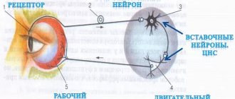
Sensory areas are functional areas of the cerebral cortex that receive sensory information from most of the body's receptors through ascending neural pathways.
Primary sensory and motor areas occupy less than 10% of the surface of the cerebral cortex and provide the most basic sensory and motor functions.
Association zones are functional areas of the cerebral cortex. They connect newly received sensory information with previously received and stored in memory blocks, and also compare information received from different receptors. Sensory signals are interpreted, comprehended and, if necessary, used to determine the most appropriate responses, which are selected in the association zone and transmitted to the associated motor zone. Thus, associative zones are involved in the processes of remembering, learning and thinking, and the results of their activity constitute what is usually called intelligence.
Individual large association areas are located in the cortex next to the corresponding sensory areas. For example, the visual association zone is located in the occipital zone, immediately anterior to the sensory visual zone, and carries out the associative functions described above associated with visual sensations. For example, the auditory association area analyzes sounds, dividing them into categories, and then transmits signals to more specialized areas such as the speech association area, where the meaning of words heard is perceived.
Motor areas are functional areas of the cerebral cortex that send motor impulses to voluntary muscles along descending pathways that begin in the white matter of the cerebral hemispheres.
Date of publication: 2015-02-03; Read: 1220 | Page copyright infringement
studopedia.org - Studopedia.Org - 2014-2018 (0.001 s)…
The highest division of the central nervous system is the cerebral cortex (cerebral cortex).