Oxygen starvation of the fetus, resulting from impaired blood circulation in the placenta, leads to serious illnesses and deviations in the development of the child. One of the disorders that occurs for this reason is a subependymal cyst of the brain, which is diagnosed in newborns due to prolonged circulatory disorders, hypoxia and lack of nutrients.
A subependymal or cerebral cyst of the brain is a benign hollow formation in the ventricles of the brain, filled with fluid, formed due to hypoxia and insufficient blood supply. Since hypoxia is often diagnosed during pregnancy, the risk of formation is very high.
An intracerebral cyst in a newborn child is a natural protective response of the body to atrophy of brain tissue.
To prevent the expansion of necrosis of brain tissue, the body replaces the damaged areas, creating a natural barrier in the form of a cystic cavity. A small cyst does not pose a threat to the normal functioning of the brain.
But if fetal hypoxia is not treated during pregnancy, the cyst begins to increase in volume, leading to such dangerous manifestations as convulsions, deterioration in well-being and health, and psycho-emotional disorders.
Causes and risk factors
Fetal hypoxia is the main cause of subependymal cyst formation and has a significant impact on the development of the infant. The main risk factors for the manifestation of a cyst are the diagnosis of moderate or acute fetal hypoxia in a pregnant woman.
There are three factors for the appearance of a subependymal cyst:
- Cerebral ischemia . The most common cause of any cyst formation. Ischemia occurs due to impaired blood supply to brain tissue. In the place where the brain matter dies, a void appears, which the body fills with cerebrospinal fluid. The small size of the cyst does not lead to serious disorders; it only requires drug treatment and monitoring over time. If it grows and puts pressure on other parts of the brain, the child experiences convulsions, regurgitation, protrusion of the fontanel, and inhibition of physical and mental development.
- Hemorrhage in the head of a newborn is the second reason for the appearance of a neoplasm. The most severe deviations in the development of the child will be if hemorrhage occurs during uterine development of the fetus, and not during passage through the birth canal. The main causes of hemorrhage are the presence of infections, acute fetal hypoxia and birth trauma.
- Acute or moderate fetal hypoxia is a disruption of the functioning of the pregnant woman’s body and placenta. The main causes of hypoxia include anemia, toxicosis in the third trimester, multiple pregnancy, Rh conflict between the child and mother, polyhydramnios, placental insufficiency, and infectious diseases.
Article on the topic: Prognosis for liver cirrhosis: life expectancy and stages of the disease
What are pseudocysts?
In articles on medical topics, you can often find the expression cerebral pseudocyst in newborns. Unlike cysts, it is not a pathological phenomenon and has a different mechanism of formation. After the birth of a child, cerebrospinal fluid enters the choroid plexuses of the ventricles of the brain, forming pseudocysts. They are small, round, do not grow, and are involved in the production of cerebrospinal fluid necessary for the normal functioning of the brain.
Subependymal pseudocyst is usually determined only using instrumental examination methods. As a rule, it does not give any clinical symptoms. Pseudocysts resolve on their own in children.
Features of the pathology
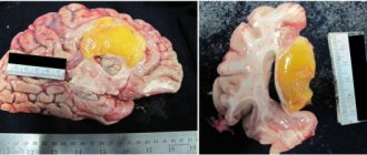
A cyst of subepinedymal localization is distinguished by the fact that it is mainly benign histologically, but there is a possibility of rapid growth and malignancy. Therefore, all children with a history of intrauterine hypoxia or birth trauma require careful dynamic monitoring of the condition and size of this formation. They are at risk.
Based on the growth pattern and histological structure, brain tumors are conventionally divided into benign and malignant. But with rapid growth, they are all malignant. After all, tumors develop in the confined space of the cranium, put pressure on the surrounding brain structures, block the circulation of cerebrospinal fluid and venous outflow, causing a clear clinical picture of malignancy.
Localization in the brain and symptoms
A subependymal cyst is localized at the site of impaired blood supply to brain tissue, most often in the ventricular region. Symptoms of the disease depend on the location and size of the cyst. The presence of a pathogenic focus in a certain area of the brain leads to disruption of the body functions for which it is responsible.
A cyst in the occipital region affects the functioning of the visual system, visual acuity decreases, double vision, etc. A cyst in the temporal lobe leads to hearing impairment, in the cerebellum – gait and coordination of movements are impaired, in the pituitary gland – hormonal imbalances.
The small size of the cyst may not manifest itself in any way on the child’s behavior. But larger ones lead to convulsions and strokes, which can result in paralysis of the body.
The main symptoms observed in the presence of a cerebral cyst in the head of a newborn child:
- sleep disturbance;
- anxious behavior;
- breast refusal;
- poor weight gain or loss;
- long, causeless crying;
- hyper- or hypotonicity of muscles;
- tremor of the chin, twitching of the limbs;
- hearing impairment;
- blurred vision, sometimes complete loss;
- regurgitation “fountain”;
- loss of consciousness, coma;
- pulsation and swelling of the fontanelle;
- convulsions.
The consequences of growth and lack of treatment of the cyst lead to mental and physical retardation in the development of the child.
Diagnosis of subependymal brain cyst
Only hardware diagnostics can confirm the presence of a pseudocyst in a baby’s head. Ultrasound examination (neurosonography) is considered an accurate, reliable way to detect abnormal tumors.
An ultrasound of the brain is informative until the baby’s fontanel is overgrown (up to one year). The method is indicated for premature babies, those who have suffered oxygen deprivation, or birth trauma.
Using neurosonography, the doctor determines the size, number and localization of cystic compactions, assesses the degree of expansion of the ventricles of the brain (which may indicate increased intracranial pressure and the risk of hydrocephalus), identifies signs of blockage of the liquor pathways and impaired outflow of cerebrospinal fluid, and studies changes in the echostructure of brain tissue.
During a second ultrasound, the specialist compares the results and sees how the pseudocyst shrinks and gradually disappears. If echo signs of a true cyst are detected in a newborn child, ultrasound will allow timely treatment to begin in order to prevent complications as early as possible.
In addition to ultrasound, for the purpose of detailed study of pathology, the following can be performed:
- Doppler encephalography to evaluate the state of blood flow and cerebral vessels.
- Two-photon or positron emission tomography. Allows you to monitor the effectiveness of prescribed conservative treatment in accordance with changes in brain tissue.
- Cerebral scintigraphy.
- Computed and magnetic resonance imaging (CT and MRI).
These tests are often performed on older children. These methods make it possible to distinguish (differentiate) pseudocyst, tumor and brain cyst, which is very important for the development of correct treatment.
If a neonatologist or neurologist prescribes an instrumental examination for a child, then refusing it to parents is irresponsible and dangerous. Early detection of any abnormal formations in cerebral tissues allows one to avoid serious consequences.
Diagnostic criteria
Using neurosonography (ultrasound) in newborns, it is easy to find cysts and other abnormalities in the brain. The fontanel, as a rule, does not close until a year, so this procedure is the fastest, easiest and most reliable.
Neurosonography must be done for premature babies, during severe pregnancy and protracted labor. If the doctor discovers a small subependymal cyst, then the brain tissue returns to normal on its own; you just need to be under the supervision of a pediatric neurologist.
If the cyst exceeds the minimum values, then additional diagnostic measures, MRI and CT are required. Diagnosis using these methods should be carried out twice a year.
Repeated examination of the cyst and recording its growth can lead to serious disturbances in the development of the child and a threat to his life.
Diagnosing a multi-chamber subependymal cyst is very problematic, since the symptoms can be easily confused with other disorders.
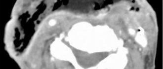
MRI of a subependymal cyst in a newborn child
Qualitative approach to therapy
Treatment of a cerebral cyst is aimed at preventing its growth. To do this, you need to find and eliminate the main cause that provokes oxygen starvation. During certain life stages of a child's growth, medical care will vary.
First aid
After birth, a neonatologist performs resuscitation care.
Article on the topic: The benefits and harms of oatmeal with milk and water: should you eat it in the morning?
Removes fluid from the respiratory tract, stimulates artificial respiration; in severe cases, an oxygen mask is used to supply pure oxygen.
The first three days after birth...
A neurologist conducts a daily examination and prescribes a restorative course of medication and rehabilitation therapy. Massage, physical therapy and sedatives are prescribed.
In childhood, the doctor observes the dynamics of the child’s development rate.
Medicines are used to stimulate speech functions and normalize the psycho-emotional state. At this stage, you may need the help of specialists such as a speech therapist and a psychologist.
...and beyond
When a child reaches adolescence, vitamin medications are prescribed to stimulate brain function and normalize metabolism. It is also necessary to take hormone replacement medications.
All physiotherapeutic and medicinal methods are prescribed only after the results of the examination and identification of deviations in the child’s development.
A cyst is a kind of cavity containing fluid, fatty deposits and other exudate. Classification of formation is carried out taking into account its location, symptoms and origin. A choroid plexus cyst in the brain in children is not an abnormality and does not require treatment. The pathology can go away on its own. At the same time, education does not have a detrimental effect on the health and development of the child. A cystic cavity is dangerous when this pathology is preceded by infectious inflammation.
Most often it is found between the head of the caudate nucleus, the optic tubercle, and the lateral angles of the lateral ventricles of the brain. It is assumed that the formation of a pseudocyst is a consequence of exposure to unfavorable factors on the fetus during embryonic development.
In newborns, cerebral pseudocysts form during fetal development. As a rule, they appear at an early stage of embryonic development, but closer to 28 weeks of pregnancy and disappear on their own. But the reason for the development of later formation of choroid plexus cysts in infants is an infection that the mother suffered during pregnancy.
Often a herpetic infection can affect the formation of a pathological process. The formation of a cyst is also influenced by the conditions under which the birth took place. As a rule, by the child’s first year the formation resolves. In order to prevent the development of other diseases against the background of this pathology, parents must be registered with a neurologist and visit him every 3 months.
If after a year the pseudocyst still does not resolve, then the doctor must decide what to do next, taking into account the results of the diagnosis, ultrasound (NSG) of the brain and the individual development of the baby.
In infants, pathology can be detected using ultrasound of the brain. Children under one year old undergo it. This allows you to timely prevent the development of cystic formations and other pathologies of the central nervous system. Ultrasound is prescribed for premature babies, as well as for children who have suffered birth trauma and hypoxia. Diagnosis is carried out through the large fontanel.
An ultrasound of a child’s brain allows one to determine the nature of the cystic formation, determine its size and shape, and distinguish it from a true brain cyst. Subependymal cysts usually form as a result of birth injuries. Pseudocysts can also occur in the choroid plexuses on the left and right. For timely diagnosis and treatment of a child under 1 year of age, you need to contact a pediatric neurologist.
In our clinic, you can make an appointment with a pediatric neurologist, perform an NSG of the child’s brain and receive adequate treatment.
Consequences and prognosis of the disease
The formation of small cysts does not have a serious impact on the quality of life, psychological and mental development of the child. Problems appear if it increases in volume. This may be affected by the presence of infections and hypoxia.
As the cyst enlarges, symptoms such as irritability, bronchopulmonary diseases, heart defects, developmental delays, and anemia begin to appear.
The first year of a child’s development does not show any serious deviations. Parents begin to notice violations at 2-3 years of age. The child has hyperexcitability, delayed speech development, and poor coordination. More serious manifestations include coma and death.
Preventive measures
In order to prevent the appearance of a cyst in the head of a newborn, it is necessary to establish the cause of its appearance. During pregnancy this is quite problematic.
It is necessary to do a genetic analysis, including amniotic fluid, but this analysis is done only in case of serious abnormalities in the development of the fetus.
Therefore, a woman needs to lead a healthy lifestyle, both before conception and during pregnancy, monitor her diet, and protect herself from intoxication and inflammatory processes.
Surgical treatment is performed very rarely, so the diagnosis of a subependymal cyst is not as scary as many people think. All that is necessary is observation by a neurologist for timely treatment of the disease and prevention of serious deviations in the development of the child.
What is the difference between a cyst and a pseudocyst?
A pseudocyst in a baby's head must be differentiated from a true cyst. The main indicator is the characteristic location. A false anomaly is formed either in the area of the bodies and lateral angles of the anterior horns of the right or left ventricle of the brain, or at the border of the thalamus (optic thalamus) and the head of the caudate nucleus.
The remaining cavity, fluid formations, if detected in other parts of the brain, are called cysts. False cysts resolve or significantly decrease in size by one year of age. True cysts in the head do not disappear, they can cause painful neurological symptoms and require constant monitoring by a neurologist.