The pituitary gland is an important regulatory center that coordinates the interaction of the endocrine and nervous systems of the human body. This organ is called the "master gland" because its hormones control the activity of other endocrine glands, including the adrenal glands, the thyroid gland, and the reproductive glands (ovaries and testes), and in some cases have direct regulatory effects in major tissues. Disruption of the pituitary gland affects the functioning of all organs and systems of the body and becomes the cause of many pathologies or deviations in human development.
COST OF SOME ENDOCRINOLOGIST SERVICES IN OUR CLINIC IN ST. PETERSBURG
| Price of a comprehensive examination for hormones (12 indicators) | from 6490 rub. |
| Endocrinologist appointment | 1000 rub. |
| Ultrasound of the thyroid gland | 1000 rub. |
| Call free: 8-800-707-1560 *The clinic is licensed to provide these services | |
What is the pituitary gland
The content of the article
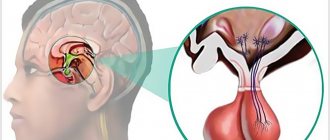
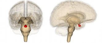
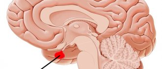
The pituitary gland is a tiny endocrine organ located at the base of the brain, in a bone formation, the so-called “sella turcica”. It is oval in shape and about the size of a pea - about 10 mm in length and 12 mm in width. Normally, in a healthy person, the weight of the pituitary gland is only 0.5-0.9 g. In women, it is more developed due to the synthesis of the hormone prolactin, which is responsible for the manifestation of the maternal instinct. The amazing ability of the pituitary gland is that it increases during pregnancy, and after childbirth the previous size is not restored.
The pituitary gland is largely controlled by the hypothalamus, which lies above and slightly behind the gland. These two structures are connected by the pituitary, or funnel-shaped, stalk. The hypothalamus is capable of sending stimulating or inhibitory (suppressive) hormones to the pituitary gland, thereby regulating its effect on other endocrine glands and the body as a whole.
The “conductor of the endocrine orchestra” consists of the anterior lobe, intermediate zone and posterior lobe. The anterior lobe is the largest (occupies 80%) and produces and releases large amounts of hormones. The posterior lobe does not produce hormones as such - this is done by nerve cells in the hypothalamus - but it releases them into the circulation. The intermediate zone produces and secretes melanocyte-stimulating hormone.
The pituitary gland is involved in several body functions, including:
- regulation of the activity of other organs of the endocrine system (adrenal glands, thyroid and gonads);
- control of growth and development of organs and tissues;
- control over the functioning of internal organs - kidneys, mammary glands, uterus in women.
The pituitary gland is a very important endocrine gland. It is located on the lower surface of the brain, in the pituitary fossa of the sella turcica of the sphenoid bone. A process of the dura mater, the diaphragm sellae, separates the pituitary gland from the cranial cavity. The infundibulum connects the pituitary gland to the hypothalamus.
Externally, the body of the pituitary gland is covered with a connective tissue capsule. The dimensions of the pituitary gland are quite individual (10-17) x 16 x (5-10) mm, the weight in men is about 0.5-0.6 g, in women - about 0.6-0.7 g. Being an anatomically single formation , the pituitary gland is divided into two lobes. The anterior lobe (adenohypophysis) is larger, it occupies 70-80% of the total mass of the pituitary gland and includes three parts: distal, tuberous and intermediate. In the posterior lobe (neurohypophysis) there is a neural part and an infundibulum.
The structure of the pituitary gland
The physiological role of the pituitary gland determines the characteristics of its blood supply. The inferior pituitary arteries arise from the internal carotid arteries. The superior pituitary arteries depart from the vessels of the arterial circle, go to the gray tubercle and funnel, where they form an anastomosis with each other and break up into capillaries that penetrate the tissue. The branches of the axons of the neurosecretory cells of the hypothalamus end on these capillaries, forming synapses. Here the neurosecret is released into the blood. From the loops of this network, portal venules are formed, running along the tubercular part to the anterior lobe of the pituitary gland, where they become wide sinusoidal capillaries. These capillaries form a secondary hemocapillary network, entwining groups of secretory cells. The capillaries of the secondary network, merging, form efferent veins, through which blood (with hormones of the anterior lobe) is removed from the pituitary gland. The posterior lobe of the pituitary gland is supplied with blood mainly by the inferior pituitary arteries. There are anastomoses between the upper and lower arteries.
The cells of the anterior pituitary gland are called adenocytes. There are 2 types of adenocides:
- chromophilic adenocytes - large cells that are well stained with dyes;
- chromophobe adenocytes are small cells, weakly stained.
Among chromophilic adenocytes, acidophilic adenocytes are distinguished - oval-shaped cells, stained pink with acidic dyes, as well as large basophilic cells, stained with basic dyes and rich in glucoprotein inclusions. The posterior part of the anterior lobe, located as a layer between it and the posterior lobe, is considered as an intermediate part. It is formed by multilayer epithelium, among the cells of which there are vesicles (pseudofollicles).
The posterior lobe is formed by ependymal cells (pituicytes), small multi-processed cells and nerve fibers, axons of cells of the supraoptic and paraventricular nuclei of the hypothalamus, the branches of which end on the capillaries of the posterior lobe. The pituitary infundibulum, connecting with the hypothalamic infundibulum, forms the pituitary stalk.
Functions of the pituitary gland
The functions of the pituitary gland are determined by the action of the hormones it secretes. Through the release of these substances, the following is carried out: regulation of the synthesis and secretion of hormones by the pituitary-dependent endocrine glands (adrenal glands, gonads); synthesis and secretion of melanins; regulation of organ growth and maturation; coordination of the functions of various organs (kidneys, uterus, mammary glands).
The anterior lobe of the pituitary gland synthesizes the so-called tropic hormones:
- somatotropin (somatotropic hormone, growth hormone);
- thyrotropin (thyroid-stimulating hormone);
- adrenocorticotropic hormone (ACTH);
- gonadotropic hormones (folliculotropin, luteotropin);
- lactogenic hormone (prolactin);
- melanocyte-stimulating hormone (melanocytotropin).
These hormones regulate the synthesis and secretion of hormones from the pituitary glands according to the feedback principle: when the concentration of a certain hormone in the blood decreases, the cells of the adenohypophysis release a signal hormone that stimulates the formation of the hormone by this gland, and an increase in its level in the blood leads to a slowdown in the secretion of the signal hormone.
In the intermediate part, lipotropic factors of the pituitary gland are produced, which influence the mobilization and utilization of fats in the body. Neurosecretory cells of the hypothalamic nuclei produce vasopressin and oxytocin, which are transported to the posterior lobe of the pituitary gland, from where they are carried by the blood.
Somatotropin is not secreted constantly, but periodically, 3-4 times a day. Its secretion increases during fasting, heavy physical labor and during sleep. With age, the production of somatotropin decreases markedly, but persists throughout life. Growth hormone has a double effect on the cells of the body: the breakdown of accumulated carbohydrates and fats in the cells is enhanced, as well as their mobilization for energy and plastic metabolism; under the influence of somatomedins produced by the liver, bone growth, protein synthesis and cell division are enhanced.
Insufficient secretion of somatotropin leads to dwarfism while maintaining a normal physique. Excessive secretion leads to gigantism. If hypersecretion begins in a person already in adulthood, then acromegaly develops. At the same time, the limbs, nose and chin, tongue and digestive organs lengthen disproportionately.
Adrenocorticotropic hormone stimulates the release of corticosteroids by cells of the adrenal cortex. The secretion of this hormone increases during certain emotional states (fear, anger, chronic stress).
Thyroid-stimulating hormone regulates the release of thyroid hormones. It activates adenylate cyclase and increases the absorption of iodine by gland cells. In addition, thyrotropin has an effect on protein metabolism - it increases the synthesis of proteins and nucleic acids, and also increases the number and size of thyroid cells.
Gonadotropic hormones stimulate the functions of the gonads. Folliculotropin regulates the development of follicles in the ovaries in women, and the formation of sperm and the development of the prostate in men. Luteotropin stimulates the production of androgens (androstenediol, testosterone, etc.) and estrogens (estradiol, estriol, etc.).
Prolactin increases the production of progesterone in the corpus luteum of the ovary and milk production (lactation). The physiological role of this hormone is not fully understood, but almost all of its known effects are related to reproduction.
Melanocytotronin regulates the distribution of the melanin pigment and thus determines the color of hair and skin. Pigment spots during pregnancy and increased skin pigmentation in older people occur as a result of hyperfunction of the intermediate lobe of the pituitary gland.
Vasopressin is involved in the regulation of urine formation, enhancing the reabsorption of water from primary urine, ensuring water-salt homeostasis of the body.
Oxytocin stimulates the smooth muscles of the uterus during labor and milk secretion. It causes contraction of the myoepithelial cells surrounding the alveoli and ducts of the mammary gland, causing milk to be released from the breast.
If you have dysfunction of the pituitary gland, contact an endocrinologist in Krasnodar.
Hormones of the anterior pituitary gland
This part of the pituitary gland is called the adenohypophysis. Its activity is coordinated by the hypothalamus. The anterior lobe of the pituitary gland regulates the activity of the adrenal glands, liver, thyroid and gonads, bone and muscle tissue. Each hormone of the adenohypophysis plays a vital role in endocrine function:
| Hormone | Target organs | Main function |
| Growth hormone (somatotropin) | Musculoskeletal tissue | Promotes body tissue growth |
| Prolactin | Mammary gland | Promotes milk production |
| Thyroid-stimulating hormone | Thyroid | Stimulates the production of thyroid hormones (triiodothyronine and thyroxine), which have an important effect on metabolic processes |
| Adrenocorticotropic hormone | Adrenal cortex | Stimulates the production of cortisol hormones from the adrenal cortex, which have anti-inflammatory and immunosuppressive effects and participates in the metabolic process |
| Follicle stimulating hormone | Ovaries and testes (testes) | Stimulates the maturation of follicles in the ovary and spermatogenesis in the testicles, the development of secondary sexual characteristics |
| Luteinizing hormone | Ovaries and testes (testes) | Ovulation, testosterone production, development of secondary sexual characteristics. |
Let us consider in more detail each hormone of the anterior pituitary gland.
Growth hormone (somatotropin)
The endocrine system regulates human body growth, protein synthesis, and cell replication. The main hormone involved in this process is growth hormone, also called somatotropin, a protein hormone produced and secreted by the anterior pituitary gland. Its primary function is anabolic: it directly accelerates the rate of protein synthesis in skeletal muscle and bone. Insulin-like growth factor is activated by growth hormone and indirectly supports the formation of new proteins in muscle cells and bone. After 20 years, every subsequent 10 years a person’s growth hormone level decreases by 15%.
Somatotropin has the effect of an immunostimulant: it is able to influence carbohydrate metabolism, increasing blood glucose levels, reduces the risk of fat deposits and increases muscle mass. The glucose-lowering effect occurs when growth hormone stimulates lipolysis, or the breakdown of fat tissue, releasing fatty acids into the blood. As a result, many tissues switch from glucose to fatty acids as their primary source of energy, meaning less glucose is supplied from the blood.
Growth hormone also initiates a diabetogenic effect, in which it stimulates the liver to break down glycogen into glucose, which is then deposited into the blood. The name "diabetogenic" comes from the similarity of elevated blood glucose levels observed between people with untreated diabetes and people suffering from excess growth hormone. Blood glucose levels increase as a result of a combination of glucose-sparing and diabetogenic effects.
The amount of growth hormone in the human body changes throughout the day. The maximum is achieved after 2 hours of sleep at night and every 3-5 hours during the day. The peak level of the hormone is observed in a child during intrauterine development at 4-6 months - 100 times more than in an adult. You can increase the level of somatotropin through exercise, sleep, and the use of certain amino acids. If the blood contains large quantities of fatty acids, somatostatin, glucocorticoids and estradiols, the level of growth hormone decreases.
Dysfunction of the endocrine growth control system can lead to several disorders. For example, gigantism is a disorder in children caused by the secretion of abnormally large amounts of growth hormone, resulting in excessive growth.
A similar complication in adults is acromegaly, a disorder that causes the bones of the face, arms, and legs to grow in response to excessive levels of growth hormone. This is reflected in the general condition by muscle weakness and pinched nerves. Abnormally low levels of the hormone in children can cause poor growth, a disorder called pituitary dwarfism (also known as growth hormone deficiency), sexual development, and mental development (significantly affected by an underdeveloped pituitary gland).
Hormone prescription
The pituitary gland produces a number of hormones that act as intermediaries between the central nervous and endocrine systems. Each biologically active substance is responsible for a specific function of the body. The table shows hormones and their purposes .
| Hormone | Function |
| Corticotropin | Controls the performance of the adrenal glands, the synthesis of steroids and cortisol. The substance helps to get out of a stressful situation and affects a person’s sexual development. |
| Thyrotropin | Regulates the activity of the thyroid gland and stimulates the formation of triiodothyronine and thyroxine. |
| Follitropin | Promotes the formation of a priority follicle, ensures its further rupture and removal from the ovary. |
| Lutropin | Responsible for the onset of ovulation, the formation of the corpus luteum and its performance within two weeks. |
| Somatotropin | Affects the growth and formation of the body. After reaching the age of 35, the level of the hormone in the body begins to decline. |
| Prolactin | In women, it supports the development of the mammary glands and controls the amount of milk during breastfeeding. In men, it is responsible for spermatogenesis. |
| Oxytocin | Helps increase potency in men, and develops maternal instinct in women. The amount of the substance increases in a good mood and decreases in stressful situations. |
| Vasopressin | Together with oxytocin, it stimulates brain activity. It increases with large blood loss and decreased blood pressure. |
| Effector | Ensure the production of melanin and protect the skin from ultraviolet rays. |
| Lipotropin | Promotes the burning of carbohydrates in the body and reduces excess fat. |
| Endorphin | Reduces the threshold of pain and stress, controls the body's reaction during shock. |
The correct functioning of hormones is disrupted when the pituitary gland is diseased.
Thyroid-stimulating hormone (TSH)
Thyrotropic hormone is intended to regulate the functions of the thyroid gland and regulates the synthesis of substances T3 (thyroxine) and T4 (triiodothyronine), associated with metabolic processes, the digestive and nervous system, as well as the functioning of the heart. With a high level of TSH, the amount of T3 and T4 substances decreases, and vice versa. The level of thyroid-stimulating hormone varies depending on the time of day, age and gender. During pregnancy in the first trimester, the TSH level decreases significantly, but in the third trimester it may even exceed the norm.
Thyroid-stimulating hormone deficiency can occur due to:
- injuries and inflammation in the brain;
- inflammatory processes, tumors and oncological diseases of the thyroid gland;
- incorrectly selected hormonal therapy;
- stress and nervous tension.
- Excessive production of TSH may occur due to:
- thyroid diseases;
- pituitary adenomas;
- unstable production of thyrotropin;
- preeclampsia (during pregnancy);
- nervous disorders, depression.
Checking the TSH level through laboratory tests must occur simultaneously with checking T3 and T4, otherwise the test result will not allow an accurate result to be established. With a simultaneous decrease in TSH, T3 and T4, the doctor can diagnose hypopituitarism, and with an excessive amount of these components - thyrotoxicosis (hyperthyroidism). An increase in all hormones of this group may indicate primary hypothyroidism, and different levels of T3 and T4 are a possible sign of thyrotropinoma.
Decoding brain MRI data
Pituitary gland dysfunction is associated with pathological processes, which result in structural changes in gland tissue. Magnetic resonance imaging will help differentiate the disease, determine the nature of the lesion and its location. The method allows you to visualize the slightest deviations from the norm; thanks to MRI, it is possible to diagnose pituitary gland diseases in the early stages.
Empty sella syndrome is characterized by a thinning gland that does not fill the pituitary fossa. On a sagittal section, the organ has a crescent shape and occupies the lower part of the bone pocket.
Empty sella turcica on MRI
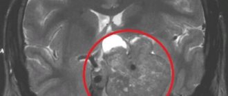
MRI of the pituitary gland also reveals tumors and cystic formations, allowing timely treatment of the pathology. An adenoma on MRI images is defined as an area with a changed structure, most often located in the anterior lobe. The following factors indirectly indicate the presence of a tumor:
- asymmetry of the gland, change in shape and size;
- violation of boundaries, deformation of the bone pocket;
- displacement of the pituitary stalk from the central axis.
Depending on the size, neoplasms are divided into:
- picoadenomas (up to 3 mm);
- microadenomas (3-10 mm);
- macroadenomas (10-30 mm);
- giant adenomas (over 30 mm).
MRI can detect small tumors that develop asymptomatically. Timely diagnosis of microadenoma is possible during a native study, but if the tumor is small, as well as if malignancy is suspected, the administration of contrast is necessary.
The nature of the blood supply to a malignant formation has significant differences from a similar benign process. A solution of gadolinium salts illuminates the vasculature, revealing the adenocarcinoma's own system. In the photographs, the tumor has a heterogeneous glandular structure, its boundaries are blurred.
Depending on the location, endosellar and endoextrasellar tumors are distinguished. The former do not extend beyond the sella turcica, the latter grow beyond the boundaries of the bone pocket.
Pituitary cysts on MRI appear as dark spots (on T1-weighted images) with clear boundaries. They need to be differentiated from macroadenomas, which are round formations with a compacted membrane.
Insufficient production of somatotropin is characterized by hypoplasia of the adenohypophysis and infundibulum, and an atypical location of the neurohypophysis. These signs help to identify the cause of hormonal disorders and correct the functioning of the gland with the help of medications, preventing possible disturbances in the growth and development of the body.
The MRI method helps to differentiate the pituitary gland lesions from pathological processes occurring in the surrounding tissues and having a similar clinical picture.
Adrenocorticotropic hormone (ACTH)
Adrenocorticotropic hormone affects the activity of the adrenal cortex, which produces cortisol, cortisone and adrenocorticosteroids, and also has a slight effect on sex hormones that control sexual development and reproductive function of the body. Cortisol is vital to processes that include immune function, metabolism, stress management, blood sugar regulation, blood pressure control and anti-inflammatory responses.
In addition, ACTH promotes fat oxidation, activates the synthesis of insulin and cholesterol and increases pigmentation. Pathological excess ACTH can trigger the development of Itenko-Cushing's disease, accompanied by hypertension, fat deposits and weakened immunity. Hormone deficiency is dangerous due to metabolic disorders and decreased ability to adapt.
The level of adrenocorticotropic hormone in the blood varies depending on the time of day.
The greatest amount of ACTH is found in the morning and evening. The production of this hormone is stimulated by stressful situations such as cold, pain, emotional and physical stress, as well as a decrease in blood glucose levels. The influence of the feedback mechanism will inhibit ACTH synthesis.
Increased ACTH levels may occur due to:
- Addison's disease (bronze disease) – chronic insufficiency of the adrenal cortex;
- Itsenko-Cushing's disease, manifested by obesity, hypertension, diabetes mellitus, osteoporosis, decreased function of the gonads, etc.;
- the presence of tumors in the pituitary gland;
- congenital adrenal insufficiency;
- Nelson's syndrome is a disease characterized by chronic renal failure, hyperpigmentation of the skin and mucous membranes, and the presence of a pituitary tumor;
- syndrome of ectopic ACTH production, the symptom of which is a rapid increase in muscle weakness and a kind of hyperpigmentation;
- taking certain medications;
- postoperative period.
The reasons for a decrease in ACTH may be:
- dysfunction of the pituitary gland and/or adrenal cortex;
- the presence of an adrenal tumor.
Prolactin
Prolactin, or luteotropic protein hormone, affects sexual development in women - it takes part in the formation of secondary sexual characteristics, stimulates the growth of the mammary glands, regulates the lactation process (including preventing the onset of menstruation and new conception of the fetus during this period), is responsible for the manifestation of maternal instinct, helps maintain progesterone. In men, prolactin regulates testosterone synthesis and sexual function, namely spermatogenesis, and also affects prostate growth. Its indicators in a woman increase during breastfeeding. Undoubted is its participation in water, salt and fat metabolism, tissue differentiation.
Excess prolactin can cause absence of menstruation in women and milk production in non-breastfeeding women. Hormone deficiency can cause problems with conception in women and sexual dysfunction in men.
It is important to note that a few days before taking a prolactin test, it is absolutely impossible to have sexual intercourse, visit baths and saunas, drink alcohol, or be exposed to stress and nervous strain. Otherwise, the test result will be distorted and show an increased level of prolactin.
Elevated levels of prolactin in the blood can be caused by:
- prolactinoma – a hormonally active benign tumor of the anterior pituitary gland;
- anorexia;
- hypothyroidism – low production of thyroid hormones;
- polycystic ovary syndrome - numerous cystic formations in the gonads.
The cause of prolactin hormone deficiency may be:
- tumor or tuberculosis of the pituitary gland;
- head injury that has a depressing effect on the pituitary gland.
Functions of the gland
Now you can more clearly understand what the pituitary gland is thanks to a description of its functions. For example, the anterior lobe produces many protein hormones. The substance prolactin is responsible for adequate milk production in nursing women. Somatotropin is needed for the growth of the body. If there is not enough of it, then the development of the body is suspended, and the person may remain a dwarf. When there is too much of the hormone, excessive growth can be observed.
To keep the thyroid gland healthy, the pituitary gland produces thyroid-stimulating hormone. If violated, the consequences can be dire. The adrenal cortex is influenced by adrenocorticotropic hormone, and the development of the genital organs and the onset of puberty depends on estrogen and testosterone - female and male sex hormones.
The posterior lobe also represents the pituitary gland. Its functions are to produce the substances already described: oxytocin and vasopressin. The first hormone takes part in the contraction of smooth muscles of the intestine, gall bladder and urinary tract. Oxytocin stimulates the contraction of the uterine muscles during labor. This hormone is also produced to contract the mammary glands, which will promote the appearance and production of milk.
The pituitary gland and the cerebral hemispheres are connected by a stalk through which small arteries pass, providing nutrition to the organ. Doctors say that not all functions of this gland have yet been sufficiently studied, and that in addition to the synthesis of chemicals, there is another role for such an element. The exact amount of hormones synthesized has not been established.
As mentioned above, the pituitary gland is responsible for reproductive function, body growth, and metabolism. Conventionally, the gland is divided into two parts - the anterior (adenohypophysis), which occupies most of it, and the posterior. Each of them is responsible for the secretion of its own group of hormones. Recently, scientists have begun to distinguish the intermediate zone, but this is simply a section of the adenohypophysis without any visual differences. However, the so-called neural infundibulum passes through this part, which connects the gland to the hypothalamus.
The pituitary gland, producing hormones, is indirectly connected with almost every system of the body. However, most of the secretions are used to stimulate metabolism (the process of digestion and assimilation of food), human growth (increase in the size of bone tissue), and reproduction (secretion of sperm, eggs). In the female body, the pituitary gland partially produces sex hormones that prepare the uterus for pregnancy and fertilization.
The pituitary gland is also responsible for the normal development of the hypothalamus and the functioning of the thyroid gland, so some of its diseases can affect the patient’s emotional mood. It’s just that at this moment there is a third-party effect on the brain and its cortex, which causes certain disturbances in the patient’s behavior.
The adenohypophysis is the main part of the pituitary gland, which scientists also divide into three parts:
- most of;
- a leaf-shaped process, the functions of which, by the way, have not yet been fully determined;
- intermediate part.
The adenohypophysis is responsible for the production of the following groups of hormones: TTC, ACTH, FSH, LH, growth hormone and prolactin (including in the male body, but in much smaller quantities).
The posterior lobe of the pituitary gland, which is also often called the neural lobe, is completely penetrated by nerve processes. Vasopressin and oxytocin are produced here (they are transported throughout the body using neural endings along with electrical impulses).
Follicle stimulating hormone and luteinizing hormone
Endocrine glands secrete a number of hormones that control the development and regulation of the reproductive system. Gonadotropins include two glycoprotein hormones:
- Follicle-stimulating hormone (FSH) – stimulates the production and maturation of sex cells, or gametes, including eggs in women and sperm in men. FSH also promotes the growth of follicles, which then release estrogens in a woman's ovaries. In the male body, FSH performs an important function - it stimulates the growth of seminiferous tubules and the production of testosterone, which is essential for spermatogenesis;
- Luteinizing hormone (LH) causes ovulation in women and the production of estrogen and progesterone in the ovaries. LH stimulates testosterone production in men. The hormone affects the permeability of testicular tissue, thereby allowing more testosterone to enter the bloodstream. By maintaining normal LH levels, favorable conditions are created for spermatogenesis.
Significant excess of normal hormone levels can be caused by:
- fasting;
- stressful condition;
- polycystic testicular syndrome;
- pituitary tumor;
- alcoholism;
- insufficient function of the gonads;
- ovarian wasting syndrome;
- excessive exposure to x-rays;
- endometriosis;
- intense physical activity;
- renal failure.
During menopause, such an analysis result is considered normal.
Reduced hormone levels can also be a physiological norm, or can be caused by:
- luteal phase deficiency;
- smoking;
- lack of menstruation;
- polycystic ovary syndrome;
- Simmonds disease – total loss of function of the anterior pituitary gland;
- growth retardation (dwarfism);
- obesity;
- systematic use of potent drugs;
- Sheehan syndrome – postpartum infarction (necrosis) of the pituitary gland;
- impaired activity of the hypothalamus and/or pituitary gland;
- Denny-Morphan syndrome;
- increased concentration of prolactin in the blood;
- pregnancy;
- cessation of menstruation after the establishment of a cycle.
An excess of FSH and LH leads to premature puberty, and a lack of hormones can cause infertility and secondary hypofunction of the gonads.
Structure and physiology
The size of the pituitary gland is small - its normal height ranges from 3 to 8 mm, width - from 10 to 16 mm, and it weighs no more than 1 gram. From below, behind and in front, the organ is protected by the bone of the sella turcica, and from above it is covered by a diaphragm, which has an opening. Through the hole, the pituitary gland connects to the hypothalamus with a fairly thin stalk, thus resembling a small cherry hanging on a stalk.
The pituitary gland has an oval shape and consists of two lobes, unequal in size, different in structure, as well as in origin - anterior and posterior.
There is also a very small middle lobe (intercalary), related in origin to the anterior lobe, in which beta-melanocyte-stimulating hormone is produced.
Adenohypophysis
The anterior lobe is called the adenohypophysis. It accounts for approximately ¾ of the total volume of the inferior cerebral appendage. The adenohypophysis consists of glandular cells that produce so-called tropic hormones that control the activity of peripheral endocrine glands.
Types of tropic hormones:
- somatotropic - affect the tissues of the body;
- thyroid-stimulating - on the thyroid gland;
- lactotropic - mammary gland;
- corticotropic - adrenal glands;
- gonadotropic - sex glands (gonads).
In addition, in the anterior lobe there are so-called zero cells that do not participate in hormone formation. Their function is not fully understood.
Neurohypophysis
The posterior lobe is called the neurohypophysis. It does not produce hormones, but accumulates neuropeptides synthesized in the hypothalamus - oxytocin and vasopressin, after which they are activated and sent into the bloodstream.
Influence of the hypothalamus
The hypothalamus scans the activity of endocrine organs and some other body processes. Then he gives the command to the pituitary gland to “spur” or “hold back” the work of the target organs. For the hypothalamus itself, the target organ is the pituitary gland.
The hypothalamus exercises its control using neurosecretory cells. They produce releasing hormones, which regulate the function of the anterior lobe, as well as oxytocin and vasopressin.
Blood vessels and nerve fibers (axons) pass through the peduncle of the pituitary gland. Releasing hormones travel from the hypothalamus to the adenohypophysis through the bloodstream. Oxytocin and vasopressin are transported to the neurohypophysis along axons through neuroimpulses.
Oxytocin
Oxytocin is a hormone that plays a vital role in labor. It stimulates uterine contractions, which promotes the birth of a child. It can be used in synthesized form as a drug that helps speed up contractions. The hormone is also responsible for the manifestation of the maternal instinct and takes part in lactation - it stimulates the release of breast milk when feeding a newborn, as a response to the sight, sounds of the child, thoughts about him, full of love. Oxytocin is produced under the influence of estrogens. The mechanism of action of the hormone on the male body is to increase potency.
Oxytocin is also known as the “love hormone” because it is released into the bloodstream during orgasms in both men and women. Oxytocin significantly affects human behavior, mental state, sexual arousal, and may be associated with improved emotions such as trust, empathy, and reduced anxiety and stress. The hormone oxytocin is a neurotransmitter: it can give a feeling of happiness and calm. There are known cases of the hormone helping people with autism in social functioning.
The only way to increase oxytocin levels is through activities that improve your mood, such as relaxing treatments, walking, making love, etc.
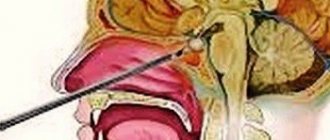
How to prepare for an MRI of the pituitary gland
The magnetic resonance imaging procedure does not require special preparation. The patient needs to warn the doctor about the presence of metal prostheses, implants, implanted electrical devices and pacemakers. Before the examination, remove outer clothing and accessories.
To increase the information content of the method, MRI is performed with intravenous administration of a contrast agent. In this case, you need to make sure that the solution used is tolerable. Patients prone to allergic reactions should warn their doctor.
Before the procedure, the doctor conducts a conversation, explains how the examination is carried out, how long it takes. Reminds you to remain still during scanning. Following the specialist’s recommendations affects the quality of the image and the diagnosis.
Antidiuretic hormone (vasopressin)
The main function of antidiuretic hormone, also known as vasopressin, is to maintain fluid balance. It increases the volume of fluid in the body by stimulating the absorption of water in the kidney channels. This hormone is released by the hypothalamus when it detects a lack of water in the blood.
Once the hormone is released, the kidneys respond by absorbing more water and producing more concentrated urine (less dilute urine). Thus, it helps stabilize the water level in the blood. The hormone is also responsible for increasing blood pressure by constricting arterioles, which is extremely important during shock blood loss as an adaptation mechanism.
The active growth of vasopressin is promoted by low blood pressure, dehydration and large blood loss. The hormone can remove sodium from the blood, saturate body tissues with fluid and, in combination with oxytocin, improve brain activity.
Low levels of vasopressin in the blood contribute to the development of diabetes insipidus, a disease characterized by polyuria (6-15 liters of urine per day) and polydipsia (thirst). Excessive production of this hormone is quite rare. It leads to Parhon's syndrome, which causes low blood density and high sodium levels. In addition, such patients will be plagued by a number of “unpleasant” symptoms: rapid weight gain, headache, nausea, loss of appetite, general weakness.
Intermediate zone of the pituitary gland
It is the smallest lobe and its function is to produce and secrete several hormones:
- melanocyte-stimulating hormone - affects pigmentation of the skin, hair and changes in the color of the retina;
- gamma-lipotropic hormone – stimulates fat metabolism;
- beta-endorphin – reduces pain and stress levels; gamma-
- met-enkephalin – regulates human behavior and pain.
The consequence of a lack of melanocyte-stimulating hormone is albinism. This is a congenital disease characterized by the absence of the pigment melanin, which colors the skin, hair and retina of the eyes. An excess of lipotropin threatens exhaustion, a deficiency – obesity.
When is a test for pituitary hormones needed?
Malfunction of the pituitary gland leads to an increase or decrease in the level of hormones in the blood, which leads to the occurrence of various diseases and abnormalities. Therefore, it is important to carry out timely diagnosis of the “master gland” of the endocrine system and correction of hormone levels. For preventive purposes, it is recommended to take tests 1-2 times a year. This will help reduce possible negative consequences for the body to a minimum.
Examination of the pituitary gland and the brain as a whole is recommended in the following cases:
- too early or delayed puberty;
- excessive or insufficient growth;
- blurred vision;
- disproportionate increase in some parts of the body;
- breast enlargement and lactation in men;
- inability to conceive a child;
- headache;
- a large amount of urine excreted with increased thirst;
- obesity;
- insomnia at night and drowsiness during the day;
- long-term depressive state that cannot be treated with medication and psychotherapeutic methods;
- feeling of weakness, nausea, vomiting (if there are no problems with the gastrointestinal tract);
- causeless fatigue;
- prolonged diarrhea.
Investigation of the pituitary gland is possible through instrumental and laboratory diagnostics.
Pathology of pituitary gland functions
Deviations in the functioning of the pituitary gland have many causes, both congenital and acquired. Loss of individual hormones (complete shutdown of gland functions) or increased secretion lead to a number of concomitant diseases.
Why does the pituitary gland enlarge in the brain? When the concentration of hormones in the blood is insufficient, the hypothalamus sends signals to the gland to stimulate secretion. The gland begins to work actively, which leads to tissue enlargement.
An increase in the size of the pituitary gland in the brain also occurs with the growth of a tumor (usually benign). The exact causes of the pathology have not been established; only provoking factors have been identified.
Hypofunction
Refers to an endocrine type of pathology. Lack of hormone secretion (or complete absence) leads to the failure of all processes in the body. All age groups can be affected by the disease.
Hyperfunction
The negative circular linkage mechanism fails. The release of excess amounts of hormones into the bloodstream leads to inhibition of the production of releasing hormones in the hypothalamus of the brain (the signal arrives through the nervous network). Thus, inhibition of secretion in the pituitary gland of the brain occurs - the production of secretion in the peripheral glands decreases.
The disruption of communication triggers the autonomous work of cells - signals from the pituitary gland to suspend work do not work, the concentration of secretion becomes excessive.
If pathological symptoms occur, it is necessary to undergo a comprehensive examination. Therapy methods are selected individually.
Pituitary gland disorders
A common disorder of the pituitary gland is the formation of tumors in it. However, such tumors are not malignant. They can be of two types;
- secretory – produces too many hormones;
- nonsecretory – keeps the pituitary gland from functioning optimally.
The pituitary gland can increase or decrease not only due to pregnancy or age-related changes, but also due to the action of harmful factors:
- long-term use of oral contraceptives;
- inflammatory process;
- traumatic brain injury;
- surgical intervention in the brain;
- hemorrhage;
- cystic and tumor formations;
- radiation exposure.
Pituitary diseases in women cause menstrual irregularities and infertility; in men they lead to erectile dysfunction and metabolic disorders.
Treatment of pituitary diseases, depending on the symptoms of the pathology, can be carried out using various methods:
- medicinal;
- surgical;
- radiation therapy.
The fight against pituitary gland dysfunction can take a significant period of time, and in most cases the patient has to take medications for life.
Symptoms of pituitary gland dysfunction
A malfunction of the pituitary gland affects the production of hormones - an excessive or insufficient amount of secretion enters the organs and glands with the blood. Signs of dysfunction of the pituitary gland may not appear immediately, but after several months.
Pathological symptoms appear depending on the cause of the disorder in the gland.
General symptoms:
- Increased fatigue (a person feels completely powerless even after a night's rest);
- Dry skin, prone to cracks;
- Minor injuries cause fractures (bone fragility), regeneration is slow;
- Rapid weight loss or sudden weight gain (without appetite);
- Impaired memory and thought processes;
- Decreased sexual desire;
- Irregular menstruation in women (or complete lack of regularity);
- Erectile dysfunction in men;
- Sudden mood swings (depression, attacks of rage).
Symptoms of pituitary gland dysfunction in the brain in women can occur during pregnancy. Cells producing the hormone prolactin grow - the symptoms are temporary and are not considered a pathology (physiological feature).
According to statistics, every tenth case of gland dysfunction has a cause - a tumor. Enlargement of the pituitary gland of the brain - the reasons are tissue proliferation under the influence of hormonal levels or other negative factors (trauma, heredity).
The general symptoms are accompanied by characteristic clinical manifestations:
- Loss of consciousness;
- Headache;
- A sharp decrease in visual acuity with a progressive course (optic atrophy).
A gradual enlargement of the pituitary gland in the brain leads to compression of surrounding tissues and the appearance of symptoms characteristic of damage to other parts of the central nervous system.
Simmonds syndrome
It is characterized by a disruption in the production of hormones in the hypothalamic-pituitary system.
Specific symptoms and neurovegetative manifestations:
- Sudden weight loss;
- Reduced excretion of biological fluids (urine, sweat);
- The skin takes on an earthy tint;
- Muscle weakness;
- Reactions are slow;
- Development of hypotension;
- Hypoglycemic syndrome;
- Joint pain;
- Convulsive syndrome.
Women of reproductive age completely lose the ability to conceive. In men, areas with hair are prone to complete baldness, and the external genitalia decrease in size.
Sheehan syndrome
Develops in women during complicated childbirth (or other conditions with massive blood loss). The development of hypotension leads to a decrease in blood supply to the gland. Lactotrophic cells are most often affected - lactation is absent or stops. The menstrual cycle is disrupted. General symptoms are similar to hypotension - weakness, dizziness, drowsiness.
Pituitary dwarfism
Insufficient production of tropic hormones leads to delayed physical development (growth, internal organs and tissues). Mental development remains within normal limits.
Diabetes insipidus
The secretion of antidiuretic hormone is reduced, which causes a disturbance in the water-salt balance in the body. Excessive urination is accompanied by intense thirst.
Acromegaly
Excessive secretion of the hormone somatotropin leads to a disproportionate enlargement of the limbs and individual parts of the face (nose, lips, lower jaw). The patient complains of joint pain.
Gigantism
Neuroendocrine pathology is common in children and adolescents. The anterior pituitary gland in the brain overproduces growth hormone. There is a violation of metabolic processes and deviations in mental development.
Itsenko-Cushing's disease
Excessive secretion of cortisol is accompanied by a symptom complex:
- Hypertension;
- Tendency to osteoporosis;
- The patient has a corpulent body with thin limbs;
- Pustular skin lesions (against the background of reduced immunity);
- Characteristic areas of pigmentation (neck, elbows);
- Striae on the skin;
- Excessive hair growth on the body and face (women develop a mustache and beard).
The skin on the face becomes purple in color.
Hyperprolactinemia
The increase in prolactin levels in the blood is due to both physiological and pathological aspects. In both women and men, colostrum begins to be released from the mammary glands. Reproductive dysfunction, emotional and personality disorders are noted.
Normal levels of pituitary hormones
| Hormone | Normal indicator |
| Thyroid-stimulating hormone | 0.6 – 3.8 µIU/ml (RIA method) 0.24 – 2.9 µIU/ml (IF method) |
| T3 – thyroxine | 2.6 – 5.7 pmol/l |
| T4 – triiodothyronine | 9 – 220 pmol/l |
| Adrenocorticotropic hormone | 0 – 50 pg/ml |
| Luteinizing hormone | 2.12 – 4 mIU/ml (in men) 18.2 – 52.9 mIU/ml (in women during the ovulation period), 3.3 – 4.66 mIU/ml (in women in the follicular phase), 1, 54 – 2.57 mIU/ml (in women in the luteal phase), 29.7 – 43.9 mIU/ml (in women during menopause) |
| Follicle stimulating hormone | 1.9 – 2.4 mIU/ml (in men), 2.7 – 6.7 mIU/ml (in women during the ovulation period), 2.1 – 4.1 mIU/ml (in women in the luteal phase) , 29.6 – 54.9 mIU/ml (in women during menopause) |
| Prolactin | 100 – 265 mcg/l (in men), 130 – 140 mcg/l (in women of childbearing age), 107 – 290 mcg/l (in women during menopause) |
| Somatropin | 0 – 10 ng/ml |
If you find an error, please select a piece of text and press Ctrl+Enter