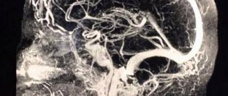
Carotid angiography is an invasive X-ray imaging procedure of the carotid arteries that is performed through a catheter inserted into a blood vessel in the arm or leg. A contrast agent is injected through the catheter, which is opaque to X-rays and therefore shows the internal lumen of the carotid arteries. This procedure is the gold standard for visualization of the carotid and cerebral vessels.
Preparing for the study
- Preparation for the study involves shaving the access site (usually the groin area).
- You should tell your doctor about any allergic reactions you have.
- If you are diabetic, tell your doctor so your medications can be adjusted on the day of the test.
- Tell your doctor if you are allergic to anything, especially iodine, shellfish, X-ray contrast, latex, or rubber products (such as rubber gloves).
- It is advisable to drink enough fluids before the examination to reduce the possible effects of contrast on the kidneys.
Types of cerebral angiography
There are several classifications of this type of examination. It is divided depending on the method of administration of the drug, as well as on the number of vessels that are included in the examination.
The following types of this examination are distinguished depending on the method of injection of the X-ray-containing substance:
- puncture or direct - contrast is injected directly into the brain vessel using a puncture;
- catheterization or indirect - contrast is administered using a catheter through the femoral artery.
Depending on the extent of the vessels that can be visualized, the following types of angiography are distinguished:
- general angiography - the entire vascular network of the brain is visible;
- selective cerebral angiography of the brain - one of the pools can be examined (in total, there are two blood supply pools in the brain: vertebrobasilar and carotid);
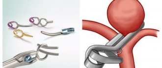
- superselective angiography - individual small-caliber vessels are visualized in one of the pools. It is used not only as a diagnostic method, but also for treatment, in which immediately after visualizing the location of the thrombus or embolus in the vessel, it is removed.
After research
Tell your doctor or nurses if you feel:
- Itching, dry throat
- Nausea
- Chest discomfort
- Vision problems
- Weakness in one or more limbs
At the end of the study, you will be transferred to a ward. It is necessary to remain in bed for several hours. The doctor will periodically examine the access site and assess the general condition. If there are no problems, then discharge can be made the next day.
Angiography of cerebral vessels
Before the examination, the patient is given premedication, which includes sedatives and antihistamines. The patient is placed on a special X-ray table. Cerebral angiography of cerebral vessels begins with catheterization of the selected vessel. Usually the carotid or vertebral artery is punctured, and the aorta is catheterized to perform panangiography (examination of all cerebral vessels). Peripheral vessels (femoral, cubital, brachial, subclavian arteries) can serve as an alternative puncture site. After anesthesia, the catheter is inserted into the mouth of the carotid, vertebral artery or aortic arch under x-ray control.
Next, X-ray contrast is slowly introduced, which weakly transmits X-rays. In the first minutes of administration of a contrast agent, the patient may experience redness of the face, a salty, metallic taste in the mouth, but these sensations, as a rule, disappear quickly. X-rays are taken in anteroposterior and lateral projections.
This procedure is repeated several times to determine the rate of venous outflow. Upon completion, the catheter is carefully removed and the puncture site is pressed for 10-15 minutes to stop bleeding. The examination is recorded on digital media or recorded on x-ray film. The manipulation itself takes about an hour, the result is given in a few hours. Within 24 hours, the doctor must monitor the patient’s condition: examine the puncture site, palpate the pulse in peripheral vessels, measure vital signs (temperature, blood pressure, heart rate, respiratory movements). When puncturing the femoral artery, after removing the catheter, the leg should be in an extended position for 6 hours. The patient is also recommended to drink large amounts of fluid to quickly remove the contrast from the body in the absence of contraindications.
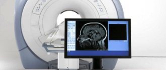
Currently, CT angiography of cerebral vessels is often used. With this method of diagnosing vascular pathology of the brain, contrast is administered through a catheter installed in the vein of the elbow. A positive point is the low radiation exposure affecting the patient’s body.
Also in neurology, MR (magnetic resonance) angiography is used, which is based on measuring the electromagnetic response of atomic nuclei in a constant magnetic field. This method practically does not require the introduction of a contrast agent and allows one to determine the function of blood flow and the structural features of cerebral vessels.
Possible complications of carotid angiography
Complications are rare - the risk is 0.5%. The most dangerous are complications from the brain. They can develop as a result of the effect of contrast or when pieces of plaque are torn off during catheter insertion. Complications include:
- Transient ischemic attack - short-term disturbance of cerebral circulation
- Ischemic stroke is a persistent disorder of cerebral circulation
- Bleeding from the arterial access site
- Carotid artery dissection with thrombosis
- Allergic reaction to contrast
Timely diagnosis of complications allows you to minimize their consequences.
The essence of the method
In order for the radiologist to see the vessels of the brain, an injection of an iodine-based X-ray contrast agent (Triiodtrust, Ultravist) is performed into one of the cerebral arteries. Injection is possible either into a vessel in the brain or through a catheter through an artery on the periphery, for example, the femoral one. Without this procedure, cerebral angiography of the cerebral vessels will be ineffective, since the arteries will be poorly visible on the image.
Next, two x-rays are taken, in frontal and lateral projection. After which the radiologist writes his report.
Features of CT angiography
Since the angiography method has been used for more than a century, it is constantly being improved. A more modern and high-quality method for visualizing cerebral vessels is cerebral CT angiography. Although in general the survey method is similar to the traditional one, there are some peculiarities:
- It is carried out not using an X-ray machine, but using a tomograph. Also based on the passage of X-rays through the human body, it takes a large number of images at once, layer by layer, which makes it possible to more accurately visualize the vessels and surrounding tissues.
- The image turns out to be three-dimensional, which allows you to view the vessel from all sides.
- The contrast injection is given into a vein, not an artery.
- There is no need to keep the patient under observation after the procedure.
CT angiography is a more effective and safe method of vascular imaging.
6. Contraindications for MRI of head and neck vessels
This diagnostic procedure is strictly contraindicated when:
- The presence in the body of foreign bodies of any origin and nature, containing metal or elements from it. These may include surgical staples, pins, pacemakers, unrecovered shot fragments, hearing aids, insulin stimulators, dentures, and even braces.
- Chronic and acute renal failure, pregnancy, during the lactation period, with identified allergies - since the study of blood vessels, arteries and veins is often carried out using a contrast agent. It is toxic in certain doses and can negatively affect the condition of a weakened body.
- For epilepsy and claustrophobia, MRI diagnostics are not advisable.
Description of diseases diagnosed using arteriography
Arteriography can only be prescribed by a specialist. Patient complaints of dizziness, migraine and tinnitus are not indications for a contrast study. With such symptoms, the patient should consult a doctor to determine the need for a procedure.
With the help of a contrast MRI study you can:
- diagnose the development of a cerebral aneurysm;
- determine blockage or degree of narrowing of blood vessels (stenosis);
- check the location of vascular clips;
- determine the presence of arteriovenous malformation;
- diagnose vasculitis.
Each of the above diseases should be considered separately.
Interpretation of MRI of cerebral arteries
After the patient has completed the examination, the results are deciphered, consisting of several stages:
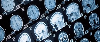
- The tomograph transfers many of the images taken to a special computer. The number of images can reach a thousand, and the slice thickness is 1-3 millimeters.
- The radiologist carefully examines the images and draws up a written report that describes in detail the anatomical structures, possible deviations in structure and functioning.
If a person does not have any problems with the arteries, then in the pictures they will be highlighted evenly, there will be no nodes or voids. If a tumor or cyst is present, dark spots will appear on the images.
The radiologist describes what he sees in the images, and the final diagnosis is made by the attending physician based on the MRI report.
At the medical office, you can undergo a magnetic resonance imaging procedure of the arteries of the brain on any day and time convenient for you. The center’s doctors have extensive experience in quickly and clearly interpreting the images received.
MRI of the cerebral arteries is a safe procedure that shows the slightest manifestations of the disease in the early stages of their occurrence. The study does not cause discomfort, is quick and painless, and its effectiveness is not comparable to any other type of study of the cerebral arteries.
Features of MR angiography
MR angiography is even more informative than CT. It allows you to see soft tissues that are difficult to visualize on CT. It is carried out using a magnetic resonance imaging scanner and is not an x-ray method, unlike other angiography methods. This avoids exposure to radiation.
Another advantage is good visualization even without the use of contrast, which is why MR angiography without contrast can be used for allergy sufferers.
The main contraindication for use is the presence of any metal objects in the body (artificial pacemakers, prostheses, implants, metal clips on blood vessels).
Perhaps selective cerebral angiography of the brain has already become commonplace and routine for doctors. It may be inferior in effectiveness to CT and MRI angiography. However, being more affordable and not requiring special high-tech equipment, it is still actively used 100 years later for diagnosing brain diseases.
How is an MRI of the cerebral arteries performed?
After the patient enters the office, he must notify the doctor about contraindications, if any. He needs to remove all metal objects. If the procedure will be carried out in clothing, you must make sure that there is no metal on it.
The patient is asked to lie on the couch and the position of the head is stabilized. This will reduce the risk of deterioration in the quality of the study, since the slightest movements of the head may result in blurry images.
Preparation for MRI of cerebral arteries
Patients who are afraid of being in a confined space can take sedatives first. In case of deterioration of health, the patient can always contact the doctor during the procedure via two-way communication.
Modern high-field tomographs produce quite loud sounds during operation. There is no need to be afraid of this - such sounds indicate normal operation of the device. To reduce discomfort in the medical room, each patient is given headphones, in which pleasant music plays during the procedure, drowning out the noise from the operation of the tomograph.
After the end of the study, the patient is given images recorded on a disk or flash drive, as well as a written report from the doctor, within half an hour.
Aneurysm
An aneurysm is a specific section of a blood vessel that is intensively supplied with blood and grows in size. The convex wall of the vessel often compresses neighboring tissues and nerve endings. Damage to the aneurysm, which results in hemorrhage, poses a particular threat.
An aneurysm is diagnosed in any part of the brain, but is more often localized in the area where branches originate from arteries. Aneurysms develop asymptomatically and only when they reach large sizes do they manifest clear symptoms. Main signs of pathology:
- deterioration in the quality of vision;
- severe headaches;
- nausea;
- sudden loss of consciousness.