Her Majesty Nature has taken care of the most reliable protection of one of the two most important organs - the brain. But the structure of the skull (which is the protective shell) is so complex that it lends itself to only one research method - radiation. Ultrasound does not allow one to determine bone pathologies; tomography does not help in analyzing the condition of the head box. Therefore, when the question is about the integrity of the protective membrane of the brain (it is important to understand that we are talking about the skull, not brain tissue), only X-rays help.
X-ray of the skull: when is it needed and how is it performed?
Radiography is a quick and reliable way to determine pathologies that are located within dense tissues.
The advantages of a skull x-ray are as follows:
- high information content of images taken in various projections;
- high efficiency;
- non-invasiveness and relatively simple technology for performing the procedure;
- accessibility - today head x-rays
can be taken in almost any clinic; - low radiation exposure.
Where to get a head x-ray
If you need to take a head photograph, contact the Central Clinical Hospital of the Russian Academy of Sciences. We guarantee prompt and accurate diagnosis through the use of advanced digital equipment that provides the lowest level of radiation. Experienced doctors interpret X-ray results. You can make an appointment on the Central Clinical Hospital website online or by phone. You can find out how much the procedure costs at the clinic. Also, current prices are indicated on our website.
You can find out prices for brain x-rays and make an appointment online on our website or by calling the clinic.
When is a skull x-ray prescribed?
Survey X-ray of the skull
in different settings can be prescribed to patients who are concerned about:
- cephalgia, or, in other words, headache, of varying localization and intensity;
- trembling in the limbs;
- the appearance of a veil before the eyes or darkness;
- nosebleeds;
- painful chewing of food;
- decreased visual and hearing acuity;
- cases of fainting for no apparent reason;
- the appearance of asymmetry of the facial bones.
X-rays of the head are also indicated for mechanical injuries - bruises, blows, falls from a height, and so on.
Various specialists can prescribe an X-ray examination of the skull: a neurologist, a surgeon, an oncologist, an ophthalmologist and others.
What does the procedure show?
X-ray images clearly visualize:
- cheek bones;
- lower jaw bones;
- bony pyramid of the nose;
- sphenoid bone;
- eye sockets;
- temporomandibular joints;
- mastoid processes of the temporal bones.
If a more accurate and detailed diagnosis of the condition of the bone tissues of the skull is necessary, targeted images are taken, which can show the following pathological conditions:
- formed calcifications - can provoke pathological development of the cranial bones;
- partial calcification of tumors;
- hemorrhages and hematomas;
- fluid in the paranasal sinuses;
- fractures of the skull bones.
Using the X-ray method, it is possible to identify congenital pathologies of the skull, as well as increased intracranial pressure. The latter may be indicated by so-called impressions - marks similar to fingerprints that are located on the inside of the bone tissue.
How harmful are x-rays?
The radiation dose received by an adult patient is a small part of the annual norm (about 4%). This value is comparable to an hour's stay on the beach. Doctors point out that this approach is incorrect. Indeed, in some cases, the study is carried out for vital reasons, in order to detect a fatal disease. Therefore, the amount of research conducted should be subordinated to the main goal - saving human life. It happens that the diagnosis is also carried out on pregnant women, carefully covering the stomach with an apron.
Preparation for the procedure
Special preparation x-ray of the head
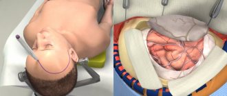
does not require. Before the procedure, the patient must remove all metal jewelry, glasses, and, if possible, dentures. If the dentures are fixed or a metal implant is installed, the radiologist must be warned about this in advance. Then, depending on the configuration of the X-ray machine, the patient takes a lying, sitting or standing position.
A lead vest or apron is placed on the patient's body to prevent exposure below the neck level. The head is secured with special clamps, since the first condition for obtaining a high-quality image is immobility.
Preparation requirements, procedure for performing skull x-rays
This type of x-ray does not require any preparatory measures. Before it is carried out, the doctor clarifies the fact that there is no pregnancy, if we are talking about a female patient, explains exactly how the procedure will take place, how many pictures will need to be taken, what is required of the patient during the process. If the procedure is prescribed for a child, the parents prepare him for diagnosis and explain in a clear manner to the child how he needs to behave. Doctors do not set any restrictions on diet or the amount of physical activity before the examination unless they are required by the patient’s general condition, regardless of the prescribed procedure.
Before starting the diagnosis, the doctor asks the patient to remove all metal jewelry and accessories from the head and neck, as they may appear on the pictures in the form of additional darkening, thereby distorting the results.
The image can be captured in different positions - the patient can lie, sit or stand, depending on which area is being examined. The body of the subject is covered with a special protective apron with lead plates. The head, if necessary, can be fixed with special belts or rollers to ensure its complete immobility while capturing the image. The doctor takes the required number of pictures. During the process, he can change the patient’s position and position.
Pictures can be taken in the following projections:
- axial;
- semi-axial;
- anterior-posterior;
- posterior-anterior;
- right lateral;
- left side.
There is also such a thing as radiography methods. It involves recording images in special projections that allow obtaining an image of a specific area. For example, the methods according to Reza, Ginzburg and Golwin differ from each other, but they all provide an overview of the optic canals and the superior orbital fissure. Images according to Schüller, Mayer and Stenvers allow you to study the condition of the temporal bones.
Most often, for a doctor to make a diagnosis, it is enough to take pictures in two projections - the front and one of the sides. The whole procedure lasts no more than 5 minutes. It is absolutely painless, and the only atypical sensation that may arise from it is a metallic taste in the mouth due to exposure to x-rays.
Technique
To take an X-ray, the patient must stand up, sit near the X-ray machine, or lie down on its work table. It is important to remain still and not breathe while filming. If you need to take a picture in several projections, the doctor will tell you how to change the position.
X-ray of the skull in 2 projections
To obtain the most detailed and complete information about the condition of the nasal bones, photographs can be taken in two projections - frontal and lateral. In the first case, the patient faces the X-ray machine, in the second - sideways (left or right).
Indications and contraindications for x-rays of the skull
Due to the fact that the procedure uses x-rays, it should be carried out only on the direction of a doctor, and only in cases where there is an objective need to obtain information about the condition of the skull bones in this way.
Among the indications for a skull x-ray:
- suspected traumatic brain injury (open or closed);
- tumor processes;
- possible developmental anomalies - congenital or acquired;
- pathologies of ENT organs, for example, sinuses;
- the presence of a number of symptoms with an unclear etiology: disturbances of consciousness, dizziness, constant severe headaches, symptoms of hormonal imbalance.
As for contraindications, they are related to the dose of radiation received during the diagnostic process. For example, examination methods involving the use of X-ray irradiation are generally not recommended for pregnant women, especially in the first trimester. If possible, the doctor prescribes diagnostic methods that are gentler on the fetus.
The second category of patients for whom skull x-rays are prescribed with caution are children. Childhood is not an absolute contraindication for the procedure; moreover, in some cases, an X-ray of the skull is an objective necessity, for example, if it is necessary to confirm the doctor’s suspicions of congenital pathologies of bone development.
It is believed that modern X-ray machines cannot significantly irradiate a child during diagnosis. Thus, the permissible radiation dose per year for a person is no more than 50 microsieverts per year, and radiography equipment “gives” the patient a dose of no more than 0.08 microsieverts per session. In this case, the problem is the fact that not every medical institution has at its disposal modern X-ray machines with dosed radiation, and in X-ray rooms there is more often outdated equipment that has been in use for decades. Nevertheless, sometimes you simply cannot refuse an X-ray of the baby’s skull. This diagnostic method is one of the most popular in pediatric neurosurgery, traumatology and neurology. If there are certain indications, skull x-rays are performed even for newborn babies.
How dangerous is the research?
X-ray is a non-invasive and painless procedure. It can also be called relatively safe, since the radiation exposure is minimal. At the same time, of course, x-rays are not a procedure that can be repeated many times in a row. There are certain norms and frequency that must be observed.
Contraindications for
An absolute contraindication for head x-rays is pregnancy. There are also relative restrictions. These are children under 15 years of age, mental illness, serious condition.
X-ray of a child's head
A different approach is used to study the skull of children:
- The child is more likely to receive a higher dose of radiation;
- Exposure to radiation can adversely affect a child's body.
For these reasons, an X-ray of a baby’s head is prescribed only as a last resort, if other diagnostic methods are not informative and the baby’s life is at risk. It is difficult to choose an alternative method, since the bones of the skull have a complex structure, and ultrasound does not recognize all pathologies.
The main reason for prescribing x-rays for a child under one year of age is injury. Radiation of babies is undesirable, but often this is the only way to identify birth injuries that threaten the child’s life. In the case of an X-ray of the head of a newborn, all other organs are protected as much as possible.
X-ray of the skull: decoding
When interpreting an X-ray of the skull in 2 projections, specialists evaluate the dimensions and features of the location of the bones, as well as the structure of the nasal sinuses. These indicators must correspond to the norm for the age category of the subject.
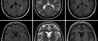
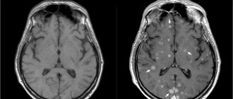
X-rays also make it possible to partially analyze the condition of the soft tissues of the brain (although for an accurate diagnosis of this organ it is better to use MRI or CT). The images can visualize tumor growths, and their location and size can be assessed. The main sign of a malignant neoplasm will be the presence of darkening of an uneven structure. If the tumor is benign, its contours will be smooth and clear.
Normal indicators
Let's figure out what a skull x-ray
in cases where there are no pathologies. When describing the images, the radiologist evaluates the size, shape, thickness and location of the skull bones, as well as the vascular system, the condition of the nasal sinuses and cranial sutures. All of the listed characteristics must correspond to the patient’s age.
X-ray of the skull for head trauma
The main questions that a specialist should answer based on an x-ray for a head injury:
- Is the integrity of the bones of the skull damaged?
- If there is a fracture, is it accompanied by the entry of bone fragments into the cranial cavity?
- Are the eye sockets, sinuses and ears damaged?
- Is there damage to the brain due to compression by the deformed bones of the skull?
The most common injuries to the calvarium are linear fractures (cracks) of its bones. In most cases, they appear in the place where the force was applied. By the way, this fact greatly facilitates the process of identifying a fracture/crack. The fracture is visualized as a sharp strip, in some places diverging in different directions, with uneven edges. Depending on the complexity, the fracture may have a different position, direction, and size. Multiple fractures can affect one or both halves of the skull. The most unfavorable situation is when the fracture passes to the cranial suture and causes its divergence.
Research methodology
The procedure is not difficult and takes place in a matter of minutes. The patient is seated on a special chair or placed on a research table-tripod. When tomography of the skull, five projections are used. To ensure a stationary state, the head is fixed in the required position. For emotional people, the clinic prepares psychotropic drugs that reduce anxiety and nervous tension.
The images are sent for development, after which they fall into the hands of the leading radiologist. The quality of the images is checked before the patient leaves the X-ray room. After carefully studying the images, proper treatment is prescribed. If complications of the disease are detected, doctors may suggest hospitalization for therapeutic treatment. The clinic will provide all treatment methods and proper care for the patient until complete recovery.
When should an x-ray be taken?
For children, this study is most often performed in cases of trauma, including birth trauma. Often this is the only method that helps identify life-threatening pathologies. The following doctors can refer you for this procedure: oncologist, neurologist, traumatologist, surgeon, ophthalmologist, endocrinologist. Symptoms and indications for examination are:
- headache;
- tremor (shaking) of hands;
- nosebleeds;
- darkening of the eyes;
- dizziness;
- decreased vision or hearing;
- fainting;
- facial asymmetry;
- suspicion of a malignant tumor;
- recent injury;
- some endocrine diseases;
- congenital pathologies.
Pictures are also taken during treatment to monitor the effectiveness of the therapy.