Vascular angiography: what it is and how it is done, indications, consequences
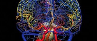
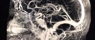
You can quite simply describe what angiography means - it is obtaining images of blood vessels. This diagnostic procedure allows you to conduct a full examination of the vessels, determine where they are blocked, identify areas of blood clots, places of narrowing and thinning of the vascular walls. For angiography
a special contrast agent is introduced, which is highlighted during scanning and helps to identify potential or existing pathologies.
Aortography at the Innovative Vascular Center
The approach to performing aortography in our clinic is based on our belief that angiography is the final stage of diagnosis and should be performed only after receiving sufficient information from other, minimally invasive treatment methods. Most often we combine angiography and endovascular surgery.
The study is performed in a specialized X-ray operating room, where a Philips Allura Xper FD20 angiographic complex is installed, equipped with a software package for high-speed recording of the movement of contrast in vessels and a number of programs that allow the calculation of many parameters. The modern method of computational (subtraction) angiography allows us to obtain additional information about hemodynamics and the condition of blood vessels.
Indications for use
The main tasks of angiography are to identify pathologies of the circulatory system and determine their nature. And this is really possible to do, since with the help of this research method it is possible to achieve ideal visualization of large veins of the extremities, arteries, aortas and pulmonary arteries. With the help of angiography, doctors have the opportunity to detect diseases such as:
- cerebral vascular sclerosis;
- congenital malformations of veins and arteries;
- hypertension of various etiologies.
There are many indications for angiography. Among the most common are:
- deep vein thrombosis with identification of the nature of the changes;
- thromboembolism (that is, blood clots in the pulmonary artery duct);
- defects of arteries and veins;
- lesions of the renal arteries;
- lesions and diseases of the aorta;
- aneurysms.
What parts of the body are being examined?
Let's look at the parts of the body that are most often subjected to angiographic examination.
Angiography of vessels of the extremities
To analyze the condition of the blood vessels of the patient’s extremities, a contrast agent is pumped into the arteries of the right or left arm. To determine the status of the leg veins, contrast is injected either through the femoral artery or through the abdominal aorta. X-ray photography is performed from different angles and positions.
Angiography of the brain
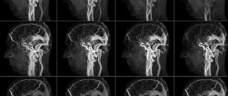
The vessels of the brain can also be examined using angiography. To make a more accurate diagnosis, a contrast agent can be injected twice, and x-rays can be taken in several projections.
Angiography of the coronary vessels of the heart
To study the condition of the coronary vessels of the heart, a contrast agent is supplied through a catheter into the inguinal or femoral artery. In this case, the catheter must be advanced to the aorta. Then the right and left coronary arteries are filled alternately with a contrast agent.
Angiography of internal organs
For angiographic examination of the liver, kidneys, or other internal organs, a contrast agent is injected through the aorta. Also, administration can be carried out into large veins that communicate with the organ being examined. Angiography of internal organs is required in cases where it is not possible to determine the nature of the pathology using non-invasive techniques or there is a suspicion that the vessels are located incorrectly.
X-ray angiography
Angiography is classified depending on the organ in which the vessels are contrasted.
For example, angiopulmonography is used to visualize the vascular network of the lungs; angiocardiography is used to visualize the atria and ventricles with large vessels.
The study requires access to a vessel that is located in close proximity to the tumor and feeds it. A catheter is inserted into the artery and, under X-ray monitoring, this catheter is brought to the vessel of interest. The contrast agent contained in the catheter allows images to be taken.
The resulting images help to see the pathological characteristics of the organ and its functions, which affect the function of the blood supply.
Stomach biopsy
X-ray allows you to contrast arteries, veins, and can also be used to obtain images of the lymph nodes of blood vessels. This study is called vasography. Study of arteries - arteriography, study of veins and lymph nodes, respectively - venography (phlebography) and lymphography.
The condition of the arterial bed in the area where the tumor is located is important for deciding the issue of surgical intervention. It is this condition that indicates the boundaries of tumor growth, as well as its growth into the artery.
The pressure that allows blood to flow through the arteries is high, and during surgical manipulations, a large volume of blood will be lost from a vessel damaged by a cancerous tumor. The speed at which blood flows through the veins is small, the pressure is insignificant, which means that it is not difficult to compress a large vein damaged by cancer without a large loss of blood.
The information obtained using angiography allows you to avoid all sorts of surprises. In addition, taking into account the results of the study, the doctor can immediately carry out a therapeutic measure, for example, installing a stent in a narrowed vessel or removing a blood clot.
Preliminary preparation
Angiography requires special preparation:
- 2 weeks before the procedure you should not drink alcoholic beverages.
- A week before the procedure, you need to exclude all blood thinners (for example, aspirin) from your medications (if any).
- Five days before the procedure, the patient must undergo additional examinations: fluorography, ultrasound of the heart
, ECG, coagulogram (blood test for clotting), blood biochemistry, blood test for Rh factor, test for HIV, hepatitis, syphilis. - 2 days before angiography, a test is performed to determine the patient’s tolerance to the components of the contrast agent (in particular iodine). If the test shows the presence of an allergic reaction, this is a contraindication and the procedure must be cancelled.
- 1 day before the examination, in the evening, you need to remove hair from the area where the puncture is expected to be made.
- At night (immediately before the procedure) you need to take a sedative. The doctor will recommend which one.
- On the morning of the procedure, the patient is given a cleansing enema for the intestines.
- On the day of the procedure, you should refrain from eating and drinking to avoid nausea and vomiting after contrast is injected into your body.
- Before starting the procedure, you must empty your bladder.
Complications of aortography
In order to avoid the development of complications, it is necessary to carefully prepare for the study and choose the least dangerous method of conducting it.
- Bleeding, hematoma, pain or swelling at the site of catheter insertion is very rare when performing arterial puncture under ultrasound guidance and using suturing devices.
- Allergic reaction to iodine, which is part of the X-ray contrast agent - it is necessary to carefully determine the history of allergies and exclude such patients.
- Damage to the vessel wall can occur during complex or rough puncture. In our clinic, if there are difficulties with arterial puncture, we use an ultrasound scanner to guide the needle into the vessel.
- The development of acute liver or kidney failure may occur due to the toxic effects of the contrast agent. With adequate fluid loading and minimal use of contrast, this complication is uncommon.
- Heart rhythm disturbances are rare and are associated with irritation of the reflexogenic zones in the aorta by the conductor or catheter.
Our specialists do everything possible to prevent these complications, and also know the necessary treatment methods in case of development.
How is the procedure done?
How angiography is done can be described as follows:
- administration of antihistamines (even if a preliminary test showed that there is no allergy to iodine);
- antiseptic treatment of the area of the body where the puncture will be made to introduce contrast;
- anesthesia (usually lidocaine is used);
- incision of the skin to provide access to a vein or artery;
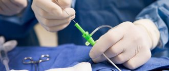
- installation of an introducer (hollow tube) to facilitate further insertion of the catheter;
- administration of a drug that prevents vascular spasm;
- inserting a catheter into a hollow tube and moving it to the beginning of the vessel to be examined;
- administration of contrast;
- shooting.
After the shooting is completed, the catheter is removed, hemostatic measures are carried out (if there is bleeding), and then a tight bandage is applied to the puncture site.
Our clinic specialists perform:
- angiography of the coronary, brachiocephalic, renal arteries, aortography, angiography of the vessels of the lower extremities, panangiography,
- permanent and temporary embolization of the uterine arteries,
- cerebral angiography,
- angioplasty and stenting of coronary, brachiocephalic, renal arteries,
- recanalization, angioplasty and stenting of the arteries of the lower extremities,
- embolization of intracranial vessels,
- implantation of stent grafts (prosthesis of the abdominal aorta),
- ventriculography,
- angiopulmonography,
- phlebography,
- cholangiography,
- drainage of bile ducts,
- targeted administration of drugs, chemoembolization,
- transesophageal electrophysiological study,
- transesophageal ischemic test,
- programming artificial pacemakers (pacemakers), cardioverter-defibrillators, devices for cardiac resynchronization therapy,
- endocardial electrophysiological study,
- implantation of a temporary and permanent pacemaker (one- and two-chamber pacemaker),
- implantation of a cardioverter-defibrillator (one- and two-chamber),
- implantation of devices for cardiac resynchronization therapy,
- catheter ablation of arrhythmogenic zones of the heart for tachyarrhythmias (radiofrequency catheter ablation),
- implantation of devices for long-term ECG recording,
- transesophageal restoration of heart rhythm during reentrant tachycardia.
Rehabilitation measures after angiography
It is important to apply a pressure bandage to the puncture site to minimize the risk of bleeding. After the procedure, the patient must be under the supervision of medical personnel for at least 6-8 hours, during which time the doctor monitors his well-being. If the condition is satisfactory, you can continue rehabilitation at home. It is necessary to refrain from physical activity for several days, and also follow all the instructions of the attending physician. In general, specific rehabilitation measures are not required during this period.
Minimally invasive (not requiring incisions) x-ray endovascular technologies are a new alternative in the diagnosis and treatment of many diseases, often superior in their effectiveness and safety to conventional surgical and therapeutic methods.
Possible complications after angiography
Angiography is valued because this examination provides a lot of information about the condition of the blood vessels. Moreover, this diagnostic procedure is, in fact, a small operation with pain relief and, like any operation, can have complications. As practice shows, after angiography the following is possible:
- Allergy. It is possible that the iodine allergy test did not show a negative reaction, since the test dose administered is quite small. Therefore, we cannot exclude the possibility that the patient will react to the contrast or to the components of the anti-blood clotting drug.
- Edema and hematomas. Minor complications may occur where the puncture was made.
- Bleeding. Since blood thinning drugs are introduced into the patient’s body, it is possible that blood clotting will be low after the procedure.
- Vascular injuries. Incorrect actions by the doctor can lead to such complications.
- Heart failure. A complication occurs in cases of violation of angiography technique.
How the angiography procedure proceeds can be affected by both a violation of the preparation rules and an incomplete examination of the anamnesis by the doctor, as well as the incorrect use of diagnostic testing techniques.
The latest angiography techniques
The development of new medical technologies today makes it possible not only to obtain angiographic images, but also to process them using a computer in order to improve their quality, and to identify certain vessels from the total mass.
Digital technologies make it possible not to insert a catheter with contrast into an artery, but to confine it to a vein. This technique is called digital subtraction.
Its use provides several advantages:
- Facilitates the work of the doctor performing angiography;
- Facilitates the patient's condition;
- Reduces the amount of contrast agent in his body.
Using digital processing techniques, the doctor receives a 3D image of the desired organ and all stages of blood flow through it.
Modern angiography makes it possible to distinguish between arterial and venous blood flows, and to more fully see the process of blood supply to the organ of interest.
X-ray angiography is an invasive method. Penetration into the body is performed in the operating room, excluding infection of the patient and the possibility of heavy bleeding.
The main advantage of X-ray angiography is that it allows for simultaneous therapeutic actions in blood vessels.
This cannot be done with angiographic studies using CT and MRI equipment. Therefore, X-ray contrast angiography is considered the “gold” standard for vascular examination.
Recovery after the procedure
How quickly a patient recovers after angiography depends largely on how extensive the study was. There are several recommendations that will help the patient recover faster:
- Follow bed rest and diet for several days after the procedure;
- eliminate stress and anxiety;
- exclude physical activity;
- Take antihistamines to minimize the likelihood of an allergic reaction.
As a rule, if the study was performed correctly, the patient's recovery takes approximately 2-3 days.
Decoding the results
There is a certain technique for deciphering angiography results. Markers are identified for each vascular pathology. For example, if the vessels have intermittent outlines in the images, we can talk about the presence of pathologies such as thrombosis or atherosclerosis. The detection of protrusions in the walls of blood vessels in the images indicates that there are either congenital changes, an aneurysm, or the same thrombosis. We can also talk about congenital pathologies in cases where the contrast agent does not enter the capillaries, but passes directly into the vein.
If, upon administration of contrast, there is a lack of its spread, this indicates the presence of thrombophlebitis, thrombosis or thromboembolism.
Angiography
What can angiography reveal?
•Vascular pathologies (atherosclerosis, aneurysm, thrombosis, malformation, etc.).
•Diseases and congenital heart defects.
•Renal dysfunction.
•New growths (tumors, cysts).
•Vascular lesions of the retina.
•Internal bleeding.
Types of angiography
•If the object of study is arteries, arteriography . In this case, the contrast agent is injected into the femoral, less commonly, into the axillary artery.
• Venography (phlebography) . It is especially relevant when diagnosing thrombosis. Direct venography involves the introduction of a contrast agent directly into a vein, indirect - into an artery, into the tissue of the organ being examined or into the bone marrow space.
•When visualization of lymphatic vessels is necessary, a method such as lymphography . This allows you to get a more complete picture of systemic diseases and tumors of the pelvis and lower extremities. The contrast agent is injected directly into the lumen of the lymphatic vessel. Currently, lymphography is in little demand - preference is given to more advanced research methods.
Preparation for the procedure
About half a month before the angiography, it is advisable to stop drinking alcohol. All other preparations are made on the eve of the study. In particular, it is necessary to shave the hair in the groin or under the armpit if you plan to puncture the femoral or axillary artery. You will also need to remove all metal jewelry. The last 4 hours before the procedure is the time when you cannot eat or drink.
Immediately before the study, when the patient is already lying on the angiographic table, premedication is performed: sedatives, anti-allergenic and analgesic drugs are infused through a catheter inserted into a vein.
How is angiography performed?
•The area where the puncture is planned is treated with an antiseptic, after which local anesthesia is administered.
•An incision of only a few millimeters is made in the anesthetized area. It is needed to easily insert a special needle with a wide lumen and pierce the artery with it.
•A metal conductor is inserted into the lumen of the needle - it is advanced to the place that needs to be examined. Then the needle is carefully pulled out and the catheter is inserted along the guidewire. When it reaches the target, the conductor is also removed.
•A contrast agent is injected into the vessel through a catheter, and an X-ray is taken at the same time. At this stage, the patient may experience dizziness, nausea, pain in the chest or head - this is how the body sometimes reacts to the contrast agent. There is nothing to worry about; the discomfort will soon go away on its own.
•After development, the resulting images are studied. If necessary, additional photography can be carried out.
•The final stage of angiography is removal of the catheter followed by application of a bandage to the puncture site. After a day it can be removed.
After the procedure
Bed rest is indicated for the 24 hours following angiography. The patient is under medical supervision and his body temperature is periodically measured. The puncture site must be inspected. The fact is that a bruise may form; in exceptional cases, the vessel is blocked by a detached blood clot.
Advanced Angiography Options
Classic angiography was described above. The development of technology has made it possible to make some adjustments to this procedure, to simplify and improve this research method. In particular, the following options appeared:
• digital subtraction angiography . The difference from the classical method is the use of a computer to enhance the resulting image. This allows you to reduce the dose of the contrast agent, as well as inject it directly into the vessel, without a catheter;
• computed tomographic angiography . In this case, it is possible to assess not only the condition of the vessels, but also the nature of the blood flow. The contrast agent is also administered intravenously, and the level of X-ray radiation is noticeably lower than with traditional angiography;
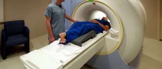
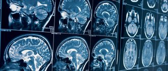
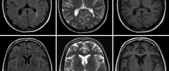
• magnetic resonance angiography . This method does not involve the use of X-rays at all - instead, a magnetic field is involved in the process. A contrast agent is also not required, although such agents are sometimes used. In this case, contrast is administered intravenously. MRA provides insight into both vascular anatomy and functionality.
