Contraindications and restrictions
Contraindications to MRI of the brain and MR angiography of intracranial arteries are:
- pregnancy in the first trimester;
- the presence in the body of metal implants, cardiac and neurostimulators, metal staples and/or clamps on the blood vessels of the head, foreign bodies not related to medicine, for example, bullets or shot;
- overweight - this is due to the fact that the tomograph table on which the patient lies has a maximum load that it can withstand.
For claustrophobia, that is, fear of closed spaces, and the inability to remain still, associated with psychological and neurological disorders, the doctor may recommend sedatives or suggest another diagnostic method.
Non-contrast MR angiography of cerebral arteries (intracranial angiography)
Types and costs
| Service | Cost, rub. | Action in the center on Musa Jalil |
| Non-contrast MR angiography of cerebral arteries (intracranial angiography) | 4950 rub. | 4500 rub. |
The prices indicated on the website are not a public offer (according to Article 435-437 of the Civil Code of the Russian Federation). You can find out the exact cost of studies and additional services from the administrators of our MRI centers by calling the numbers listed on the website or using the feedback form.
The Moscow MRI Center performs complex magnetic resonance angiography of the cerebral arteries. We use modern equipment and the latest diagnostic techniques that guarantee the reliability of the results.
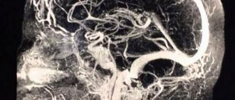
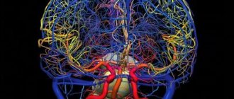
Magnetic resonance angiography of cerebral vessels is one of the most promising and rapidly improving methods of modern diagnostics of the vascular bed, which does not require direct puncture. When performing MRA, the doctor can examine various structural and pathological changes in the arteries of the brain tissue, assess the nature of physicochemical and pathophysiological processes. The method allows you to obtain a series of thin sections, simulate a three-dimensional reconstruction of the vascular network in the desired area, and also highlight individual trunks in the projection of the corresponding part of the brain.
The appointment of magnetic resonance angiography is possible:
- for cerebral ischemia, strokes;
- vertebrobasilar insufficiency;
- cerebrovascular accidents;
- dizziness;
- aneurysms of cerebral arteries (including mycotic ones);
- arteriovenous malformations (AVMs, angiomas, hemangiomas) of blood vessels;
- tinnitus;
- intracranial pressure and hydrocephalus;
- headaches;
- concussion and bruise of the brain;
- encephalopathy;
- nosebleeds.
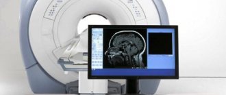
Our centers have modern diagnostic equipment, staffed by qualified personnel. This allows us to guarantee the reliability and accuracy of the results. The patient is given a scan, a digital recording and a description of the study with recommendations. As a rule, the diagnostic procedure takes up to 10 minutes, depending on the scope of the study. It is possible to perform MRI using contrast agents, which increases the information content. Diagnostics are carried out using a modern digital system PHILIPS ACHIEVA 1.5T, which ensures high accuracy of results.
Survey results
The results of the examination are:
- images obtained during diagnostics are saved in digital format and can be recorded on a CD or flash drive;
- a written opinion prepared by a highly qualified specialist.
All materials are given to the patient. They need to be given to a doctor who has issued a referral for an MRI of the brain and MR angiography of the intracranial arteries. He uses this data to establish or clarify the diagnosis and suggest optimal treatment.
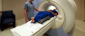
Methodology and preliminary preparation
The technique is based on the influence of a magnetic field and electromagnetic waves. As a result of the study, multidimensional images of the vessels of a certain area are obtained. Particularly valuable is the diagnosis of blood vessels in the neck and brain. The duration of MR angiography is 30-40 minutes.
The procedure can be performed with or without the administration of a contrast agent. Before angiography, an allergy test for a contrast agent is performed. It is not recommended to eat food 8 hours before the procedure. The doctor must be warned in advance about taking medications. Before the procedure, you must remove all metal-containing products.
Among the preparatory activities:
- eliminate alcohol 15 days before the procedure;
- the woman needs to make sure that she is not pregnant;
- take blood clotting tests;
- do an ultrasound of the heart;
- do an enema.
The study is carried out with an empty bladder.
Preparation for MRI of cerebral vessels
The study does not require special preparation. You should notify your doctor in advance about the presence of:
- claustrophobia;
- allergies to drugs;
- chronic pathologies;
- metal implants in the body.
Before entering the diagnostic room, you must remove all jewelry and metal accessories and leave your mobile phone outside the door.
You can download the document for review: MRI studies (general preparation rules)
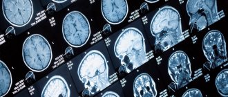
Differences from simple MRI of the brain
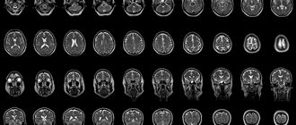
Using standard magnetic resonance imaging, the doctor can obtain information about the condition of soft structures, sinuses, and cerebral fluid. This type of study makes it possible to diagnose various inflammatory processes, tumors, pathologies of the pituitary gland, congenital developmental anomalies, etc. MRI of brain vessels visualizes only the vascular system. Using the resulting images, the doctor can examine in detail the condition of the veins, arteries, capillaries, and also diagnose the slightest changes associated with them.
MRI of the head and neck with a vascular program can detect the following common pathologies:
- thrombosis;
- congenital anomalies of the connection of arteries and veins;
- intracranial hemorrhages arising against the background of oncological and other processes, severe injuries of the skull;
- aneurysms;
- dissection of vascular walls;
- venous stagnation;
- angioma;
- hemorrhagic and ischemic stroke;
- atherosclerosis;
- areas of narrowing, formation of bends, loops.
The procedure also allows us to identify indirect signs of increased ICP, inflammatory processes in the vascular walls, and structural pathologies.
MRI or CT angiography, which is better?
MRI and CT of vessels are the two main non-invasive methods for diagnosing angiopathologies at an expert level. The information content of magnetic resonance and computed tomography is very high, but each of them has its own disadvantages. The main disadvantage of vascular CT is the presence of radiation exposure to the body. This diagnostic method cannot be considered completely safe. The average radiation dose per scan can reach 5-7 mSv.
It is difficult to examine the vessels of the lower extremities using MRA. For this area, ultrasound of the vessels of the lower extremities or CT is better suited. Due to the low sensitivity of the MRI machine to weak blood flow, doctors may falsely diagnose pseudostenosis or occlusion during MR angiography. CT angiography of the vessels of the head will show an aneurysm better than MRI of the vessels of the brain.
With computed tomography, doctors have the opportunity to scan the vessels of almost the entire body at one time, which is impossible during MRA, where the examination is limited to certain areas of the body.
MR angiography, compared to computed tomography, has a longer duration of examination, which does not always allow examining patients in serious condition or young children who will require general anesthesia to remain immobile.
The advantage of MRI using the angiography protocol compared to CT is the absence of harmful effects on the body due to the absence of ionizing radiation and toxic contrast agent containing iodine.
The final decision about whether MRI or CT angiography is better should be made by the attending physician. His knowledge of the medical history, patient's health status and diagnostic goals will allow him to make the right choice between magnetic resonance or computed tomography angiography of blood vessels. If the patient himself decides that he needs to undergo vascular diagnostics for preventive purposes, then it is better to start his diagnostic path with a safe MRI of the vessels of the brain and neck or ultrasound of the vessels of other parts of the body.
Previous Next
Preparing for magnetic resonance angiography
Before undergoing magnetic resonance angiography, the patient receives a questionnaire in which he must indicate whether there are metal objects in his body, such as artificial joints, artificial pacemakers, intravenous ports, intrauterine devices, metal plates, etc. This is done because metal objects may interfere with obtaining a clear image. Before the examination, the doctor will ask you to remove all metal objects, jewelry, clothing with metal buttons, a wig, glasses and dentures. The red dye used for tattooing also contains iron salts, but their concentration has virtually no effect on the results of the method. Before the study, you should inform your doctor about any allergies you have to any drugs.
You can usually eat before the test (unless otherwise indicated). If you have taken any medications or drugs before, you can also take them. Typically, magnetic resonance angiography is performed in a specialized chamber shaped like a cylinder. Some patients may experience claustrophobia when in confined spaces. Therefore, if necessary, they are given a sedative.
What will MR angiography show?
MRI of arteries, sinuses and veins reflects a wide range of vascular abnormalities. Tomography in angio mode is capable of well visualizing:
- arteriovenous malformations;
- stenosis of the vascular bed
- thrombosis;
- vascular occlusion;
- bifurcations and kinks of blood vessels;
- vascular parietal tumors;
- acute circulatory disorder.
MR angiography allows you to find the cause of swelling of the legs and distinguish varicose ulcers from thrombotic ulcers. A non-invasive method for diagnosing blood vessels in medical centers in St. Petersburg is used quite often for planning surgical interventions.
Indications for the study
Most often, vascular MRI angiography is used to diagnose blood vessels in the brain, heart and neck and search for pathologies.
The study is indicated if there is a history or suspicion of the following ailments:
- Traumatic brain injury;
- Neoplasms;
- Increased thrombus formation;
- Stroke;
- Heart attack.
You should also be alarmed if your memory and attention are deteriorating, or you often feel dizzy or sore. This may be a sign of pathological conditions.
Also, this type of diagnosis can be prescribed if it is necessary to perform surgical intervention in the area of the location of blood vessels. Often, MRI angiography of blood vessels is performed after treatment in order to assess the dynamics of treatment.
The patient's condition after the procedure and possible complications
Bed rest is recommended during the day. The attending physician monitors the patient’s general condition (examination of the intervention area, temperature measurement). If the person’s condition is satisfactory, the bandage is removed on the second day and sent home. The risk of complications is minimal. For most people, the study is not dangerous.
Possible problems:
- allergy to contrast agent, anesthesia;
- in the presence of chronic diseases there is a risk of acute renal failure and myocardial infarction;
- bleeding from the place where the puncture was made.