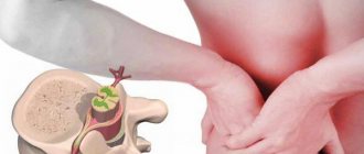
Paroxysmal disorders of consciousness in neurology are a pathological syndrome that occurs as a result of the course of the disease or the body’s reaction to an external stimulus. Disorders manifest themselves in the form of attacks (paroxysms) of various types. Paroxysmal disorders include migraine attacks, panic attacks, fainting, dizziness, epileptic seizures with and without convulsions.
Neurologists at the Yusupov Hospital have extensive experience in treating paroxysmal conditions. Doctors know modern effective methods for treating neurological pathologies.
Disorder of consciousness
Paroxysmal disorder of consciousness manifests itself in the form of neurological attacks. It can occur against the background of apparent health or during an exacerbation of a chronic disease. Often, paroxysmal disorder is recorded during the course of a disease that is not initially associated with the nervous system.
The paroxysmal state is characterized by the short duration of the attack and the tendency to recur. The disorder has different symptoms, depending on the provoking condition. Paroxysmal disorder of consciousness can manifest itself as:
- epileptic seizure,
- fainting,
- sleep disorder,
- panic attack,
- paroxysmal headache.
The causes of the development of paroxysmal conditions can be congenital pathologies, injuries (including at birth), chronic diseases, infections, and poisoning. Patients with paroxysmal disorders often have a hereditary predisposition to such conditions. Social conditions and harmful working conditions can also cause the development of pathology. Paroxysmal disorders of consciousness can cause:
- bad habits (alcoholism, smoking, drug addiction);
- stressful situations (especially when they are repeated frequently);
- disturbance of sleep and wakefulness;
- heavy physical activity;
- prolonged exposure to loud noise or bright light;
- unfavorable environmental conditions;
- toxins;
- sudden change in climatic conditions.
Paroxysmal myoplegia
At the present time, both the mechanism of development of myoplegic attacks and the pathogenesis of the entire disease remain the subject of study by physicians, biochemists and physiologists around the world. Previously, there was Conn's hypothesis, according to which the basis of the disease is periodically occurring increased excretion of aldosterone - intermittent hyperaldosteronism. This was confirmed by the positive effect of therapy with aldosterone antagonists. However, it has recently been proven that the shifts in aldosterone concentration that accompany paroxysmal myoplegia are secondary.
There are suggestions about a genetically determined violation of the permeability of the membranes of myofibrils and cell membranes of other structural elements in striated muscle tissue, as a result of which sodium ions and water pass into the myofibrils, where they accumulate in vacuoles. A change in sodium content in the blood serum leads to a corresponding change in potassium concentration. The consequence of this imbalance is a disruption of intracellular metabolic processes (including glycogen exchange) in muscle tissue and myoneural synapses, neuromuscular transmission is blocked, myofibrils lose their ability to contract. When ionic balance returns to normal, the function of myofibrils and neuromuscular synapses is restored and the muscles regain the ability to contract.
Hypokalemic paroxysmal myoplegia
It occurs more often than others. It is an autosomal dominantly inherited disease with incomplete gene penetrance. However, sporadic cases of myoplegia are not uncommon. The peak incidence occurs in the age group of 10-18 years, although cases of periodic paralysis have also been observed in people over 30 years of age. It occurs more often in men than in women. Outside of paroxysm, neither subjective nor objective signs of the disease are determined. An attack can be triggered by overeating, consuming excessive amounts of carbohydrates or table salt, physical overexertion, and drinking alcohol. In women, paroxysmal myoplegia is usually associated with the menstrual cycle - attacks are often observed on the 1st day of menstruation or 1-2 days before its onset.
Myoplegic paroxysm usually begins in the morning or at night. Patients wake up to find themselves paralyzed. Myoplegia spreads to the skeletal muscles of the entire body, limbs and neck, and in severe cases - to the respiratory and facial muscles. Severe muscle hypotonia and loss of all tendon reflexes are typical. The attack is accompanied by vegetative symptoms - tachycardia, facial hyperemia, hyperhidrosis, polydipsia, rapid breathing. Its duration varies from an hour to 1-2 days, but more often it is 2-4 hours. Towards the end of the paroxysm, there is a gradual increase in muscle strength, starting from the distal parts of the limbs. Active movements of the patient contribute to the rapid restoration of lost motor functions.
The frequency of myoplegic paroxysms varies significantly. In severe cases, attacks occur daily, myoplegic status or moderate muscle weakness of a permanent nature develops, worsening in the morning. This can lead to the formation of a myopathic syndrome with muscle wasting, most pronounced in the proximal parts. In some cases, paroxysms are abortive in nature, when paralysis covers only the legs, one limb, the right or left half of the body.
Hyperkalemic paroxysmal myoplegia
The hypokalemic form is much less common. Autosomal dominant inheritance with high penetrance is characteristic. In some families, paroxysmal myoplegia can be traced in 4 generations. People of different sexes get sick with approximately the same frequency. The typical age of manifestation of the disease is the first decade of life.
Paroxysm of myoplegia is often provoked by hunger or occurs in a state of rest after physical activity. It begins with paresthesia in the face and limbs, then muscle weakness occurs in the distal arms and legs, which quickly spreads to all muscle groups, including the facial muscles. The process is accompanied by tendon areflexia and total muscle hypotonia. Severe vegetative symptoms are typical - palpitations, profuse sweating, arterial hypertension, severe thirst. The duration of myoplegic paroxysm, as a rule, does not exceed 1-2 hours.
Normokalemic paroxysmal myoplegia
It is observed extremely rarely; only 4 families with a similar form of myoplegia are described in the literature. It is inherited in an autosomal dominant manner, and the onset of the disease occurs mainly in the first 10 years of life. Normokalemic paroxysmal myoplegia is characterized by significant variability in the severity of paroxysms - from moderate generalized muscle weakness to complete paralysis, including the facial muscles. Some patients experience muscle hypertrophy with the formation of an athletic physique.
Typically, paroxysm is provoked by hypothermia or develops at rest after intense physical activity. Characterized by a slow increase in muscle weakness and an even slower decrease at the end of the attack. A long duration of paroxysms is typical - from 1-2 days to several weeks.
Epilepsy disorders
In epilepsy, paroxysmal conditions can manifest themselves in the form of convulsive seizures, absence seizures and trances (non-convulsive paroxysms). Before a grand mal seizure occurs, many patients feel a certain type of warning sign - the so-called aura. There may be auditory, auditory and visual hallucinations. Someone hears a characteristic ringing or feels a certain smell, feels a tingling or tickling sensation. Convulsive paroxysms in epilepsy last several minutes and may be accompanied by loss of consciousness, temporary cessation of breathing, involuntary defecation and urination.
Nonconvulsive paroxysms occur suddenly, without warning. With absence seizures, a person suddenly stops moving, his gaze is directed ahead, he does not react to external stimuli. The attack does not last long, after which mental activity returns to normal. The attack goes unnoticed by the patient. Absence seizures are characterized by a high frequency of attacks: they can be repeated tens or even hundreds of times per day.
Symptoms
Paroxysmal tachycardia occurs quite often. This disturbance of heart rhythm is provoked by various types of myocardial lesions.
This disease most often affects those people who have additional pathways in the myocardium. After all, this contributes to the appearance of vicious circulation of excitation in this part of the heart.
It is important to note the vivid nature of the clinical picture of such attacks. First, the patient feels a spontaneous heartbeat, the rate of which constantly increases.
A paroxysm of tachycardia can end as spontaneously as it began. However, even after the normal rhythm of the heart muscle is restored, the person continues to feel extrasystoles.
It is also worth emphasizing that some people are not at all worried about such symptoms, perceiving them as moderate, but during an attack they experience weakness, dizziness and nausea.
Panic disorder (episodic paroxysmal anxiety)
Panic disorder is a mental disorder in which the patient experiences spontaneous panic attacks. Panic disorder is also called episodic paroxysmal anxiety disorder. Panic attacks can occur from several times a day to once or twice a year, while the person is constantly expecting them. Severe anxiety attacks are unpredictable because their occurrence does not depend on the situation or circumstances.
This condition can significantly impair a person's quality of life. The feeling of panic can be repeated several times a day and last for an hour. Paroxysmal anxiety can occur suddenly and cannot be controlled. As a result, a person will feel discomfort while in society.
Sleep disorders
The manifestations of paroxysmal sleep disorders are very diverse. These may include:
- nightmares;
- talking and screaming in sleep;
- sleepwalking;
- motor activity;
- night cramps;
- shuddering when falling asleep.
Paroxysmal sleep disorders do not allow the patient to regain strength or rest properly. After waking up, a person may feel headaches, fatigue and weakness. Sleep disorders are common in patients with epilepsy. People with this diagnosis often have realistic, vivid nightmares in which they run somewhere or fall from a height. During nightmares, your heart rate may increase and you may perspire. Such dreams are usually remembered and can be repeated over time. In some cases, during sleep disorders, breathing disturbance occurs; a person may hold his breath for a long period of time, and there may be erratic movements of the arms and legs.
Treatment
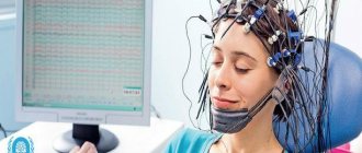
To treat paroxysmal conditions, consultation with a neurologist is necessary. Before prescribing treatment, the neurologist must know exactly the type of attacks and their cause. To diagnose the condition, the doctor clarifies the patient’s medical history: when the first episodes of attacks began, under what circumstances, what their nature is, and whether there are any concomitant diseases. Next, you need to undergo instrumental studies, which may include EEG, EEG video monitoring, MRI of the brain and others.
After performing an in-depth examination and clarifying the diagnosis, the neurologist selects treatment strictly individually for each patient. Therapy for paroxysmal conditions consists of medications in certain doses. Often the dosage and the drugs themselves are selected gradually until the required therapeutic effect is achieved.
Typically, treatment of paroxysmal conditions takes a long period of time. The patient should be constantly monitored by a neurologist for timely adjustment of therapy if necessary. The doctor monitors the patient’s condition, assesses the tolerability of the drugs and the severity of adverse reactions (if any).
The Yusupov Hospital has a staff of professional neurologists who have extensive experience in treating paroxysmal conditions. Doctors are proficient in modern effective methods of treating neurological pathologies, which allows them to achieve great results. The Yusupov Hospital performs diagnostics of any complexity. Using high-tech equipment, which facilitates the timely start of treatment and significantly reduces the risk of complications and negative consequences.
The clinic is located near the center of Moscow and receives patients around the clock. You can make an appointment and get advice from specialists by calling the Yusupov Hospital.
Mental disorders of the neurotic register are widespread in the Ukrainian population. Neurotic, stress-related and somatoform disorders (F4 according to ICD-10) often occur in the form of paroxysmal states. Approximately 10–25% of the world's population suffers from panic and somatoform disorders. These conditions occur mainly in young, socially active ages (20–45 years), and are more often observed in women than in men. Basically, psychogenic paroxysmal conditions have a chronic course with a tendency to relapse, although there are patients with long-term remissions. Disability develops in 50% of those suffering from these disorders. When examined after 20 years, the same symptoms can be found in 73–94% of patients. In psychogenic paroxysmal disorders, depressive layers complicate the clinical picture in approximately 70% of cases, accordingly increasing the risk of suicidal behavior. In 20% of cases, addiction to alcohol and other psychoactive substances is observed
In the specialized literature there are many definitions of paroxysmal states and this is explained to some extent by the difference in methodological settings. According to I.P. Pavlov, these are “periodic explosions of excitation in the central nervous system, which form the basis of a paroxysmal attack.” Paroxysmal activity is characterized by the suddenness of its appearance and cessation, this is especially demonstratively observed in the paroxysmal manifestations of brain biopotentials - paroxysmal depolarizing postsynaptic potentials (V. Sh. Okudzhava, 1966). In the broad sense of the word, paroxysmal manifestations of activity are inherently adaptive and occur in various forms of neurotic disorders, however, they have acquired clinical polymorphism (N. A. Maruta, 1997, 2000; S. P. Kolyadko, 1997).
Today there is no single typology based on the etiology, pathogenesis and clinical variants of psychogenic paroxysmal conditions. The first descriptions of vegetative crises appeared at the end of the last century. An example is De Costa syndrome, which includes a number of symptoms from the cardiovascular and respiratory systems. Then vagal crises of Govers, sympathetic crises of Barre were described, N.I. Grashchenkov designated these conditions as diencephalic, or hypothalamic, crises. Mental phenomena during vegetative crises were described a very long time ago and were part of such generalized syndromes as vagotonia and sympathicotonia. The study of the symptoms of psychogenic paroxysmal disorders is not fundamentally new for domestic psychiatrists. These disorders were considered within the framework of borderline neuropsychic disorders, mainly neuroses, using the term “fear neurosis” (V. A. Gilyarovsky, 1935; M. O. Gurevich, M. Ya. Sereysky, 1946). Neuropathologists have been studying the problem of diagnosing such conditions for many years (A. M. Vein et al., 1981, 1988, 1994, 1997, etc.), considering these conditions as “vegetative crises” with an emphasis on the vegetative component of the attack. The introduction of this phenomenon in the classification of psychiatric diseases (DSM, ICD-10) and in replacing the term with “panic attacks” emphasized the leading role of mental manifestations of paroxysm (anxiety, fear). However, the incompleteness and limitations in this sense of both terms are obvious. Panic sensations are not the only and obligatory manifestation of mental disorders during a crisis. In psychogenic paroxysmal states, phobic attacks with a specific plot of fear may come to the fore; cognitive disorders (“lightheadedness”, “feeling of derealization”, “presyncope”, etc.); conversion crises with functional neurological symptoms (“amaurosis”, “mutism”, “feeling of numbness or weakness in the limbs”, “feeling of a lump in the throat”; individual hyperkinesis, convulsive and muscular-tonic phenomena, etc. may occur); hyperventilation attacks with a specific triad - increased breathing, paresthesia and tetany.
It is known that the pathogenesis of psychogenic paroxysmal disorders involves both genetic and environmental factors, as well as dysfunctional cerebral ones, which cause the launch of a whole complex of various pathological mechanisms. Many facts have been obtained indicating dysfunction of nonspecific integrative systems of the brain. Increased brain activation was revealed, which is evidence of the involvement of the ascending reticular systems of the brain, as well as the hypothalamic-limbic activation system. The main pathophysiological mechanism is the presence of disintegration between synchronizing and activating systems, cortical, subcortical and brainstem systems, left and right hemispheres.
However, works on the study of psychogenic paroxysmal disorders do not have a systematic approach; they most often solve individual aspects of the clinic and diagnosis of this type of disorder, without deeply considering the connection between the clinic and pathogenesis, and therefore have limited significance in solving the problem of diagnosis and treatment of these conditions. Diagnostic criteria that would be based not only on the data of clinical-psychopathological studies, but also on neurophysiological methods that objectify them have not been sufficiently developed.
The purpose of this study was to identify neurophysiological characteristics during paroxysmal states of the neurotic register and their correspondence to the clinical and psychopathological features of these disorders.
Materials and research methods
The department examined 23 patients (15 women and 8 men, aged from 20 to 45 years). We considered patients with psychogenic paroxysmal disorders, which can be classified in accordance with ICD-10 under heading F4: neurotic, stress-related and somatoform disorders: panic disorder - 5 patients; agoraphobia with panic disorder - 2 patients; anxiety-phobic disorders - 1 patient; somatoform-autonomic dysfunction - 8 patients; dissociative disorder - 2 patients; hypochondriacal disorder - 2 patients; neurasthenia - 3 patients. Thus, patients with endogenous mental, organic neurological, as well as severe somatic pathology that could explain their condition were excluded. During the study, clinical indicators were analyzed that reflected the severity of symptoms and features of paroxysm; and also computer electroencephalographic mapping of the brain was carried out using a 17-channel EEG system DX-5000P, unified according to European standards. The frequency-amplitude and topographic characteristics of the alpha rhythm, as well as the localization and representation of slow rhythms were assessed.
Research results and discussion
When conducting an electroencephalographic study of patients with neuroses with paroxysmal conditions, three groups of patients were identified. The first group included 5 patients whose EEG showed a medium- or high-amplitude (from 30 to 90 μV) alpha rhythm; with a weak reaction to closing and opening the eyes; smoothed zonal differences, weak or paradoxical increase in alpha rhythm during photostimulation. During hyperventilation, an increase in the disorganization of the main rhythm was recorded in the form of an increase in the density of slow waves, predominantly in the theta range, up to 12%, with an amplitude not exceeding the main rhythm. The clinical picture of such patients was asthenic, astheno-hypochondriacal symptoms with a vegetative component. Patients complained of paroxysmal states of “feeling of faintness”, “feeling of possible loss of consciousness”, accompanied by irritable weakness, increased fatigue, dizziness, nausea and fluctuations in blood pressure.
In the second group of patients (6 people), a medium-amplitude EEG with a dominant alpha rhythm in the background recording, amplitude from 30 to 70 μV, with a decrease in its density, was electroencephalographically recorded. During hyperventilation, an increase in diffuse island-wave fast waves of the beta range with periods of bilateral synchronization of theta waves, a frequency of 6–7 oscillations per second, and an amplitude of 40–50 μV was recorded. The clinical picture in this group of patients was dominated by paroxysmal states of the “panic attack” type with a pronounced vegetative component, as well as subdepressive, depressive, depressive-hypochondriacal symptoms, and agoraphobia.
The third group (9 people) included patients in whom a low-amplitude or even “flat” EEG (from 10 to 20 μV) was encephalographically recorded. Due to the low alpha rhythm, there was a predominance of pathological rhythms in the beta range and theta range, 12% of the total recording, which were not organized in a bilaterally synchronous manner, without clear localization. Reaction to functional loads is reduced. The clinical picture in this group of patients was characterized by intense emotions, the severity of paroxysms of anxiety and fear, quite strong in strength and depth of affect, irritability up to aggressiveness.
An electroencephalographic study in a psychosomatic hospital in three previously unexamined patients, in whose clinical picture autonomic symptoms predominated, changes were found that were indicative of organic brain damage. An EEG was recorded with a high-amplitude (more than 100 μV), peaked, low-modulated alpha rhythm with a frequency of 16 Hz throughout the study. Zonal differences are weakly expressed. Quite often, amplitude asymmetry of the alpha rhythm was observed, or slow activity predominated in one of the hemispheres. During photostimulation, there was no suppression of the alpha rhythm, but a sharp, high-amplitude rhythm arose, which was subjectively accompanied by a feeling of “lightness” and the onset of an “attack.” These conditions should be classified as simple partial seizures with predominantly vegetative manifestations.
conclusions
Thus, psychogenic paroxysmal states are quite common in neuroses and are characterized by an interest in the diencephalic structures of the brain. Correspondences were found between the electroencephalographic characteristics and the clinical features of paroxysmal disorders and three groups of patients were identified:
- The medium- or high-amplitude alpha rhythm is hypersynchronous, characteristic of asthenic conditions.
- Paroxysmal phenomena against the background of a medium-amplitude alpha rhythm, characteristic of neuroses against the background of minimal microorganic symptoms.
- Low-amplitude, characteristic of “pure” neuroses.
EEG mapping also makes it possible to differentiate neurotic paroxysmal states and epileptiform vegetative paroxysms, which have a similar clinical and psychopathological picture, but fundamentally different approaches to therapy.
Literature
- Dyukova G. M., Vorobyova O. V., Storozhakova Ya. A. Clinical polymorphism of vegetative crises (panic attacks) // Journal of Neurology and Psychiatry named after. S. S. Korsakova. - 1992. - T. 92, issue. 5. - pp. 37–42.
- Autonomic disorders: clinical picture, treatment, diagnosis / Ed. A. M. Veina. - M.: Medical Information Agency, 1998. - 752 p.
- Vein A. M. Panic attacks // International Medical Journal. - 1997. - T. 3, No. 3. - P. 75–79.
- Vein A.M., Vorobyova O.V. Universal cerebral mechanisms in the pathogenesis of paroxysmal states // Journal of Neurology and Psychiatry named after. S. S. Korsakova. - 1999. - T. 99, issue. 12. - pp. 44–46.
- Maruta N. A., Vorobyova T. M., Kolyadko S. P. Systemic biological approach to the problem of paroxysmal states in neurotic disorders // Ukrainian Bulletin of Psychoneurology. - 2000. - T. 8, VIP. 3. - pp. 64–67.
- Reminyak I. B. Structural and functional characteristics of the brain of patients in syncope (differential diagnosis, treatment). — Author's abstract. dis. ...cand. honey. Sci. - Kharkiv, 1997. - 18 p.
- Solovyova A.D., Loseva M.M. The importance of activating and deactivating mechanisms of the brain in autonomic regulation // Journal of Neuropathology and Psychiatry named after. S. S. Korsakova. - 1990. - T. 90, issue. 6. - pp. 26–28.
- Diagnostic Criteria from DSM-IV. — 4th ed. - Washington, DC, 1994.
- Hirschfeld RM Panic disorders: diagnosis, epidemiology and clinical course // Journal of Clinical Psychiatry. - 1997. - Vol. 57, No. 10. - P. 3–8.