Fourth ventricle of the brain
The fourth ventricle (ventriculus quartus) develops from the cavity of the rhombencephalon. At the top it communicates with the III ventricle using the cerebral aqueduct, and at the bottom - with the central canal of the spinal cord, through the median aperture (apertura mediana ventriculi quarti), paired lateral apertures (aperturae laterales) of the IV ventricle communicate with the subarachnoid space of the brain (cisterna cerebello-medullaris) . The lateral openings of the fourth ventricle are located near the flocculus of the cerebellum in the lateral corners of the roof of the rhomboid fossa (Fig. 466). The fourth ventricle is filled with cerebrospinal fluid and the choroid plexus. Consists of top and side walls; its bottom is formed by a diamond-shaped pit. The upper wall of the fourth ventricle is limited by the superior medullary velum, which is attached to the cerebellar uvula (lingula cerebelli). The lower medullary velum (velum medullare inferius), which is a thin epithelial plate, begins here (Fig. 467). Adjacent to it is the vascular base of the fourth ventricle (tela choroidea ventriculi quarti) - the tegmentum. The junction of the superior and inferior medullary sails forms the apex of the tent (fastigium). The lateral border of the fourth ventricle is the superior cerebellar peduncle.
466. Rhomboid fossa and cranial nerve nuclei. 1 - nucl. n. oculomotorii; 2 - nucl. n. trochleares; 3 - nucl. tractus mesencephalic! n. trigemini; 4 - n. motorius n. trigemini; 5 - nucl. sensorius principalis n. trigemini; 6 - nucl. n. abducentis; 7 - nucl. n. facialis; 8 - nucl. n. vestibularis; 9 - nucl. cochleares; 10 - n. facialis; 11 - nucl. salivatorii superior et inferior; 12 - nucl. n. hypoglossi; 13 - nucl. ambiguus; 14 - n. tractus spinalis n. trigemini; 15 - n. tractus solitarii; 16 - n. accessorius; 17 - nucl. dorsalis n. trigemini; 18 - nucl. spinalis n. accessorii; 19 - tuberculum n. gracilis; 20 - tuberculum n. cuneati; 21 - trigonum n. vagi; 22 - colliculus facialis; 23 - eminentia medialis; 24 - trigonum lemnisci; 25 - colliculus inferior; 26 - colliculus superior
The rhomboid fossa represents the floor of the fourth ventricle. Develops from the isthmus of the rhombencephalon, hindbrain and medulla oblongata. It has upper, lower and lateral angles. The rhomboid fossa is divided by brain stripes (striae medullares), which are located in the middle of the bottom of the rhomboid fossa and extend transversely to the lateral angles. The superior angle of the rhomboid fossa is bounded by the superior cerebellar peduncles, the inferior angle by the inferior cerebellar peduncles, which diverge to the sides at an angle of 50-85°, and the lateral angles by the inferior and middle cerebellar peduncles.
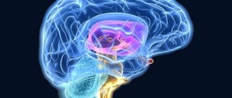
467. The brain on a sagittal section. 1 - sulcus centralis; 2 - lobus paracentralis; 3 - precuneus; 4 - sulcus parietooccipitalis; 5 - cuneus; 6 - sulcus calcarinus; 7 - gyrus occipi totem poralis; 8 - corpus pineale; 9 - cerebellum; 10 - medulla oblongata; 11 - ventriculus quartus; 12 - pons; 13 - colliculi superior et inferior; 14 - pedunculi cerebri; 15 - aqueductus cerebri; 16 - hypophysis; 17 - chiasma opticum; 18 - commissura anterior; 19 – fornix
The diamond-shaped fossa is divided into two symmetrical parts by the median groove (sulcus medianus). In the upper corner of the fossa, above the brain stripes, which are lighter in color, lies the paired medial eminence (eminentia medialis). Its lower part ends with the facial tubercle (colliculus facialis). The facial tubercle is represented by the convexity of the facial nerve, which in this place bends around the nucleus of the abducens nerve (nucl. abducens). Closer to the ventral surface from the nucleus of the abducens nerve lie the solitary tract (nucl. tr. solitarius), the parasympathetic nucleus - the superior salivary nucleus (nucl. salivatorius superior), the motor nucleus (nucl. motorius) of the facial nerve (VII pair). The medial eminence contains the motor nucleus of the trigeminal nerve (V pair).
Lateral and above the medial eminence there is a bluish place (locus ceruleus), where the sensitive nucleus (nucl. sensorius) and the spinal tract of the trigeminal nerve (tr. spinalis n. trygemini) (V pair) are located.
In the lateral corners of the rhomboid fossa there is a vestibular area (area vestibularis). In this area there are 4 nuclei of the vestibular nerve (nucl. vestibulares): medial, lateral, superior, inferior and two nuclei of the auditory nerve: anterior cochlear (nucl. cochlearis anterior) and posterior cochlear (nucl. cochlearis posterior) (VIII pair). In the lower corner of the rhomboid fossa near the median sulcus there is a triangle of the hypoglossal nerve (trigonum n. hypoglossi), onto which the nucleus of this nerve is projected (XII pair). Lateral to the triangle of the hypoglossal nerve there is a darker triangular hillock (ala cinerea), which is the location of the parasympathetic nucleus of the vagus nerve (nucl. dorsalis n. vagi). Somewhat higher, on the line ala cinerea, lies the large motor double nucleus (n. ambiguus), belonging to the IX and X pairs of cranial nerves. Lateral to this nucleus is the lower salivary nucleus - parasympathetic (nucl. salivatorius inferior), from which part of the fibers of the glossopharyngeal nerve emerges. Next to it is the sensitive nucleus of the IX and X pairs (nucl. tr. solitarii).
Thus, summing up the topography of the nuclei of the bottom of the rhomboid fossa, it should be noted that the motor nuclei are located closer to the midline, the sensitive ones lie more laterally, and the autonomic ones are distributed between them.
Anatomy of the Human Fourth Ventricle - information:
IV ventricle, ventriculus quartus , is a remnant of the cavity of the posterior medullary bladder and is therefore a common cavity for all parts of the hindbrain, constituting the rhombencephalon (medulla oblongata, cerebellum, pons and isthmus). The IV ventricle resembles a tent, in which a bottom and a roof are distinguished.
The bottom, or base, of the ventricle has the shape of a rhombus, as if pressed into the posterior surface of the medulla oblongata and the pons. Therefore it is called the rhomboid fossa, fossa rhomboidea. The central canal of the spinal cord opens into the posteroinferior corner of the rhomboid fossa, and in the anterosuperior corner the fourth ventricle communicates with the aqueduct. The lateral angles end blindly in the form of two pockets, recessus laterales ventriculi quarti, curving ventrally around the inferior cerebellar peduncles.
The roof of the IV ventricle, tegmen ventriculi quarti, has the shape of a tent and is composed of two brain sails: the upper one, velum medullare superius, stretched between the upper cerebellar peduncles, and the lower one, velum medullare inferius, a paired formation adjacent to the peduncles of the cerebellum. The part of the roof between the sails is formed by the substance of the cerebellum. The lower cerebral velum is supplemented by a sheet of soft membrane, tela choroidea ventriculi quarti, covered from the inside with a layer of epithelium, lamina choroidea epithelialis, representing a rudiment of the posterior wall of the posterior cerebral bladder (a plexus is associated with it - plexus choroideus ventriculi quarti).
The body choroidea initially completely encloses the cavity of the ventricle, but then, during development, three openings appear in it: one in the region of the lower angle of the rhomboid fossa, apertura mediana ventriculi quarti (the largest), and two in the region of the lateral recesses of the ventricle, aperturae laterales ventriculi quarti. Through these openings, the fourth ventricle communicates with the subarachnoid space of the brain, due to which cerebrospinal fluid flows from the cerebral ventricles into the interthecal spaces. In the case of narrowing or fusion of these holes due to inflammation of the meninges (meningitis), the cerebrospinal fluid accumulating in the cerebral ventricles does not find an outlet into the subarachnoid space and hydrocele occurs.
Diamond-shaped fossa, fossa rhomboidea , has four sides corresponding to the diamond shape - two upper and two lower. The upper sides of the rhombus are bounded by the two superior cerebellar peduncles, and the lower sides by the two inferior cerebellar peduncles. Along the rhombus, along the midline, from the upper to the lower corner there is a median groove, sulcus medianus, which divides the rhomboid fossa into the right and left halves. On the sides of the furrow there is a paired elevation, eminentia medialis, caused by the accumulation of gray matter.
Inferiorly, the eminentia medialis gradually narrows, turning into a triangle onto which the nucleus of the hypoglossal nerve, trigonum nervi hypoglossi, is projected. Lateral to the lower part of this triangle lies a smaller triangle, noticeable by its gray color, trigonum nervi vagi, which contains the autonomic nucleus of the vagus nerve, nucleus dorsalis nervi vagi.
At the top, the eminentia medialis has an elevation - the facial tubercle, colliculus facialis, caused by the passage of the facial root and the projection of the abducens nerve nucleus. In the area of the lateral angles, the vestibular field, area vestibularis, is located on both sides; the nuclei of the VIII pair are located here. Some of the fibers emerging from them run across the rhomboid fossa from the lateral angles to the median sulcus in the form of horizontal stripes, striae medullares ventriculi quarti. These stripes divide the rhomboid fossa into superior and inferior halves and correspond to the boundary between the medulla oblongata and the pons. Topography of the gray matter of the rhomboid fossa.
The gray matter of the spinal cord directly passes into the gray matter of the brain stem and partly spreads along the rhomboid fossa and the walls of the aqueduct, and partly is divided into separate nuclei of cranial nerves or nuclei of bundles of pathways. To understand the location of these nuclei, it is necessary to take into account, as noted, that the closed neural tube, during the transition from the spinal cord to the medulla oblongata, opened on its posterior side and unfolded into a rhomboid fossa. As a result, the posterior horns of the gray matter of the spinal cord seemed to diverge to the sides. The somatic-sensitive nuclei located in the posterior horns were located laterally in the rhomboid fossa, and the somatic-motor nuclei corresponding to the anterior horns remained located medially.
As for the vegetative nuclei located in the lateral horns of the spinal cord, then, according to the position of the lateral horns between the posterior and anterior, these nuclei, during the unfolding of the neural tube, turned out to lie in the rhomboid fossa between the somatic-sensitive and somatic-motor nuclei. As a result, in the region of the rhomboid fossa, unlike the spinal cord, the nuclei of the gray matter are not located in the anteroposterior direction, but lie in rows - medially and laterally. So, for example, the somatic-motor nuclei of the XII and VI pairs lie in the medial row, the vegetative nuclei of the X, IX, and VII pairs are in the middle row, and the somatic-sensitive nuclei of the VIII pair are lateral.
Projection of the cranial nerve nuclei onto the rhomboid fossa:
XII pair - hypoglossal nerve, n.hypoglossus, has a single motor nucleus, located in the lowest part of the rhomboid fossa, in the depth of trigonum n. hypoglossi.
XI pair - accessory nerve, n. accessorius, has two nuclei (both motor): one is located in the spinal cord and is called nucleus n. accessorii, the other is a caudal continuation of the nuclei of the X and IX pairs of nerves and is called nucleus, ambiguus. It lies in the medulla oblongata dorsolateral to the olivary nucleus.
X pair - vagus nerve, n. vagus, has three nuclei:
- the sensitive nucleus, nucleus solitarius, is located next to the nucleus of the hypoglossal nerve, in the depths of trigonum n. vagi;
- vegetative nucleus, nucleus dorsalis n. vagi, lies in the same area;
- the motor nucleus, nucleus ambiguus (double), common with the nucleus of the IX pair, is embedded in the formatio reticularis, deeper than the nucleus dorsalis.
IX pair - glossopharyngeal nerve, n. glossopharyngeus, also contains three nuclei:
- the sensitive nucleus, nucleus solitarius, lies lateral to the nucleus of the hypoglossal nerve;
- vegetative (secretory) nucleus, nucleus salivatorius inferior, lower salivary nucleus; its cells are scattered in the formatio reticularis of the medulla oblongata between n. ambiguus and olive kernel;
- motor nucleus, common with n.vagus and n.accessorius, nucleus ambiguus.
VIII pair - vestibulocochlear nerve, n. vestibulocochlearis, has multiple nuclei projecting onto the lateral corners of the rhomboid fossa, in the area vestibularis. The nuclei are divided into two groups, corresponding to the two parts of the nerve. One part of the nerve, pars cochlearis, the cochlear nerve, or the auditory nerve itself, has two nuclei: the posterior one, nucleus cochlearis dorsalis, and the anterior one, nucleus cochlearis ventralis, located lateral and anterior to the previous one. Another part of the nerve, pars vestibularis, is the nerve of the vestibule, or gravitational nerve, has four nuclei (nuclei vestibulares):
- medial is the main thing;
- lateral;
- top;
- lower.
The presence of four nuclei in humans reflects the early stages of phylogenesis, when fish had several separate gravitational-sensing apparatuses.
VII pair - facial nerve, n. facialis, has one motor nucleus located in the formatio reticularis partis dorsalis of the pons. The nerve fibers extending from it on their way through the thickness of the bridge form a loop that protrudes on the rhomboid fossa in the form of a colliculus facialis.
Intermediate nerve, n. intermedins, closely connected in its course with the facial nerve, has two nuclei:
- vegetative (secretory), nucleus salivatorius superior (superior salivary nucleus), embedded in the formatio reticularis of the pons, dorsal to the nucleus of the facial nerve;
- sensitive, nucleus solitarius. VI pair - abducens nerve, n. abducens, has one motor nucleus located in the loop of the facial nerve, therefore the colliculus facialis on the surface of the rhomboid fossa corresponds to this nucleus.
V pair - trigeminal nerve, n.trigeminus, has four nuclei:
- sensitive, nucleus pontinus n. trigemini, projected in the dorsolateral part of the upper part of the bridge;
- the nucleus of the spinal tract, nucleus spinalis n.trigemini, is a continuation of the previous one along the entire length of the medulla oblongata to the cervical spinal cord, where it comes into contact with the substantia gelatinosa of the dorsal horns;
- motor nucleus, nucleus motorius n. trigemini (chewing), located medial to the sensitive one;
- nucleus of the midbrain tract, nucleus mesencephalicus n. trigemini, lies lateral to the aqueduct. It represents the core of proprioceptive sensitivity for the muscles of mastication and for the muscles of the eyeball. It is possible that this nucleus reflects the independent development of the first branch of the trigeminal nerve (n. ophthalmicus), called n in animals. ophthalmicus profundus and related to the organ of vision, which explains the location of the nucleus in the midbrain.
Fourth (IV) ventricle of the brain, its walls, cerebrospinal fluid outflow tracts
Fourth (IV) - ventricle, ventriculus quartus,
is a derivative of the cavity of the rhombencephalon.
The medulla oblongata, pons, cerebellum and isthmus of the rhombencephalon take part in the formation of the walls of the fourth ventricle. The cavity of the IV ventricle is formed by the posterior (dorsal) surfaces of the medulla oblongata and the pons. The border between the medulla oblongata and the pons on the surface of the rhomboid fossa is the medullary striae (IV ventricle), striae medullares (ventriculi quarti).
They originate in the area of the lateral corners of the rhomboid fossa and plunge into the median sulcus.
Roof of the IV ventricle, legmen ventriculi quarti,
hangs over the diamond-shaped fossa.
the medullary velum, velum medulldre craniale,
take part in the formation of the anterior superior wall of the roof The posterior inferior wall is more complex. It is composed of the inferior medullary velum, velum medullare caudale,
which is attached to the legs of the shred on the sides.
, the vascular base of the IV ventricle, tela choroidea (ventriculi quarti),
is adjacent to the inferior medullary velum The vascular base forms the choroid plexus of the fourth ventricle, plexus choroidea (ventriculi quarti).
In the posteroinferior wall of the fourth ventricle there is an unpaired
median aperture, apertura medina.
In the lateral sections there is a paired
lateral aperture, apertura laterallis.
All three apertures connect the cavity of the fourth ventricle with the subarachnoid space of the brain.
Spleen: development, topography, structure, blood supply and innervation.
The spleen, lien , performs the functions of immune control of the blood. It is located on the path of blood flow from the main vessel of the systemic circulation - the aorta - to the portal vein system, which branches in the liver. The spleen is located in the abdominal cavity, in the left hypochondrium, at the level of the IX to XI rib.
The spleen has two surfaces: diaphragmatic and visceral. The smooth convex diaphragmatic surface, fades diaphragmatica, faces laterally and upward towards the diaphragm. The anteromedial visceral surface, faces visceralis, is uneven. On the visceral surface, the gate of the spleen, hilum splenicum, and the areas to which neighboring organs are adjacent are distinguished. The gastric surface, faces gdstrica, is in contact with the fundus of the stomach. The renal surface, faces rendlis, is adjacent to the upper end of the left kidney and to the left adrenal gland. The colonic surface, fades colica, is located below the gate of the spleen, closer to its anterior end.
The spleen has two edges: upper and lower, and two ends (poles): posterior and anterior.
The spleen is covered on all sides by peritoneum. Only in the area of the gate, where the tail of the pancreas faces, is there a small area free of peritoneum.
From the fibrous membrane, tunica fibrosa, located under the serous cover, connective tissue crossbars extend into the organ - trabeculae of the spleen, trabeculae splenicae. Between the trabeculae there is parenchyma, the pulp (pulp) of the spleen, pulpa splenica. There is red pulp, pulpa rubra, located between the venous sinuses, sinus venularis, and white pulp, pulpa alba.
Development and age-related characteristics of the spleen. The spleen anlage appears at the 5-6th week of intrauterine development in the form of a small accumulation of mesenchymal cells in the thickness of the dorsal mesentery. In the 2-4th month of development, venous sinuses and other blood vessels are formed. In a newborn, the spleen is round and has a lobular structure.
Vessels and nerves of the spleen . The artery of the same name (splenic) approaches the spleen, which is divided into several branches that enter the organ through its gate. The splenic branches form 4-5 segmental arteries, and the latter branch into trabecular arteries. Pulp arteries with a diameter of 0.2 mm are directed into the parenchyma of the spleen, around which lymphoid periarterial couplings and the periarterial zone of splenic lymphoid nodules are located. Each pulp artery is ultimately divided into brushes - arteries with a diameter of about 50 microns, surrounded by macrophage-lymphoid couplings (ellipsoids). The capillaries formed during the branching of the arteries flow into the wide splenic venular sinuses, located in the red pulp.
Venous blood from the splenic parenchyma flows through the pulpal and then trabecular veins. The splenic vein formed at the portal of the organ flows into the portal vein.
Innervation of the spleen is carried out through sympathetic fibers approaching the spleen as part of the plexus of the same name. Afferent fibers are processes of sensory neurons located in the spinal ganglia.
Ticket No. 19