Ependymoma is a pressing problem in oncology, neurology and neurosurgery. The most complex organ of the human body is the brain, which consists of tissue called ependyma. All the internal cavities of the brain are filled with this tissue. Under the influence of various factors, ependyma can change - grow, transform into a tumor of ependymocytes (epithelial-like cells). The location of emendioma is usually the posterior cranial fossa. People of all ages are susceptible to the disease. Mostly the neoplasm is benign. It is more common in pediatric patients: 60% of patients with ependymoma are children under 5 years of age. Moreover, unlike adults, in children the tumor is in most cases malignant. Low-grade ependymoma can metastasize and treatment should begin immediately. Removal of the ependymoma is generally recommended.
Causes of ependymoma formation
What serves as the driving force for tumor development is not known exactly. Oncologists identify only factors that precede the appearance of a tumor of this type:
- heredity: if close relatives have a history of cancer, the person is at increased risk;
- harmful working conditions: regular exposure of the body to carcinogenic, chemical, nuclear substances contributes to the occurrence of various kinds of diseases, including central nervous system tumors;
- radioactive radiation;
- infection with oncogenic viruses such as herpes, papilloma, etc.;
- poor ecological environment;
- bad habits.
People with reduced immunity and survivors of cancer are also at risk of developing ependymoma.
What are the histological types of ependymomas?
The properties of tumor tissue in ependymomas are very diverse. That is, ependymomas within themselves differ in the structure of the tumor tissue (experts talk about the histological variant of the tissue). There are low-grade ependymomas. This means that their cells divide slowly, and the tumor itself grows slowly. Cells of ependymomas of higher degrees of malignancy divide very quickly, the tumors themselves have different qualities and they grow aggressively.
The World Health Organization (WHO) divides ependymomas according to their histological tissue structure into the following types (WHO classification):
- Subependymomas, WHO grade I: low-grade tumors that grow slowly.
- Myxopapillary ependymomas, WHO grade I: tumors of low malignancy, grow slowly, most often appear in the spinal canal.
- WHO grade II ependymomas: usually grow slowly, with a slight tendency to grow aggressively. Depending on how most cancer cells look under a microscope, they will be divided into several variants within the group.
- Anaplastic ependymomas of WHO grade III: these are tumors that have all the signs of aggressive growth.
It is impossible to draw an exact boundary between grade II and grade III ependymomas based solely on histological analysis of the tumor tissue. Therefore, it is impossible to say in advance whether the tumor is grade II or grade III. Only by observing how the tumor actually grows can we talk about its degree of malignancy.
Classification of ependymomas
Today, doctors distinguish four types of tumors of this type according to their etiology and degree of malignancy:
- Mixopapillary type.
A benign neoplasm is located in the lower parts of the spinal cord. It occurs more in adult patients. The prognosis after treatment is favorable. - Sudappendymoma.
A benign formation is located inside the ventricles of the brain, grows slowly, and there are practically no symptoms. Relapses are rare. - Classic type ependymoma.
The most common type of pathology. Recurs frequently and progresses to the next stage. - Anaplastic type - ependymoblastoma.
This type is diagnosed in a quarter of all cases of ependymoma. The malignant tumor grows rapidly and metastasizes in all cases.
In most cases, removal of brain ependymoma is performed.
Ependymoma Clinic
The clinical manifestations of ependymoma directly depend on where exactly it is located.
| Localization of ependymoma | The symptoms it provokes |
| Lateral ventricles of the brain | Increased intracranial pressure, which manifests itself:
|
| Fourth ventricle of the brain | The patient tries to keep his head in a certain position to reduce headaches, which is the most common symptom of ependymoma. It develops due to increased intracranial pressure and in the initial stages occurs suddenly or after physical exertion, coughing, or a sudden change in body position. In addition, the patient suffers from vomiting, dizziness, convulsions and mental disorders. |
| Spinal cord |
|
| Horsetail area |
|
The complex of symptoms inherent in the disease
The following signs of pathology are distinguished:
- severe and chronic headaches;
- dizziness;
- nausea, vomiting;
- impaired coordination of movements;
- unsteadiness of walking;
- difficulty swallowing;
- impairment of auditory and visual functions;
- epilepsy attacks;
- mental retardation (in a child);
- increased intracranial pressure;
- irritability;
- constant feeling of fatigue;
- impaired fine motor skills;
- decreased concentration.
If the tumor has involved nearby tissues, the following symptoms appear:
- sharp and severe back pain;
- gastrointestinal disorders;
- urinary incontinence.
Symptoms vary depending on the location of the tumor.
What treatment methods for ependymoma in Israel have proven effective?
Surgery is usually the most appropriate treatment option for ependymoma. But there are cases when a large tumor in the lower part of the spinal column cannot be removed due to the large number of nerve endings in this section, so other methods of therapy are used.
Innovative techniques that are used in the treatment of cancer in Israel (fractionated radiotherapy, stereotactic radiosurgery) reduce the radiation dose and allow control of the pathological process. Only the bed of the eliminated tumor is irradiated, as a result, side effects are reduced and the best effect is achieved.
Chemotherapy is increasingly prescribed as one of the methods of palliative treatment, helping to reduce pain and improve the patient’s quality of life.
Diagnosis of the disease
In order to determine the history of the origin of the pathology, the stage of the disease, the size of the tumor, the neurologist prescribes:
- Computed tomography.
— Magnetic resonance imaging.
- Puncture of cerebrospinal fluid to confirm the diagnosis.
These methods allow you to examine the tumor as accurately as possible. After making a preliminary diagnosis, it is recommended to:
- angiography;
- stereotactic biopsy;
- ultrasonography;
- ventriculoscopy;
- electroencephalography.
MRI and CT are the most modern and effective methods for diagnosing tumors.
Diagnostics
To diagnose brain ependymoma, you need to consult a neurologist. The specialist will examine the medical history in detail and prescribe a number of tests and studies:
- MRI is an informative examination method that allows you to accurately determine the size and location of the tumor, its structure, and shape.
- CT scan – determines the focus of the disease, reveals whether there are metastases to other organs and systems.
- Electroencephalography – examines the functioning of the blood vessels in the brain.
- Ophthalmoscopy - the procedure consists of examining the fundus, assessing the optic nerve and retina.
- Angiography is performed using contrast to assess the condition of the blood vessels in the brain.
- A biopsy is a procedure that allows you to determine the type of tumor. During the process, tumor tissue is collected and then tested in the laboratory.
The examination results allow the doctor to obtain a complete picture of the disease. If necessary, the doctor prescribes a consultation with doctors from other areas of medicine: ophthalmologist, otolaryngologist, psychologist. Comprehensive diagnostics is the basis for making a final diagnosis and determining further treatment tactics.
Treatment method for ependymoma
Today, the following methods of treating the disease are used:
- Operation
. Surgical removal of brain ependymoma is the main method of treating the pathology. If the formation is malignant, then even complete excision gives a survival rate of no more than five years. The most disappointing prognosis for epindymoblastoma. In our clinic, neurosurgical operations are performed by experienced doctors using new generation equipment and technology.
If the tumor is localized in the posterior cranial fossa, access to it is limited and complete excision is impossible. After surgery, radiation therapy is performed. If the patient is a child, radiation exposure is minimized or replaced with chemotherapy.
- Stereotactic radiosurgery
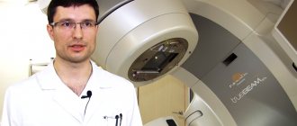
. Used as an alternative to surgery to treat patients over 14 years of age. Irradiation of the tumor is performed using a CyberKnife. The radiosurgery method is effective for the treatment of myxopapillary tumor and subepindymoma. Impairment of neurological processes may persist after treatment.
- Radiation therapy
. The technique involves influencing the formation of radiological radiation. The main indication for use is localization of the tumor in a hard-to-reach place, and also as an addition to a set of measures after surgery. The advantage of the method is that the radiation is directed strictly to the affected tissues, without the risk of damaging healthy cells, unlike surgery.
You can make an appointment at our clinic using the contact number. Diagnosis, treatment and monitoring of the patient’s condition are carried out by qualified doctors.
Description and causes of cerebral ependymoma
Brain ependymoma is usually called a tumor pathology that develops in the tissues of the ventricular structure of the brain. Ependymocytes are cells that form the inner layer of the ventricular system and the spinal canal. Such tumor formations account for 7-8% of all detected neoplasms in the intracranial region. The age preference for the disease is childhood (up to five years), more than half of the patients belong to this category. The prevalence of the pathology is frequent, ranking third among all childhood tumor formations. Often develops into a malignant form.
The location of the anomaly varies, but is most often detected in the occipital region (cranial cavity). It is characterized by slow growth and does not penetrate into adjacent areas. The main danger of the disease is the appearance of a compressive effect. Cancerous growths (metastases) do not go beyond the central nervous system, as they spread through the cerebrospinal fluid channels. The most common form of metastasis is the penetration of processes into the spinal cord.
The causes of the pathology have not been fully identified. It is generally accepted that pathogenic factors include general unfavorable conditions, exposure to radioactive elements, carcinogens, toxins, oncogenic viruses, and increased solar activity in the patient’s place of residence. Often, laboratory tests reveal the presence of the aggressive SV40 virus in the cellular layer of ependymomas. Medical experts do not exclude the influence of genetic disorders or the presence of concomitant congenital disorders.
Experts have identified 4 main types of disease, differing in the speed of progression and level of benignity:
- Myxopapillary tumor. A benign form localized in the lower region of the spinal cord. Detected in older patients.
- Subependymoma. It is characterized by slow growth and absence of symptoms. When removed, it rarely recurs.
- Classic shape. The most common option. A complication is blockage of the liquor channels. When removed it often recurs. Capable of transforming into 4th form.
- Anaplastic formation. It is characterized by rapid growth, produces a large number of processes, and has the highest stage of malignancy.


