Phakomatosis - main symptoms:
- Skin rashes
- Convulsions
- Skin spots
- Impaired movement coordination
- Insomnia
- Epileptic seizures
- Hearing loss
- Visual impairment
- The appearance of a tumor formation
- Baldness
- Mental retardation
- Mental development disorder
- Facial skin pigmentation
- Daytime sleepiness
- Dementia
- White eyelash color
- Antisocial behavior
- White hair color
- Premature aging
- White eyebrow color
What is phakomatosis
Phakomatosis is a multicomponent concept that unites a number of congenital pathologies that affect the eyes, nervous system and human skin. The type of inheritance of the disease is autosomal dominant. Phakomatoses provoke the formation and progression of various tumors and hematomas - benign neoplasms that develop in organs.
Phakomatosis is a very serious congenital disease that is diagnosed in children at an early age. The symptoms are multifaceted, however, rash and tumor-like formations are its main characteristic. If the tumor is localized in the brain, then with its development the patient may experience psychological abnormalities.
Phakomatoses are also distinguished by the presence of hemianopsia - a person partially sees the space around him. Scientists can accurately diagnose the problem only by the age of 7-10 years, despite the fact that the general clinical picture of phakomatosis in children is reflected in the first years of life. However, cases have been recorded where the pathology did not manifest itself for decades.
In the International Classification of Diseases (ICD-10), phakomatosis is coded Q85.
Clinical manifestations
All phakomatoses are characterized by pronounced polymorphism of skin manifestations, damage to various structures of the nervous system and visceral organs.
It is characteristic that at an early age, mainly dermatological symptoms are detected, and neurological and others are detected later. In some cases, phakomatosis is combined with congenital deficiency of the immune system (Louis-Bart disease), premature development of aging and (or) the risk of tumor malignancy.
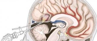
Damage to the central nervous system in this pathology morphologically looks like a proliferation in the membranes and brain tissue of nodules, cystic cavities, neurofibromatous formations, areas of demyelination, atrophy and calcification, congenital vascular anomalies such as aneurysms, hemangiomas.
In the clinic there is a picture of convulsive syndrome with a different nature of the course. Children often experience epileptic seizures, as a result of which phakomatosis is often accompanied by delayed neuropsychic development, mental retardation, and antisocial behavior. The severity of mental retardation is directly proportional to the severity and frequency of epileptic seizures.
The group of diseases is characterized by lesions of different pairs of cranial nerves with the development of oculomotor, auditory disorders, paresis and other symptoms.
There may be pyramidal insufficiency (often unilateral), extrapyramidal disorders (brady and oligokinesia, hyperkinesia, tonic muscle spasms), cerebellar dysfunction (coordination problems), and sleep function disorders.
Dermatological manifestations are usually detected in the first weeks or months of life. Elements of the rash can be single or widespread, of different colors and sizes, often asymmetrical.
Pigment spots of different shades of brown, areas of reduced pigmentation, nodular elements, neurofibromas, papillomas, shagreen skin with plaques, hemangiomas are considered typical.
In some cases, phakomatosis occurs with endocrine disorders such as obesity, diabetes insipidus, and disorders of normal sexual development. There are symptoms of a vegetative-trophic nature (hair loss, brittle nails, dryness and flaking of the skin).
Quite often there is eye damage, which manifests itself quite early after the birth of the child. Upon examination, retinal hemangiomatosis and persistent pathological dilatation of conjunctival vessels are revealed. It may be asymptomatic and manifest only in the form of reduced visual acuity.
Lesions of internal organs are structurally and functionally caused by tumors developing in them, which often have a benign growth pattern, but are prone to frequent recurrence and progression.
In addition, reduced immunity in children with phakomatosis contributes to the tendency to acquire various infections that worsen the course of the underlying disease.
Causes of phakomatosis
The main cause of phakomatoses is considered to be a mutation in the TSC1 and TSC2 genes. Most often, phakomatoses occur spontaneously: the development of the proteins tuberin and hamartin, which are responsible for proper cell growth and division, is disrupted. Many tumors and neoplasms begin to develop in the body in those people who have insufficient quantities of such proteins.
Phakomatoses are inherited, however, due to the autosomal dominant type of pathology, the probability of its occurrence in the next generation ranges from 0% to 50%.
Classification
Currently, the classification includes about thirty different forms of phakomatosis.
The most common and well-studied forms are the following:
- tuberous sclerosis;
- neurofibromatosis;
- phakomatosis fifth;
- encephalotrigeminal phakomatosis;
- Ito hypomelanosis;
- Louis-Bar syndrome;
- Klippel syndrome;
- Bourneville disease;
- Sturge-Weber syndrome;
- albinism;
- neurofibromatosis type 2.
In tuberous sclerosis, unexpected gene mutations in chromosomes can be observed.
This form has three obvious signs:
- epilepsy attacks from birth, convulsions;
- abnormalities of mental and mental development;
- the appearance of colorless spots on the skin from birth.
Changes are recorded in the brain from birth, since some convolutions can be distinguished from healthy ones.
Defects in the internal organs of a sick child are also observed: a benign neoplasm in the heart muscle, which leads to death, as well as tumors of other organs.
↑ Tuberous sclerosis
The characteristic manifestations of tuberous sclerosis are considered to be a triad of symptoms - mental retardation, epileptic seizures and vesiculopapillary skin rashes in combination with brain tumors.
Tuberous sclerosis is accompanied by the development of multiple tumors. The disease is inherited in an autosomal dominant manner. However, in at least 75% of cases, this pathology owes its origin to spontaneous mutations. The full syndrome never appears in two successive generations. The disease is accompanied by at least two different locations of the gene - on chromosome 9 (9d34) and chromosome 11 (11c22-c23). Diagnostic criteria
The diagnosis of tuberous sclerosis can be made if one main symptom or two minor ones are present.
Main features
- Periungual fibroma (Fig. 24.3).
- Retinal hamartomas (more than one).
- Angiofibromas of the face (Fig. 24.4).
- Subependmal glial nodules (detected by neurological imaging studies).
- Multiple tubercles in the cerebral cortex (Fig. 24.5).
- Bilateral renal angiomyolinomas.
The diagnosis of tuberous sclerosis can also be established if the patient has one of the following symptoms:
- shagreen thickening of the skin;
- fibrous plaques in the forehead;
- multiple cardinal rhabdomyomas;
- giant cell astrocytoma;
- isolated retinal phacoma;
- isolated tubercle in the cerebral cortex.
Secondary signs If there are no first-order signs, but the patient is found to have two of the following distinctive features, then a diagnosis of tuberous sclerosis can be made:
- depigmented spots;
- bilateral polycystic kidney disease;
- cardiac rhabdomyoma;
- angiomyolipoma of the kidney;
- pulmonary lymphangiomyomatosis;
- widespread hypomnelization of the cortex and subcortex.
General characteristics Changes in the skin Angiofibromatosis
Rarely occurs before 2 years of age, but during puberty it is observed with a frequency of 85%. The pathognomonic location of the lesion is in the form of a butterfly on the skin of the nasobuccal region, without affecting the upper lip, but involving the chin in the process.
Hypopigmented spots
Most often they take the form of “ashy foliage” and appear as elongated whitish spots, usually along the line of the dermatome. They can appear as early as the first year of life (you may need a Wood ultraviolet lamp to see them). The symptom is not considered a natognomonic sign of tuberous sclerosis.
Shagreen spots
In 40% of patients with tuberous sclerosis, depigmented skin thickenings are found, usually localized in the lumbar region (Fig. 24.6).
Fibrous plaques in the forehead area
This pathognomonic symptom can be observed already at birth, but in general it is observed only in 25% of cases. Plaques look like raised red “waxy” growths on the skin.
Fibroids
Periungual and subungual fibromas arising from the fingernails and toenails. Gingival fibroids are less common. Skin growths appear on the back of the neck and shoulders. Fibroids occur in 50% of patients with tuberous sclerosis.
Changes in internal organs
In patients with tuberous sclerosis, the pathological process may involve the kidneys, heart, lungs and other internal organs.
Kidneys
Multiple angpomnolinomas and benign renal cysts.
Heart
Rhabdomyomas, usually multiple, but usually asymptomatic.
Lungs
Subpleural cysts, fibrosis and secondary emphysema.
Damage to the nervous system
- Seizures:
- A. during the neonatal period and early childhood - infantile convulsions;
- b. in older children - of the grand mal type.
- Mental retardation is observed in approximately 60% of patients.
- Intracranial injuries.
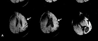
- Formation of neoplasms in the cortex and subcortex of the brain. The neoplasms are hamartomas and are detected by CT and MRI. In some cases, malignancy occurs, which is the cause of 25° cases of early death in tuberous sclerosis.
Ophthalmological manifestations
The most characteristic manifestations of tuberous sclerosis on the part of the organ of vision are retinal hamartomas and other damage to the posterior segment of the eye.
Phacoma (retinal hamartoma)
It occurs in approximately 50% of patients and is bilateral in 50% of cases. It may appear as a flat, smooth, and translucent mass, or it may have the appearance of a raised, calcified “mulberry.” Combined forms are also observed, combining features of both types. These damages. as a rule, they are not prone to pronounced growth (Fig. 24.7).
Vascular changes in the retina
Accompany tumors. Vascular aneurysms and malformations are characteristic, which can cause hemorrhages into the vitreous body (Fig. 24.8).
Pathology of the pigment epithelium
Changes in the pigment epithelium are described that occur in the middle periphery of the retina and create a picture of a “perforated” surface due to the appearance of zones of depigmentation (Fig. 24.9).
Papilledema
Papilledema or optic atrophy may occur. Occasionally, oblique position of the optic disc and coloboma occur.
Extraretinal disorders
Other features of the state of the visual organ in tuberous sclerosis have been described, including:
- megalocornea;
- keratoconus;
- posterior embryotoxon;
- glaucoma;
- cataract.
Neurofibromatosis
Neurofibromatosis is characterized by the appearance of neurofibromas under or on the skin - tumors that can reach a meter in diameter.
The formations affect nerve tissue and cause dangerous consequences, such as:
- breathing problems;
- swallowing disorder;
- blurred vision;
- disruption of the functioning of vital organs.
Mental disabilities are also present.
Symptoms of fifth phakomatosis are most often observed in children aged about ten years. Small tumors appear, which over time develop into malignant neoplasms accompanied by ulcers.
Encephalotrigeminal angiomatosis is characterized by the presence of vascular tumors on the back of the head and face of a person. On the face, tumors appear as burgundy-colored spots located on one side of the face. From birth, convulsions on the opposite side of the face, as well as epileptic seizures, are noted.
Ito's hypomelanosis is often accompanied by dementia, baldness, and light spots on the skin that resemble marble stains.
Symptoms of Ito hypomelanosis.
Severe immune disorders, vasodilation and cerebellar pathology are characteristic of Louis-Bar syndrome. About 15% of people who have a history of such phakomatoses often suffer from various forms of cancer.
Klippel syndrome can be diagnosed by the following signs:
- the presence of flat tumors on the leg;
- the presence of a flaming nevus on the lower limb;
- aneurysms and vascular dilations appear at the site of the lesion;
- the limb becomes enlarged and lengthened.
Bourneville disease can also be called tuberous sclerosis. Characterized by pronounced signs of dementia, it is the most severe form of phacomatous disease. The presence of adenoma of the sebaceous glands is noted.
Sturge-Weber syndrome is manifested by a delay in human intellectual development and the appearance of characteristic pigment spots in the face.
Albinism as a form of phakomatosis is caused by a defective gene, due to which melanin is not observed in the body. It is characterized by the presence of pale white skin, white eyelashes, hair and eyebrows.
Symptoms of albinism.
Neurofibromatosis type 2 is an extremely rare type of phakomatosis. It is characterized by an asymptomatic course, accompanied by the appearance of slowly growing tumors.
Symptoms of the disease
Phakomatoses of any type have fairly similar clinical symptoms, which are expressed in the presence of a characteristic rash and defects in the central nervous system and internal organs of the patient.
The clinical picture is as follows:
- convulsive epileptic seizures, starting in childhood;
- antisocial behavior;
- visual impairment;
- deterioration of hearing function;
- impaired coordination of movements;
- insomnia at night, drowsiness during the day.
Premature aging is periodically observed in people diagnosed with phakomatosis.
Diagnostics and therapy
Any suspicion of phakomatosis requires consultation with specialists from various branches of medicine - pediatrics, genetics, neurology, endocrinology, psychiatry, dermatology, ophthalmology.
Examinations include genealogical and DNA analysis, biochemistry of blood and urine, a wide range of instrumental and neuropsychological studies - EEG, echography of the brain and internal organs, MRI, angiography, x-ray studies of the kidneys, stomach and intestines. If necessary, other types of examinations are prescribed to clarify the diagnosis.
There is no specific treatment for phakomatoses. Only symptomatic therapy is carried out according to indications for each patient with a specific form of the disease. The following groups of drugs are prescribed:
- anticonvulsants (Carbamazepine, Topalepsin, Epitropil and others);
- diuretics (for example, Diacarb);
- neurometabolic (vitamins B12, B6, Nootropil, Encephabol, Aminalon).
If conservative therapy is ineffective, tumors are removed surgically, which is especially indicated if the tumor is suspected of being malignant, with a sharp progression of clinical symptoms, or the development of brain compression syndrome.
Along with medications, psychocorrective measures aimed at restoring or developing the child’s impaired intellectual abilities, relieving identified mental disorders, training and social adaptation play an important role in the treatment of phakomatosis. Comprehensive psychological assistance to families is practiced.
Diagnostics
The following specialists can diagnose phakomatoses:
- neurologist;
- pediatrician;
- ophthalmologist;
- gastroenterologist;
- nephrologist;
- cardiologist;
- geneticist;
- endocrinologist
The clinician must:
- study the clinical picture of the patient;
- conduct a thorough examination for symptoms of phakomatosis;
- study the patient's complaints.
In almost every case, the doctor prescribes the following tests:
- electroencephalography;
- magnetic resonance therapy;
- CT scan;
- angiography;
- echocardiography;
- blood chemistry;
- Analysis of urine;
- genetic testing;
- ophthalmoscopy;
- ultrasonography;
- electrocardiogram.
Reverse ophthalmoscopy
Phakomatoses have different symptoms at different ages, which significantly complicates the diagnosis of the disease, however, after passing all the necessary examinations, the doctor will be able to confirm or deny the presence of pathology in the patient.
Treatment of phakomatosis
Treatment of phakomatosis is divided into:
- medicinal;
- surgical;
- psychocorrective.
Drug treatment is aimed at removing visible symptoms.
Various drugs are used:
- anticonvulsants (Levetiracetam, Carbamazepine);
- dehydrating (Acetazolamide);
- neurometabolic (group of vitamins B).
In the presence of epileptic seizures, taking neurometabolics that have a stimulating effect is not allowed.
Surgical treatment of this pathology is used to excise the malignant neoplasm. If the tumor is in the brain, then the operation is performed by neurosurgeons.
Psychocorrection occupies not the last position in the list of treatment methods for getting rid of phakomatoses. It helps the child adapt socially, as well as develop intellectual and mental abilities.
Phakomatoses have an unfavorable prognosis. The result of therapy depends on the form of the disease, its severity and the age of the patient. Injury or infectious disease can contribute to the development of complications.
If you think you have Phakomatosis
and symptoms characteristic of this disease, then doctors can help you: neurologist, pediatrician, ophthalmologist.
Source
Did you like the article? Share with friends on social networks:
Treatment
There is currently no special treatment for the group of phakomatous diseases. Treatment is mainly aimed at reducing the symptomatic manifestations of a particular form.
The following groups of drugs are prescribed:
- Anticonvulsants (Topalepsin);
- Diuretics, for example Diacarb.
- Nootropic drugs (Vitamins B6, B12, nootropil).
In the absence of a positive effect from conservative treatment, a surgical method of therapy is prescribed.
The purpose of this method is to remove a developing malignant tumor, with an increase in its clinical symptoms and its rapid growth.
In addition to drug treatment, mental correction of the patient is included in the treatment. This method aims to:
- Improving mental and mental indicators in the child;
- Correction of existing deviations;
Depending on the type and severity of mental disorders, the following measures are recommended:
- Classes with a psychologist;
- Game therapy;
- ABA therapy (behavior correction).
Psychological consultation of parents is also mandatory.