Find out more about other diseases starting with the letter “Y”: Juvenile parkinsonism; Juvenile absence epilepsy; Juvenile myoclonic epilepsy.
Not all diseases can be completely cured. We are talking, first of all, about those that arise due to genetic conditioning. It is especially sad when it comes to childhood diseases. But medicine does not stand still, and offers treatment options that allow you to maintain an acceptable quality of life for the baby. Such a rare disease as juvenile parkinsonism, although today cannot be completely cured, is successfully corrected by medical treatment.
What does it represent?
While “classic” Parkinson’s disease can appear for a variety of reasons, childhood parkinsonism always occurs due to failures in genetics. Scientists have found that it affects not only children, but also people under 25 years of age. Most often it attacks girls and young girls. If there are several cases in one family, the spread of the disease is considered sporadic.
The disease usually manifests itself at the end of the first decade of life, or at the beginning of the second. An earlier age of development of pathology is extremely rare (less than 1% of all cases).
Juvenile parkinsonism differs from the “adult” version of the disease in that even with a long illness, the patient retains reason and intellectual abilities. There is a complete absence of dementia or mental disorders.
There are cases of increased tendon reflex or other pyramidal symptoms.
Medical Internet conferences
Early-onset Parkinson's disease is usually called cases of primary parkinsonism that develops before the age of 45 years. Among the above patients, a subgroup with juvenile (juvenile) parkinsonism is considered separately. These include individuals with the onset of primary parkinsonism at the age of 20-25 years. Early parkinsonism is a fairly heterogeneous disease. Thanks to the development of molecular genetics, it has been established that genetic factors play a major role in the development of early onset Parkinson's disease.
Improving the quality of diagnosis, the use of modern neuroimaging methods to identify pathological changes in the basal ganglia has made it possible to identify the disease in the early stages, right up to the “pre-disease”, and to standardize the diagnosis of this disease.
A clinical case of juvenile (juvenile) parkinsonism with onset at the age of 17 years is presented.
Patient F, 32 years old, was admitted to the neurological department of the Municipal Clinical Hospital No. 9. At the time of admission, he complained of stiffness throughout the body, slowness of movements, gait disturbance, tremor of the hands, more pronounced on the right, and memory impairment.
From the anamnesis of the disease, it is known that he has considered himself sick since 1997, when he first began to notice stiffness of movements and trembling of the hands, more pronounced on the right side, and noticed that his gait was gradually changing: it was difficult to start moving, his hands were pressed to his body, his head was tilted forward . In 2001, he was hospitalized in the neurogenetic department of the Research Institute of Neurology in Moscow, where he was diagnosed with juvenile parkinsonism and was prescribed treatment with madopar 250 mg/day, cyclodol 1/2 tablet 3 times a day. Against this background, stiffness and slowness of movements decreased. However, after some time, involuntary movements appeared in the form of twitching of the shoulders. I took Pronoran 1 tablet. 3 times a day, without a positive effect. In August 2011, he was hospitalized in the neurological department of the Municipal Clinical Hospital No. 9 with a diagnosis of juvenile parkinsonism, predominantly rigid form, grade 3 severity according to Hoehn-Yahr, progressive course, with severe postural instability and walking impairment, on-off phenomenon with motor fluctuations and dyskinesias Treatment was adjusted in the form of adding Midantan 1.5 tablets twice a day, continuing to take Madopar 1/4 t 5 times a day, against which there was an improvement in the condition in the form of cessation of twitching in the shoulders. Upon discharge, it was recommended to continue taking the prescribed therapy.
Neurological status: clear, in contact. The patient is adequate, oriented in his own personality, consciousness and surrounding environment, place and time. The sense of smell is preserved. Pupils S=D, reaction to light is lively, palpebral fissures S=D, movement of the eyeballs in full. Convergence and accommodation are preserved. Horizontal medium-wide nystagmus when looking to the left. Sensitivity on the face is preserved, corneal and conjunctival reflexes are preserved. The face is relatively symmetrical. The hearing is preserved. Swallowing is difficult, mainly of solid food, the reflex from the soft palate and the back wall of the pharynx is preserved. Tongue in the midline No significant sensitivity disorders were identified. Active movements in full, muscle strength 5 points, muscle tone changed like a “gear wheel”, more pronounced in the right hand. Rest tremor of the hands, more on the right side, worsens with excitement. There is no muscle atrophy. Tendon and periosteal reflexes S=D are reduced. Abdominal S=D. There are no pathological reflexes. Hypokinesia in the form of slow gait, hypomimia, acheirokinesis. He is not stable in the Romberg position and performs coordination tests with uncertainty. Stiffness in gait. Mild cognitive impairment. There are no urinary disorders. No meningeal signs were detected.
While in the hospital, the patient continued taking Midantan 1.5 tablets 3 times a day, Madopara ½ tablet 5 times a day, and received antioxidant therapy (Mexidol).
During the treatment, improvements were noted in the form of a decrease in complaints of stiffness and tremors in the hands
Conclusion: It is necessary to take into account the fact that one of the features of juvenile parkinsonism is the early onset of dyskinesias associated with taking levodopa even in low doses. In this regard, special attention should be paid to the prescribed therapy and dosage adjustment of antiparkinsonian drugs taken.
Causes
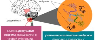
Juvenile parkinsonism is always genetically determined. There is a malfunction in the functioning of dopaminergic neurons, which should normally release dopamine. But due to a “breakdown” in the genes, this process becomes difficult or stops completely. This is the cause of external motor dysfunction. It should be taken into account that such a genetic error is inherited in an autosomal recessive manner.
It happens that both parents are healthy, but the damaged gene is present in their chain. That is, they transmit the possibility of illness to the born baby. In this case, in the future, symptoms of parkinsonism are found in no more than 25% of grown-up children. If there is a defect in the genome of only one parent, then the percentage of transmission of the disease to the offspring is practically reduced to zero.
Classification
Juvenile parkinsonism in most cases is accompanied by three manifestations: hypokinesia, tremor and muscle rigidity. Depending on how this triad is composed, the following types of disease can be distinguished:
- trembling;
- trembling-rigid;
- rigid-trembling;
- akinetic-rigid;
- mixed.
In the first case, there is a small amplitude trembling of the head, jaw and tongue. Tremor is also present in the limbs. The trembling does not stop either at rest or in motion. At the same time, due to stress or a small emotional outburst, the rate of trembling increases significantly. Normal motor activity is also maintained, and muscle tone is within normal limits.
In tremor-rigid parkinsonism, the disease manifests itself by the appearance of tremor. Over time, the picture is complemented by a disorder of muscle tone, and hypertonicity may occur. Bradykinesia is absent or not clearly expressed.
The rigid-tremorous “variant” of the disease always begins with a sharp increase in muscle tone in the upper and lower extremities. Only then is tremor observed. Due to hypertonicity, the limbs take a forced position (flexion), then hypokinesia is expressed.
With akinetic-rigid parkinsonism, hypokinesia with increased muscle tone is observed. There is no trembling, as well as involuntary movements.
If features of each of the listed types of disease are present simultaneously, we are talking about mixed juvenile parkinsonism.
Parkinson's disease (parkinsonism)
Definition.
Parkinson's disease (PD) is a chronic brain disease. The main manifestations (symptoms) of the disease are associated with a decrease in the number of nerve cells that produce the substance dopamine. Dopamine is involved in the transmission of nerve impulses to ensure normal motor activity. The lack of dopamine in the parts of the brain involved in the regulation of movements is the main cause of slowness and limitation in the execution of habitual movements.
Parkinson's disease refers to primary or idiopathic parkinsonism, which accounts for 80-90% of the total number of patients with parkinsonism syndrome. Secondary parkinsonism is much less common and accounts for 10-15%, for example: vascular, drug-induced, toxic, post-encephalitic, post-traumatic, parkinsonism due to tumors and hydrocephalus, against the background of metabolic encephalopathy. Parkinsonism within the framework of neurodegenerative diseases is 8-12% (atypical parkinsonism, “parkinsonism-plus” - progressive supranuclear palsy, multiple system atrophy, diffuse Lewy body disease, corticobasal degeneration).
Historical reference.
Parkinson's disease was described about 200 years ago (1817) by the English physician James Parkinson in his famous “Essay on the Shaking Palsy,” in which he summarized the results of observations of six patients. Since then, PD has been studied in detail, all its symptoms have been specified, but the portrait of the disease compiled by J. Parkinson remains accurate and capacious.
Epidemiology.
PD ranks fourth in prevalence after dementia, epilepsy, and cerebrovascular pathology in patients of the older age group. Thus, about 1-2% of people over 65 years of age suffer from PD. Depending on the age at which the first signs of PD are detected, the following forms are distinguished: juvenile form - onset before 20 years of age; early onset – before 40 years of age; typical onset is over 60 years of age.
Etiology.
The possibility of a genetic predisposition has been discussed for many years, but the genetic factor does not always play a major role, because familial forms of parkinsonism account for only about 5-10%, while sporadic parkinsonism occurs in 80-90% of cases. Genetic factors play the greatest importance at the early onset of the disease, and in a later age group other factors, such as involutional changes in the brain and environmental or toxic factors, become more important.
Pathogenesis.
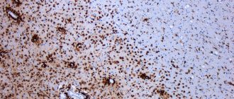
The pathogenetic basis of PD is a sharp decrease in the amount of dopamine in the substantia nigra and striatum. Dopamine synthesis occurs in the bodies of dopaminergic neurons located in the compact zone of the substantia nigra. Here it is formed in the form of small vesicles - granules, which are transported along the axons of nigrostial neurons to the axon endings in the caudate nucleus and accumulate in front of the presynaptic membrane. Under the influence of incoming nerve impulses, transmitter quanta are released into the synaptic cleft, which affects the dopaminergic receptors of the postsynaptic membrane and causes its depolarization. It is estimated that about 80% of dopamine is reabsorbed back into the presynaptic space or inactivated by the enzyme COMT (catechol O-methyltransferase) or MAO-B (monoamine oxidase type B). The most likely mechanisms of neuronal damage in PD are: oxidative stress; increased content of iron ions; specific enzymatic defects of complex I of the respiratory chain were identified in the mitochondria of the substantia nigra; induction of apoptosis processes due to the excitotoxic effect of glutamate and aspartate. But the real triggering factor for these processes has not been fully determined. In addition to dopaminergic neurons of the substantia nigra, other groups of neurons undergo degeneration in Parkinson's disease, including neurons of the dorsal nucleus of the vagus nerve, neurons of the olfactory bulb, noradrenergic neurons of the locus coeruleus, serotonergic neurons of the raphe nuclei, cholinergic neurons of Meynert's nucleus, as well as neurons of the cerebral cortex and some autonomic plexuses. Because of this, in addition to dopamine deficiency, dysfunction of the serotonergic, noradrenergic and cholinergic systems occurs. Disease manifestations such as anosmia, autonomic failure, depression, and dementia are associated with damage to extra-nigral structures.
Clinical manifestations.
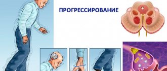
Parkinson's disease begins gradually and progresses slowly. It takes 10-15 years from the moment of the first nonspecific (premotor manifestations such as: constipation, sleep disturbances, loss of smell, depression, behavior disorder in the REM phase) to the main motor symptoms. The first motor symptoms of PD appear when more than 50% of the neurons of the substantia nigra die, and the decrease in dopamine levels is up to 80%.
| Premotor symptoms | Connection with brain structures | Braak stage |
| Constipation (constipation) | Dorsal vagal nucleus; neurons of the abdominal plexus. | 1 |
| Sleep disturbance or loss of smell | Olfactory bulb; anterior olfactory nucleus | 1 |
| Depression | Bluish area, suture nuclei. | 2 |
| Rapid eye movement disorder (REM) sleep behavior disorder | Dorsal midbrain and pons; bluish place. | 2 |
| Akinesia | Black substance. | 3 |
The main ones in the clinical picture are motor symptoms:
- Decreased motor activity (hypokinesia). The main manifestations of hypokinesia are difficulties in performing “small” movements (fastening buttons, tying shoelaces, cutting food, brushing teeth); shuffling gait, difficulty turning in bed, getting out of a chair; change in speech (decreased modulation and emotional coloring of the voice, which becomes inexpressive, monotonous, fading); change in handwriting - micrography (handwriting becomes small, letters are connected, their size decreases towards the end of the sentence); amimia – mask-like face, poor facial expressions; “Presiding pose” - the neck, torso and limbs at the joints are slightly bent, pressed to the body; propulsion, retropulsion, lateropulsion - irresistible acceleration of the patient’s movement forward, backward, to the side when walking or after a slight push, the inability to stop the movement started or change direction; acheirokinesis – absence of friendly arm movements when walking; paradoxical kinesia - in patients who can barely move with assistance and are bedridden - the possibility of rapid movements (running, jumping, waltzing, etc.) under the influence of emotional factors.
- Increased muscle tone (rigidity) – felt as “stiffness”, muscle tension (the “wax doll” phenomenon, the “cogwheel” phenomenon - occurs when a tremor is superimposed on rigidity)
- 3. Rest tremor (in 80% of patients) of the “rolling pills”, “counting coins” type - rhythmic trembling of the distal parts of the hands, especially the fingers, of low amplitude and frequency, stopping during purposeful movements and disappearing during sleep.
- Balance imbalance (postural instability)
PD is clinically manifested not only by motor disorders, but also has a whole range of non-motor manifestations that occur in all patients, regardless of the age of onset of the disease and the stage of the disease:
- Cognitive impairment
- Affective disorders (depression, anxiety, apathy, obsessive-compulsive syndrome)
- Behavioral disorders (addiction to gambling, compulsive shopping, hypersexuality, bulimia)
- Mental disorders (hallucinations, delusions, delirium)
- Sleep disorders (insomnia, hypersomnia, parasomnia)
Autonomic disorders in PD:
- Orthostatic hypotension
- Greasy scalp, increased formation of earwax.
- Sialorrhea
- Sweating
- Distal hyperhidrosis
- Urinary dysfunction
- Constipation
- Vagoinsular crises
Classification.
Parkinson's disease is classified according to the form, stage and rate of progression of the disease. Forms of the disease. Depending on the predominance of a particular symptom in the clinical picture, the following forms are distinguished: mixed (akinetic-rigid-tremor), akinetic-rigid and trembling. The mixed form is detected in 60-70% of cases of Parkinson's disease, akinetic-rigid - in 15-20% of cases of Parkinson's disease, trembling - in 5-10% of cases of Parkinson's disease. As the disease progresses, its shape may change.
Stages of the disease. The classification of the stages of Parkinson's disease proposed by Hoehn and Yarh (1967) is generally accepted: stage 1 - Hemiparkinsonism (unilateral manifestations) Stage 1.5 - unilateral manifestations involving the axial muscles. Stage 2 – Bilateral disorders without imbalance. Stage 2.5 – Bilateral disorders with initial manifestations of postural instability, but with independent restoration of balance during the push test. Stage 3 – Bilateral disorders with the addition of postural disorders. Stage 4 – Severe impairment, but can stand and walk without assistance. Stage 5 – complete disability. The patient is bedridden or wheelchair-bound.
Rate of progression.
There are three options for the rate of progression of Parkinson's disease (subject to adequate treatment):
- rapid rate of progression, in which a change in stages of the disease (first - second or second - third) occurs within 2 or less years;
- moderate rate of progression, in which the change of stages occurs in more than 2 years, but not more than 5 years;
- slow rate of progression with changes in stages after more than 5 years.
Diagnostics.
Currently, for the clinical diagnosis of Parkinson's disease, the brain bank criteria of the UK Parkinson's Disease Society are most often used (A. Hughes et al., 1992). Hypokinesia in combination with at least one of the following symptoms:
- muscle rigidity;
- resting tremor with a frequency of 4-6 Hz;
- postural instability.
Exclusion criteria for Parkinson's disease:
- history of repeated strokes with stepwise progression of parkinsonian symptoms;
- history of repeated traumatic brain injury;
- encephalitis in anemnesis;
- oculogyric crises;
- treatment with antipsychotics at the time of symptoms;
- family nature of the disease (more than 1 relative with a similar disease);
- the presence of long-term remission;
- strictly unilateral symptoms for more than 3 years;
- downward gaze paralysis;
- early rapidly progressing autonomic failure;
- cerebellar signs;
- early-onset dementia with memory, speech, and praxis impairments;
- Babinski's symptom;
- presence of cerebellar atrophy or communicating hydrocephalus on computed tomography;
- lack of response to high doses of levodopa (with the exception of malabsorption);
- contact with toxic substances that cause parkinsonism.
Criteria confirming the diagnosis of Parkinson's disease (at least 3):
- unilateral onset;
- rest tremor;
- progressive course;
- persistence of symptom asymmetry with predominance on the initially involved side;
- high effectiveness of levodopa drugs (reduction of symptoms by 70–100%);
- severe choreiform dyskinesias induced by levodopa;
- persistence of response to levodopa for 5 years or more;
- the duration of the disease is 10 years or more.
Differential diagnosis of Parkinson's disease is carried out with the following diseases: essential tremor, juvenile (juvenile) parkinsonism, drug-induced parkinsonism, vascular parkinsonism, progressive supranuclear palsy (Steele-Richardson-Olszewski syndrome), multiple system atrophy, diffuse Lewy disease, hepatolenticular degeneration.
Additional research methods.
- Structural neuroimaging - CT, MRI - are not very informative for diagnosing PD and are used to exclude secondary parkinsonism.
- Functional neuroimaging (fluorodopa PET, SPECT) the potential of these methods is high, because make it possible to detect changes in dopamine metabolism 4-6 years before the onset of clinical symptoms, but at the moment this is a technically complex and expensive research method, which does not allow its use in routine practice to identify PD.
- Transcranial sonography reveals increased hyperechogenicity of the substantia nigra, due to the increased content of ferric iron, which is detected in 80-90% of patients with PD.
- Olfactory tests (Pennsylvania test) Olfactory dysfunction is the earliest premotor symptom of PD. This test is most often used if tremor is predominant in the clinic, making a differential diagnosis between essential tremor and PD, because only idiopathic parkinsonism will be characterized by changes in olfactory tests.
Principles of treatment.
There are three main areas in the treatment of Parkinson's disease:
- Neuroprotective therapy that aims to slow or stop the degeneration of neurons in the brain. In recent years, several dozen agents have undergone experimental and clinical trials that are potentially capable of influencing various stages of the neurodegenerative cascade of cell death (antioxidants, glutamate antagonists, calcium channel blockers, anti-inflammatory drugs, trophic factors, etc.), but so far their effectiveness has not been proven managed.
- symptomatic therapy, which allows to reduce the main symptoms of the disease by correcting the neurochemical and neurophysiological imbalance that occurs in the brain;
- physical and socio-psychological rehabilitation.
Drug treatment.
Currently, treatment is carried out focusing mainly on the symptomatic effect of the drugs. Currently, antiparkinsonian drugs used for Parkinson's disease include 6 groups of drugs:
- drugs containing levodopa (a precursor of dopamine) - increase the synthesis of dopamine in the brain;
- dopamine receptor agonists – stimulation of dopamine-sensitive receptors;
- tricyclic antidepressants – inhibition of the process of reabsorption (capture) of dopamine by presynaptic structures;
- amantadine preparations – stimulation of the process of release (release) of dopamine from the presynaptic terminal;
- monoamine oxidase type B inhibitors;
- catechol-O-methyltransferase inhibitors – inhibition of dopamine catabolism (breakdown);
Since the mechanism of action of drugs in these groups is different, if necessary, they can be combined as part of combination therapy.
Drug therapy for patients with Parkinson's disease is selected individually for each clinical case and depends on a number of factors, including the severity of functional impairment, the form of the disease, the patient's age, individual sensitivity to drugs, and pharmacoeconomic considerations.
The goal of therapy is to restore impaired motor functions using minimal effective doses of drugs. During the first months of the disease, when manifestations of the disease are minimal and do not interfere with daily social or household activities, drug therapy may not be prescribed. At the beginning of treatment, monotherapy is usually used, and if its effectiveness decreases as the disease progresses, combination therapy is used.
Non-drug treatment methods.
Treatment of chronic diseases, along with the prescription of medications, requires the implementation of measures for socio-psychological support of patients. Educational programs should provide patients with selective information about the disease necessary to develop a sense of control over the disease. The treatment package should include neuropsychological training, speech therapy, and therapeutic exercises. Regular physical exercise, including aerobic exercise, isometric exercise, stretching and coordination, can improve the mobility of patients without reducing parkinsonian symptoms. Good nutrition is important to prevent weight loss and muscle loss. To implement the entire range of measures for the medical and social rehabilitation of patients, it is advisable to create a special service. It is necessary to prepare specially trained paramedics who would coordinate the work of social services, teach relatives how to care, and educate patients.
Neurosurgical treatment.
If conservative therapy is ineffective, especially when severe dyskinesias do not allow prescribing doses of levodopa necessary to correct the symptoms of Parkinson's disease, the question of surgical treatment is raised (pallidotomy, thalamotomy, implantation of intracerebral stimulators into the globus pallidus, thalamus, subthalamic nucleus, intracerebral transplantation of embryonic adrenal tissue or substantia nigra).
Symptomatic picture
Most often, the disease manifests itself clearly after 10 and before 25 years of age. Symptoms develop bilaterally, which distinguishes juvenile parkinsonism from known Parkinson's disease. A delay or other disruption in motor skills appears, and gesticulation worsens. Facial expressions may be weakened, and in general, the motor amplitude when performing everyday activities and movements decreases.
If previously the child could easily fasten a button or lace his shoes, then after the manifestation of the disease these actions cause great difficulty.
In general, the child becomes slower, movements become “poorer”, becoming more constrained.
The gait becomes “senile”: the patient shuffles, taking small steps. Synchronicity in the movement of arms and legs when walking disappears.
Then speech disorders arise. Some people's speech becomes angular, while others may experience a sharp slowdown in the rate at which they pronounce words.
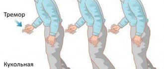
Any muscle tension causes trembling in the head and limbs. The tremor persists even at rest. Most affected children have muscle rigidity. Due to constant muscle tension (hypertonicity), arms and legs can bend, taking a static forced position.
Symptoms may become more or less severe during the day. So, after sleep, the symptoms practically disappear, and then in the evening they increase again. Dystonic symptoms may manifest themselves - most often in the feet.
Juvenile parkinsonism is characterized by preservation of intellectual abilities and normal functioning of the cognitive sphere. Mental development is carried out according to age norms.
Modern diagnostic measures
It is important to detect the disease in time, so at the first signs of pathology you should consult a doctor. Usually the diagnosis is made during the first examination by a neurologist. A competent doctor will immediately note bradykinesia, increased muscle tone and tremor. The tendon reflex will then be tested. In childhood parkinsonism it is also significantly increased. The child must be placed in a special position to check the so-called pyramidal signs (Babinsky reflexes).
Next, electrophysiological examinations are carried out:
- EEG;
- electromyography.
Based on the results of these procedures, this diagnosis is confirmed. Usually the picture is expressed by a decrease in the bioelectrical activity of the cerebral hemispheres. This suppression is especially pronounced in akinetic-rigid juvenile parkinsonism.
An MRI is also required. But, as a rule, with such a disease, no serious changes are visible. Such an examination is prescribed in order to exclude some “counter” diseases of the central nervous system. Only late changes can be recorded on tomography:
- on the caudal nucleus;
- on the globus pallidus;
- black substance.
These changes are already considered basal and manifest themselves at critical stages of the disease.
To find out how dopamine synthesis proceeds and how difficult it is, the patient is referred to a functional neuroimaging procedure. To do this, a special substance is injected, which should normally concentrate in a special area of the brain.
Additionally carried out:
- PET-CT brain;
- SPECT;
- MR spectroscopy.
Since the disease occurs for genetic reasons, the child is sent for examination to a geneticist. The specialist identifies the specific gene in which there is a malfunction, and also finds out the percentage of its heritability in the future.
The doctor also needs to distinguish juvenile parkinsonism from that of the secondary type. It occurs as a result of drug addiction or a malfunction in the endocrine system.
Elena Anatolyevna Efet: “The opinion that Parkinson’s disease threatens only older people is wrong.”
On April 11, 1755, James Parkinson, an English doctor who first described the “shaking palsy,” which is now called Parkinson’s disease, was born.
At the initiative of the World Health Organization, Parkinson's Disease Day is held annually to mark the birthday of the outstanding doctor. The head of the psychoneurological department of St. Petersburg State Pediatric Medical University, neurologist, candidate of medical sciences, spoke about what parkinsonism is and whether children and adolescents suffer from it. Elena Anatolyevna Efet.
Elena Anatolyevna, please tell us what the disease is and what Parkinson’s syndrome is?
Parkinson's disease is a chronic, progressive disease of the central nervous system primarily associated with the degeneration of dopamine-producing neurons. The interaction of unfavorable genetic and environmental factors plays an important role in the etiology of the disease. Among environmental factors, the greatest importance is given to the population's exposure to potential neurotoxins (pesticides, herbicides); there is also evidence of an increased risk of Parkinson's disease due to contact with heavy metals.
Parkinsonism syndrome or symptomatic parkinsonism develops as one of the clinical manifestations (complications) of a number of independent diseases and lesions of the central nervous system. The most well-known variants of secondary parkinsonism are vascular, toxic (including medicinal), post-traumatic, post-infectious.
What structures and cells of the brain are affected by this disease?
This disease causes the death of neurons in the substantia nigra of the brain, which belongs to the estrapyramidal system. This system is responsible for correcting muscle tone, coordination and accuracy of the movement performed.
What is the mechanism of disease development?
The development of Parkinson's disease is based on the progressive death of dopamine-containing neurons in a number of structures of the brain stem (dopamine is a chemical substance involved in the transmission of nerve impulses that control movement and coordination function), which is accompanied by a sharp decrease in dopamine content and chronic dysfunction of dopaminergic pathways of the central nervous system. As the disease progresses, the amount of dopamine produced decreases, and the patient loses the ability to control his own movements.
Pathologically, normal aging is accompanied by a decrease in the number of neurons in the substantia nigra of the brain and the presence of Lewy bodies (pathological aggregates of the α-synuclein protein) in them, as well as a decrease in dopamine content and a decrease in the number of dopamine receptors. It has been proven that the rate of neuronal death in Parkinson's disease is several times higher than in normal aging.
The key process leading to the death of dopaminergic neurons in Parkinson's disease is the accumulation of α-synuclein protein in them and the formation of aggregates and Lewy bodies from it. These bodies are found not only in the middle part of the brain, but also in its trunk, as well as in the olfactory bulbs. Despite the fact that these areas of the brain are not motor areas, they are also important because they regulate the sense of smell and the process of sleep. The presence of Lewy bodies in these areas of the brain may well explain the non-motor symptoms that accompany some Parkinson's patients. The intestines also contain dopamine cells that degenerate and lead to disturbances in the gastrointestinal tract characteristic of this disease.
Cell death presumably occurs due to the activation of a genetically programmed mechanism (apoptosis). The patient begins to feel the first symptoms of the disease only when about 70% of these cells have already died.
Is Parkinson's syndrome only a disease of older people? Does it occur in children and adolescents?
The idea that Parkinson's disease affects only older people is wrong. There are early and juvenile forms of Parkinson's disease. Early appears after 20 years, juvenile - before 20 years. According to statistics, every tenth patient is diagnosed with the disease before the age of 40. Among the causes of Parkinson's disease at a young age, hereditary factors predominate, followed by trauma, intoxication, viral and bacterial infections, and metabolic disorders.
Is this a hereditary disease?
Despite progress made in recent years in understanding the biochemical and molecular mechanisms of Parkinson's disease, the true etiology of sporadic forms of this disease remains unknown. The share of hereditary forms accounts for no more than 10–15%. The interaction of genetic predisposition and environmental factors triggers the death of brainstem neurons. Once it occurs, this degenerative process becomes irreversible and begins to spread expansively throughout the brain.
How often do children and adolescents get juvenile parkinsonism?
The disease is very rare among these groups of patients (less than 1% of all forms of parkinsonism).
What symptoms should parents be wary of?
The earliest sign of juvenile Parkinsonism is considered to be bradykinesia—slowing and decreasing motor activity, decreased gesticulation, and small range of movements. In children it often appears symmetrically. There may be a change in speech in the form of slowing down. At the initial stage, difficulties arise with the rapid execution of motor acts that require the participation of fine motor skills. The child has difficulty fastening buttons, tying shoelaces, and cannot assemble a mosaic. Diagnosing the disorder is simple: just try quickly tapping your fingers on any surface.
The second sign is the unnatural position of the lower limbs while walking. Outwardly, it looks like half-bent, twisted legs, often feet, and stiffness of movement is noted.
The third symptom is tremor-shaking that occurs in any part of the body. Tremor in children can even affect the vocal cords, which is expressed in changes in the timbre of the voice, lower and upper extremities. In the early stages, trembling occurs at rest; as the pathology progresses, it increases and manifests itself during movement.
Characterized by a sharp deterioration of symptoms in the evening, improvement in the morning or after a nap.
Does the clinical picture differ between children and the elderly?
In children, there is usually no clear staging of clinical manifestations than in adults. The disease occurs without dementia (at least clinically in children it is difficult to confirm the fact of dementia). Akinetic-rigid syndrome is often associated with juvenile parkinsonism, Sekawa syndrome. Several cases of the development of parkinsonism syndrome after tick-borne encephalitis in the form of its meningoencephalitic form - postencephalic parkinsonism - have been described. The bulk of childhood parkinsonism is reliably described as genetic (damaged genes encoding the receptor apparatus of dopaminergic receptors, or genes encoding the synthesis of dopamine itself). The picture of akinetic-rigid syndrome itself is similar to that in older people.
How are children with juvenile parkinsonism treated?
Juvenile parkinsonism responds well even to small doses of dopamine. To delay taking more serious medications in the future, the child’s dose of the vital hormone is constantly increased.
The main place in the treatment of juvenile parkinsonism is given to drug therapy. The following groups of drugs are prescribed:
· ADR (dopamine receptor agonists);
Dioxyphenylalanine preparations;
· central group anticholinergics;
· metabolites.
The classic treatment option always begins with dopamine receptor agonists. If necessary, they can be supplemented with other medications. If significant movement disorder persists, small doses of dihydroxyphenylalanine may be necessary.
Central anticholinergics are used to reduce tremor. Very young patients are prescribed minimal doses of these drugs. But therapy should continue continuously for at least 3–5 years.
Metabolic drugs include B vitamins, coenzyme Q10, L-carnitine, magnesium and aminophenylbutyric acid. The course of taking such drugs must be repeated 3 times a year. They are designed to support the functioning of the nervous system as a whole, as well as help maintain healthy muscle tone.
New promising methods are also used in the treatment of juvenile parkinsonism. For example, electrical stimulation of the basal ganglia.
The possibility of introducing dioxyphenylalanine into the body through a special duodenal pump is also currently being studied.
Parkinsonism in old age is an incurable disease, is this true in the case of juvenile parkinsonism in children and adolescents?
Compared to typical Parkinson's disease, the juvenile variant has a more benign course with slow progression. A good response to therapy, the absence of pronounced atrophic changes, and cognitive disorders allow patients to develop normally and maintain mobility for a long time. Severe disability can be observed several decades after the clinical debut.
Publication date: 04/11/2020